Contents
Download PDF
pdf Download XML
703 Views
386 Downloads
Share this article
Research Article | Volume 4 Issue 2 (July-Dec, 2023)
Nippel Discharge as Presentation of Underlingbreast Diseases
 ,
 ,
1
M.B.Ch.B, Mc.S in General Surgery at Baqubah Teaching Hospital, Diyala Governorate, 32001 Iraq
2
M.B.Ch.B, F.I.B.M.S in General Surgery at Baqubah Teaching Hospital, Diyala Governorate, 32001 Iraq
Under a Creative Commons license
Open Access
Published
Sept. 10, 2023
Abstract

The nipple discharge is the release of any fluid from the nipple of breast and represent a common symptomatic problem that causes many women discomfort and anxiety and may be a first complain of a patient. Nipple discharge can be the earliest presenting symptom of malignant breast diseases. Patients & methods A prospective study of (86) patients at Baqubah teaching hospital from 1st October 2013 till 31st March 2014. Only females included in the study. The chief complain of all patient included in our study was nipple discharge. Clinical examination, mammography, ultrasound, and cytology was done for all patients. Fine Needle Aspiration Cytology (FNAC), and definitive histological examination was done for patients presented with nipple discharge associated with breast lump. Result: The common age group of women complaining from nipple discharge was from (30-39) 32.5% , and the most common type of nipple discharge were bloody nipple discharge (40.8%) and, abnormal nipple discharge are more common with benign than malignant breast diseases. The direct cytology from nipple discharge had a good efficacy in diagnosis of malignant breast diseases (79.1%). The best modality for diagnosis of malignant breast diseases was the histopathology (100%) and the other diagnostic tests were the FNAC(78.5%). Conclusions: Any type of nipple discharge mainly bloody and purulent should not be neglected because it may be the presentation of benign or malignant breast diseases. Direct cytology from nipple discharge have a good value in diagnosis of malignant breast diseases. The histopathology has the difentive diagnosis of malignant breast diseases when there is associated mass. Aim of The Study: The present study was constructed to: 1.Determine the importance of nipple discharge as a presenting symptom of breast diseases. 2. Determine the most common type of nipple discharge that related to breast diseases and age. 3. Know the relationship between nipple discharge and presence of breast mass in breast diseases. 4. Know the relationship between nipple discharge and malignant breast diseases.

Keywords
Important Note

Key findings:

The study at Baqubah Teaching Hospital examined 86 female patients with nipple discharge, finding the most common age group to be 30-39 years. Bloody discharge was prevalent (40.8%), and direct cytology was effective in diagnosing malignancies (79.1%). The study underscores the importance of investigating all types of nipple discharge for potential breast diseases, especially malignancies.


What is known and what is new?

What is known: Nipple discharge is a common symptom that can cause discomfort and anxiety in women and may be the first sign of breast disease, including malignancies. New is in this study highlights the prevalence of nipple discharge, especially bloody discharge, among women aged 30-39. It also emphasizes the importance of direct cytology in diagnosing malignant breast diseases in patients with nipple discharge.


What is the implication, and what should change now?

Implication is the study suggests that any type of nipple discharge, particularly bloody or purulent, should not be ignored as it may indicate benign or malignant breast diseases. Healthcare providers should consider nipple discharge, especially bloody or purulent, as a potential indicator of breast disease and perform thorough evaluations, including direct cytology, to ensure early detection and appropriate management.

Introduction

Nipple discharge

It is the release of any fluid from the nipple of breast, and it is a common presenting symptom among women. [1]


The reported incidence of discharge from the nipple as a symptom of breast disease of all types varies from 5 to 10 per cent. Beasley stated that dis- charge can be milked from at least 90 per cent of apparently normal breasts, while Jackson and associates were able to obtain secretions from43 percent of patients having some type of complaint referable to the breast. [2]


Age. The average ages of patients reported in most series were in the forties. River found the average age of 52 patients with discharge from the nipple to be 40.2 years, with range from 17 to 78 years. In the group having bloody discharge who were studied by Fits, Maxwell and Horn. [3]


The average age of those with benign disease was 42years, and of those with carcinoma 54 years. In 59 cases of intraductal papilloma, Chester and Bell found an average ageof 49.4 years with range from 29 to 80 years. [4].


The breast is a group of large glands derived from the epidermis. It lies in a network of fascia derived from the dermis and the superficial fascia of the ventral surface of the thorax. The nipple itself is a local proliferation of the stratum spinosum of the epidermis. [5]


The mature breast of the female extends from the level of the second or third rib to inframammary fold at approximately the sixth or seventh rib. Transversely, it extends from the lateral border of sternum to the anterior axillary or midaxillary line. The axillary tail (of spence) extends superolaterally into the anterior axillary fold. The upper half of the breast, and particularly the upper outer quadrant, contains a greater volume of glandular tissue than do other sectors. [6]


The resting breast is defined as a postpubertal breast in an inactive state, not stimulated to become secretory by adequate hormone levels. The lactiferous sinus constitutes a lobe, with numerous lobules per lobe. It develops as a downgrowth of surface epithelium. Growth continues through the dermal papillary layer and into the reticular layer as the duct system is formed. [7]


Milk ejection occurs only during nursing. The sucking stimulation of the breast sends neural impulses to the hypothalamus. The hypothalamus stimulates oxytocin secretion by paraventricular nuclei cells, and suppresses the release of prolactin-inhibiting hormone (PIH). Oxytocin stimulates myoepithelial cells of secretory alveoli of the breast, effecting milk passage through the lactiferous ducts. Prolactin secretion is maintained when the PIH is suppressed and lactation continues. [8]


The diagnosis of ND begins with its characterization as either a physiological or pathological condition. Physiological discharge, often a manifestation of breast manipulation, is usually bilateral, is white or green, and emanates from many ducts [9].


A pathological discharge is generally unilateral, spontaneous, persistent, clear, watery, serous or bloody in appearance, and emanates from a single duct [10].


The nipple, which is well innervated by the 4th intercostal nerve, is surrounded by the areola. The pigmentation of the areola increases with pregnancy. Within the nipple and areola are some dermal papillae, and a very thin epithelium. [11]


Approximately 20 ducts open onto the surface of each nipple. The main ducts are called the lactiferous ducts. Stratified squamous keratinizing cells make up the epithelium of the ducts near their surface. Deeper in the ducts, the epithelium becomes a double layer of columnar cells. Smooth muscle is embedded within the dense connective tissue which supports the parenchymatous tissue. [8].


Some muscle is oriented circularly around the ducts; other muscle is longitudinally parallel with the ducts. Lactiferous sinuses are the dilated terminal portions of the lactiferous ducts, capable of storing milk during milk ejection. [8].


Due to loss of the placenta at birth, nipples can suffer from estrogen deficiency for a transient period until the ovaries increase hormone production. [12].


Dermis tissue surrounding the duct system is more cellular than in typical dermis and becomes the intralobular connective tissue. Less cellular dermis forms a coarser tissue which separates the lobules; it is thus interlobular connective tissue, with regions of adipose tissue contained within it. Some of the larger of these interlobular support structures form the suspensory ligaments (of Cooper). [13].


Intralobular ducts are lined with cuboidal epithelium: narrower ones have a simple cuboidal epithelium, larger ones a double layer of cuboidal epithelium. [14].


With pregnancy, the duct system completes its development. Many intralobular ducts form extensively during the proliferative phase. Secretory alveoli form at the tip of the smallest intralobular ducts and complete the lobules. Each secretory alveolus forms a small cul de sac (secretory lobule) lined with columnar epithelium. Myoepithelial cells are related to the basal side of each secretory alveolus. [8].


The secretory phase is brought on by changed levels of progesterone, lactogenic hormones (maternal and placental), and estrogen. Colostrum is secreted during the third trimester, milk shortly after birth. Not all lobules secrete at the same level. Therefore, under the microscope some lobules are distended, others are not. [12].


Interlobular partitions of connective tissue become considerably thinned as the secretory portions of the breast become more active and enlarged. Secretory cells produce numerous lipid droplets of widely varying sizes. [15]. While intracellular, lipid droplets are not surrounded by a membrane, but during exocytosis, they become enwrapped by cell membrane. Electron-dense secretory granules containing milk proteins also are found in secretory cells. These, too, undergo exocytosis. Columnar cells with sparse populations of microvilli are another component of secretory lobules. [15].


Nipple discharge

Most cases of ND are physiological in nature and are multi-ductal in origin. In these instances and particularly when associated with a creamy yellow, white or green coloured discharge, further investigations are rarely required.The term pathological nipple discharge (PND) has been introduced to describe a bloody, serous or clear discharge which is spontaneous,unilateral, uniductal and persistent. [16].


Discharge can occur from one or more lactiferous ducts.Management depends on the presence of a lump (which should always be given priority in diagnosis and treatment) and the presence of blood in the discharge or discharge from a single duct. Mammography is rarely useful except to exclude an underlying impalpable mass. Cytology may reveal malignant cells but a negative result does not exclude a carcinoma or in situ disease. [17].


  • A clear, serous discharge may be ‘physiological’ in a parous woman or may be associated with a duct papilloma or mammary dysplasia. Multiduct, multicoloured discharge is physiological and the patient may be reassured. [17].

  •  

  • A blood-stained discharge may be caused by duct ectasia,a duct papilloma or carcinoma. A duct papilloma is usually single and situated in one of the larger lactiferous ducts; it is sometimes associated with a cystic swelling beneath theareola. [17].

  •  

  • A black or green discharge is usually the result of duct ectasia and its complications [17].


The important characteristics of the discharge and some other factors to be evaluated by history and physical examination are as follows:


Nature of the discharge (serous, bloody, or other), association with a mass, unilateral or bilateral, single or multiple duct discharge, discharge is spontaneous (persistent or intermittent) or must be expressed, discharge is produced by pressure at a single site or by general pressure on the breast, relation to menses, premenopausal or postmenopausal, and patient is taking contraceptive pills or estrogen. [8].


Spontaneous, unilateral, serous, or serosanguineous discharge from a single duct is usually caused by an intraductal papilloma or, rarely, by an intraductal cancer. A mass may not be palpable. The involved duct may be identified by pressure at different sites around the nipple at the margin of the areola. Bloody discharge is suggestive of cancer but is more often caused by a benign papilloma in the duct. [18].


Cytologic examination may identify malignant cells, but negative findings do not rule out cancer, which is more likely in women over age 50 years. In any case, the involved duct—and a mass if present—should be excised. A ductogram (a mammogram of a duct after radiopaque dye has been injected) is of limited value, since excision of the suspicious ductal system is indicated regardless of findings. Ductoscopy, evaluation of the ductal system with a small scope inserted through the nipple, has been attempted but is not effective management. [18].


In premenopausal women, spontaneous multiple duct discharge, unilateral or bilateral, most noticeable just before menstruation, is often due to fibrocystic condition. Discharge may be green or brownish. Papillomatosis and ductal ectasia are usually detected only by biopsy. If a mass is present, it should be removed. [19].


A milky discharge from multiple ducts in the nonlactating breast may occur from hyperprolactinemia. Serum prolactin levels should be obtained to search for a pituitary tumor. Thyroid-stimulating hormone (TSH) helps exclude causative hypothyroidism. Numerous antipsychotic drugs and other drugs may also cause a milky discharge that ceases on discontinuance of the medication. [19].


Oral contraceptive agents or estrogen replacement therapy may cause clear, serous, or milky discharge from a single duct, but multiple duct discharge is more common. In the premenopausal woman, the discharge is more evident just before menstruation and disappears on stopping the medication. If it does not stop, is from a single duct, and is copious, exploration should be performed, since this may be a sign of cancer. [12].


Hormonal shifts from sexual stimulation, pituitary tumors, and the use of certain medications like birth control pills, blood pressure medications, major tranquilizers, antidepressants, or ulcer medications can cause an imbalance in the hormone prolactin which stimulates milk production.


Breast Infection: A spontaneous pus-like discharge from the nipple due to a mastitis (breast infection) or breast abscess and may require an antibiotic to treat. [12].


Duct ectasia: Multi-colored discharge from one nipple that occurs most often in a milk duct that is clogged and swollen. Although it usually clears up by itself, it should be monitored. [12].


Intraductal papilloma: This is a small benign (not cancer) wart-like growth in a duct lining near the nipple, which may become irritated and bleed, producing a pinkish, brown or bloody discharge. It is the most common cause of bloody nipple discharge and should be removed. [12].


History of breastfeeding: Galactorrhea is a term used to describe a milky discharge from both breasts. It occurs most often in non-breastfeeding women after pregnancy and can last a year or two. [12].


Stimulation: Squeezing or expressing the breast or nipple can produce a nipple discharge.

Fibrocystic breast changes: Cysts (sacs filled with fluid) and changes in the fibrous tissue of the breast can cause nipple discharge that is often greenish. [12].


Breast cancer: This can very rarely cause a bloody or clear nipple discharge. [12].


Workup

The standard workup for patients presenting with nipple discharge includes a thorough history and physical examination in addition to a complete breast imaging evaluation. Imaging studies allow potential localization and characterization of the lesion in question, with the option of percutaneous image-guided biopsy to achieve a tissue diagnosis. [20]. Typically, mammography and ultrasound are used to identify mass lesions responsible for nipple discharge. Additionally, magnetic resonance imaging (MRI) may be useful in the workup of pathologic nipple discharge when lesions cannot be localized with mammography or ultrasound. [20]. While MRI has been preliminarily studied for this indication at a few centers, it is not generally part of the workup. When nipple discharge is determined to be of benign etiology, duct excision may also be indicated to eliminate discharge when bothersome to the patient. [21].


In the absence of a mass or other lesion identified with breast imaging, the conventional surgical approach has been to perform major duct or lacrimal probe-guided excision. However, a histopathologic etiology is not always found on major duct or lacrimal probe-guided excision, which raises the possibility that the causative lesion might have been left in situ. [21]. Diagnostic ductography allows preoperative determination of the number, location, and extent of any underlying lesions. Preoperative use of ductography with methylene blue injection to localize lesions has been shown to increase the likelihood that a specific pathologic lesion will be found at surgery. [22] Ultrasound imaging uses high frequency sound waves to produce a picture of the internal structures of the breast. A small handheld probe is pressed gently against the skin surface. It both generates inaudible sound waves and detects any echoes reflected back off the surfaces and tissue boundaries within the breast. [23] From these reflected sound waves the computer generates a real time picture which is displayed on the monitor. The probe is moved across the skin to view the breast from different angles. Breast ultrasound is used to help clarify breast abnormalities felt by a doctor during a physical examination and to characterize potential abnormalities identified on mammography. [24]. Ultrasound is particularly good at determining if a lump is solid (which may be a non-cancerous lump or a cancerous tumour) or fluid-filled (such as a benign cyst). Ultrasound is also used to guide aspiration or biopsy of a lump.


[24]. Mammography is a specific type of imaging that uses a low-dose x-ray system to examine breasts. A mammography exam, called a mammogram, is used to aid in the early detection and diagnosis of breast diseases in women. [25].


An x-ray (radiograph) is a noninvasive medical test that helps physicians diagnose and treat medical conditions. Imaging with x-rays involves exposing a part of the body to a small dose of ionizing radiation to produce pictures of the inside of the body. X-rays are the oldest and most frequently used form of medical imaging.Three recent advances in mammography include digital mammography,computer-aided detection and breast tomosynthesis. [26]. Digital mammography, also called full-field digital mammography (FFDM), is a mammography system in which the x-ray film is replaced by solid-state detectors that convert x-rays into electrical signals. These detectors are similar to those found in digital cameras. The electrical signals are used to produce images of the breast that can be seen on a computer screen or printed on special film similar to conventional mammograms. From the patient's point of view, having a digital mammogram is essentially the same as having a conventional film mammogram. [25]. Computer-aided detection (CAD) systems use a digitized mammographic image that can be obtained from either a conventional film mammogram or a digitally acquired mammogram. The computer softwa3re then searches for abnormal areas of density, mass, or calcification that may indicate the presence of cancer. The CAD system highlights these areas on the images, alerting the radiologist to the need for further analysis. [25]. Breast tomosynthesis, also called three-dimensional (3-D) breast imaging, is a mammography system. [27].


Where the x-ray tube moves in an arc over the breast during the exposure. It creates a series of thin slices through the breast that allow for improved detection of cancer and fewer patients recalled for additional imaging. [27]. Fine-needle aspiration (FNA) of the breast is an established diagnostic method in the evaluation of palpable breast masses. [28]. Its reliability has also been demonstrated in the diagnosis of radiologically imaged, nonpalpable lesions. [29]. Fine needle aspiration (FNA) cytology has been widely used for the diagnosis of palpable breast masses. [30]. The technique involves the insertion of a fine needle (between 21 and 25 gauge) into a lesion and the extraction of a small sample of cellular material which is smeared onto glass slides. [31]. The cells are stained and examined morphologically by cytopathologists. Fine needle aspiration is simple, fast, and can be performed as an office procedure, since it requires no special equipment, causes minimal morbidity, and has high patient acceptance. [32].

Patients and Methods

 Samples

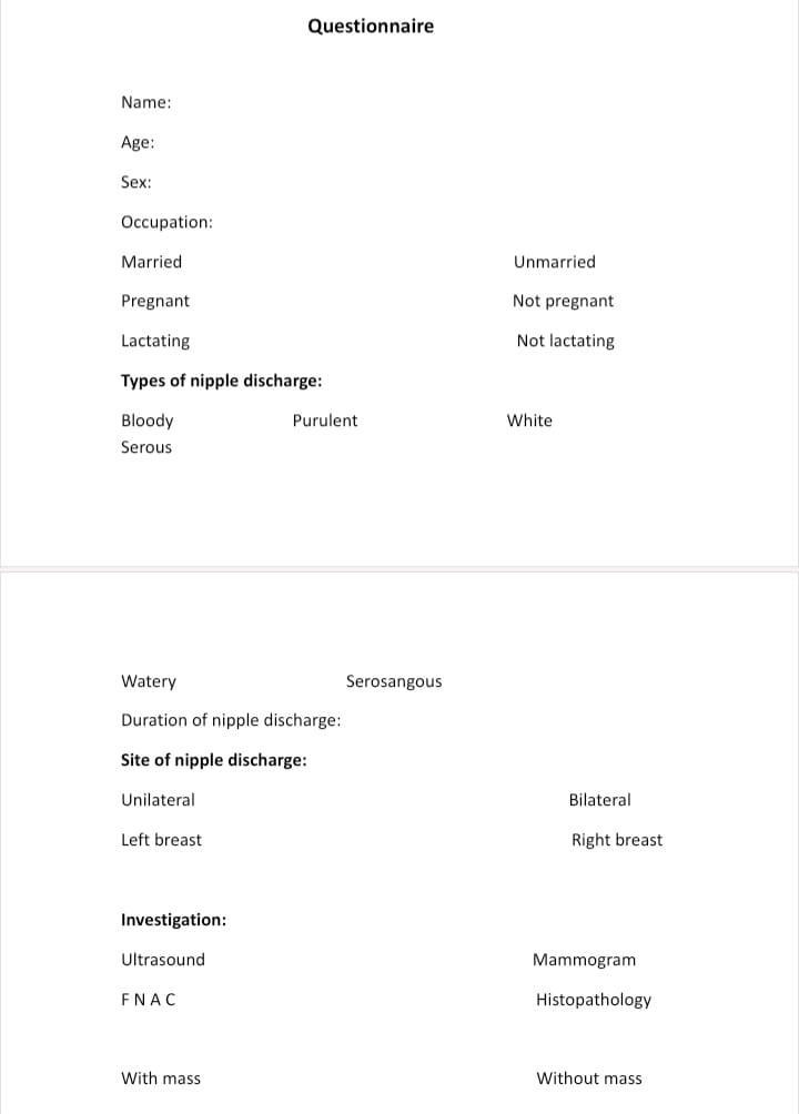
A prospective study was done for ( 86) patients at Baquba teaching hospital during the period from 1st October 2013 till 31st March 2014 .All patients were selected from breast clinic of Baquba teaching hospital . All femal patients were presented with nipple discharge involved in our study .Clinical examination, mammography,ultrasound, and cytology was done for all patients.Fine needl aspiration cytology (FNAC), and definitive histological examination was done for patients presented with nipple discharge associated with breast lumps.


Clinical evaluation:

History Detailed history including age of patient, duration of symptoms, reproductive history, marital status, pregnancy, lactation, chief complaint, associated symptoms, breast lump, previous breast diseases , and previous breast oprations. Physical examination:which done by sttaf of breast clinic and my supervisior. Color of nipple discharge, change in the skin color or thikness, nipple retraction , presence of lump (site, size,and mobility), regional lymph nodes, and examination of cotralatral breast. Ultrasound examination: done by staff of ultrasound department of Baqubah teaching hospital .Mammography examination: done by sttaf of mammogram of Baqubah teaching hospital. Cytological examination and FNAC and histopathological examnation done by staff of histopathological department of Baqubah teaching hospital.


Description: IMG-20230927-WA0000.jpg

Statistical analysis:

Data analysis was done by using Chi _ square test and X2 (P – value) less than 0.05. Number and percentage for selected variable were done.

Results

Total number of studied cases were (88), 86 of them were females and other 2 were males, (male cases not involved in our study).


Age distribution:

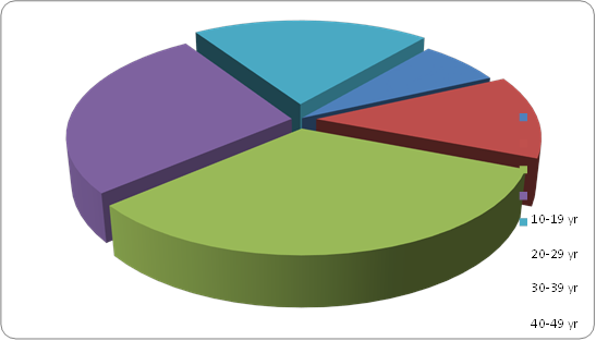
The common age group of women complaining from nipple discharge was from (30-39) and second common group was(40-49). (Table 1), (Figure 1).


Table (1): Age distribution of patient complaining from nipple discharge

AgeNumber of patient

%

10 – 19

6

6.9

20 – 29

11

12.7

30 – 39

28

32.5

40 – 49

23

26.8

>50

18

20.1

Total

86

100


Figure (1): The age distribution of patient complaining from nipple discharge.


The types of nipple discharge:

The most common type of nipple discharge was bloody nipple discharge and perulant nipple discharge and less common types were watery and serosangous nipple discharge. (Table 2).


Table (2): Types of nipple discharge

Types of nipple


discharge

Number

%

Bloody3540.8
Purulant2428
White1112.7
Serous89.3
Watery55.8
Serosangous33.4
Total86100


The relationship between nipple discharge and breast masses:

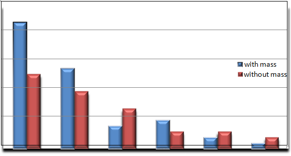
There is a strong relationship between nipple discharge and breast masses. High number of bloody and perulant nipple discharge were associated with breast masses. In the other hand the other types of nipple discharge were associated with breast masses but in less number. (Table 3), (Figure 2).


Table (3): The relationship between types of nipple discharge and presence of brast mass


Types of nipple discharge

With mass


n (%)

Without mass


n (%)

Total

P


Value

Bloody22 (45.8)13 (34.1)35

P > 0.05

Purulent14 (29.1)10 (26.2)24

P > 0.05

White4     (8.3)7     (18.4)11

P > 0.05

Serous5     (10.4)3     (7.8)8

P > 0.05

Watery2 (4.2)3      (7.8)5

P > 0.05

Serosangous1 (2.1)2      (5.2)3

P > 0.05

Total483886


Figure (2): The relationship between type of nipple discharge and presence of breast masses.


The relationship between types of nipple discharge and breast diseases (benign and malignant):

Abnormal nipple discharge are more common with benign than malignant breast diseases.

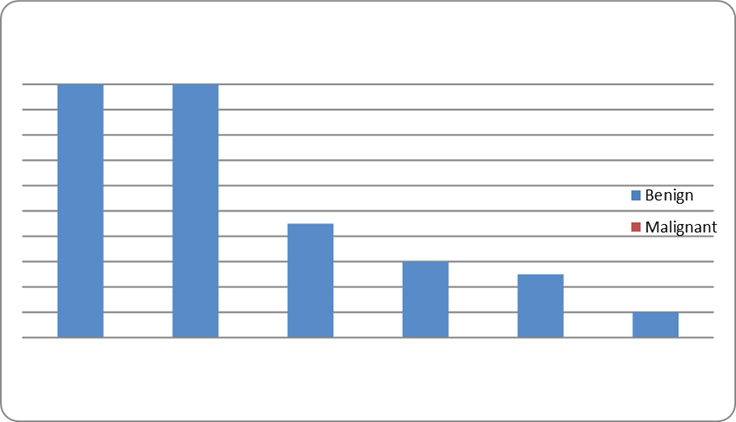
The bloody nipple discharge is strongly related with malignant breast diseases. The perulant, serous, serosangous types are also associated with malignant breast diseases but less common than bloody nipple discharge. Watery and white nipple discharge had no relation with malignant breast diseases. ( Table 4),(Figure 3).


Table (4): The relationship between the types of nipple discharge and the breast diseases (Benign or malignant).

Type of nipple discharge

Benign         breast diseases

n (%)

Malignant      breast diseases

n (%)

Total

P


value

Bloody

20 (32.2)

15 (62.5)

35

P < 0.05

Purulent

20 (32.2)

4 (16.6)

24

P > 0.05

White

9 (14.6)

2 (8.3)

11

P > 0.05

Serous

6 (9.7)

2 (8.3)

8

P > 0.05

Watery

5 (8.1)

0 (0)

5

P > 0.05

Serosangous

2 (3.2)

1 (4.1)

3

P > 0.05

Total

62

24

86


Figure(3): The relationship between the type of nipple discharge and the breast diseases ( benign and malignant). 


The relationship between the site of nipple discharge and benign and malignant diseases.

Unilateral nipple discharge with benign and malignant breast diseases are more common than bilateral nipple discharge

.

Table(5): The relationship between the site of nipple discharge and benign and malignant diseases.


ND withbenign n (%)ND withmalignant n (%)


P


value

Unilateral Breast


44(70.9)


21 (87.5)


P >0.05

Bilateral breast


18 (29.1)


3 (12.5)


P >0.05


Total


62


24


The efficacy of direct cytology of nipple discharge:

The direct cytology from nipple discharge had a good efficacy in diagnosis of malignant breast diseases that it detrmind the malignant cell in many cases.


Table(6): The relationship between effectiveness of cytology from nipple discharge and malignant diseases


No malignant


%

Malignant


%

Total

Cytology        from nipple discharge


5


20.8


19


79.1


24

The effectiveness of diagnostic tests to diagnose malignant breast diseases:

The beast modialty for diagnosis of malignant breast diseases was the histopathology and the less beast diagnostic tests were the F N A C and direct cytology from nipple discharge. While we couldnot dignosed the malignant breast diseases by the clinical examnation and radiology including (breast ultrasound and mammogram)


Table (7) : The effectiveness of diagnostic tests to diagnose malignant breast diseases


Number of malignant cases


%

Clinical examination

4

16.6

Radiology      (ultrasound                         and mammogram)

9

37.5

Cytologyfrom nipple discharge

19

79.2

F N A C

21

78.5

Histopathology

24

100


The relationship between duration of nipple discharge and bloody and non-bloody nipple discharge:

The common period of duration of bloody nipple discharge to time of examination was (0-13) days and less common duration was (2-4) weeks.


While for the non-bloody nipple discharge the common duration was (2-4) weeks and the less common duration was (0-13) days.


Table(8): Relationship between duration of nipple discharge and bloody and non-bloody nipple discharge

DurationBloody discharge%

Non             bloody discharge

%

At         time             of examination00

5

9.8

0-13 days1851.5

12

23.5

2-4 weeks1028.5

15

29.4

1-3 mounth38.5

8

15.7

4-6 mounth25.7

3

5.9

7 mounth-1 year12.8

2

4

Unknown12.8

6

11.7

Total35100

51

100

Discussion

The appearance of spontaneous nipple discharge in non lactating breast is an abnormal clinical signe that should be investigated. [33]


The type and character of the discharge have a good prognostic value and often reflect the type of underlying lesion. [34].


In our study we have (88) patient, only (2) of them were male with age of (16 and 14 year) because that we collect our patient from the breast clinic of Baquba teaching hospital; so the male not visit this clinic due to social habbit and consult out clinic ; therfor male patient not involved in our study and our patient become (86) female patient.


1: Age distribution:

Regarding age of patient in our study, the common age groups were (30-39) year (32.5% ) and( 40-49) year (26.8%) and this is similar to (Paterok EM et al;2003) [34].


2: The types of nipple discharge:

The most common type of nipple discharge represented in our study was bloody discharge (40.8 %) and this result is similar to ( Paul R .et al 2000) [35]. .study which show same result.


3: The relationship between nipple discharge and breast masses:

Regarding the type of nipple discharge of patient in our study the largest number of cases of bloody and purulent discharge were associated with breast mass and other less number of cases without mass , and this result not agree with ( Chrles W.et al 2001) [36] study , in which majority of cases were not associated with breast mass, and this is due to defeciancy in health education in our country about the importance of breast diseases and their symptoms , also due to limited number of special breast clinic in all our cities.


4: The relationship between types of nipple discharge and breast diseases (benign and malignant):

Abnormal nipple discharge regardless the types more common symptom with benign breast diseases than in malignant breast diseases, and bloody nipple discharge was most common type in malignant breast diseases rather than other types of nipple discharge (P- value < 0.05 which is significant) , and this result strongly similar to (Caleffi M, et al 2004) [38].


5: The relationship between the site of nipple discharge and benign and malignant diseases.

Regarding the site of nipple discharge in our study , in both benign and malignant breast diseases, the unilateral nipple discharge(75.5%) are more common than bilateral nipple discharge(24.5%), and this result similar to (Rosen PP,et al ;2001) [39].


6: The efficacy of direct cytology of nipple discharge:

Regarding the direct cytology from nipple discharge in our study,it had a good effectiveness in diagnosis of malignant breast diseases(79.1%) by determined the malignant cell, and this result similar to (Sheen-Chen,S,M et al;2001) study.[33]


7: The effectiveness of diagnostic tests to diagnose malignant breast diseases:

The effectiveness of diagnostic test for nipple discharge are arrenge according to their accuracy in dignose the malignant breast diseases as histopathology(100%), FNAC (78.5%), direct cytology (79.1%) and this result similar to (Caleffi M,et al;2004) [38].


8: The relationship between duration of nipple discharge and bloody and non-bloody nipple discharge:

Regarding the duration of bloody nipple discharge to the time of examination in our study the majority of cases with bloody discharge were represented with duration of (0-13) days (51.5%), this result not similar to (Herbert E, et al;2002) [39] study. ,in which the common duration was (2-4) weeks, and this is due to in our community the bloody nipple discharge is more serious and alarm symptyome that related to malignant breast diseases.

Conclusion

The nipple discharge is very important early presentation of breast diseases. -1 Most cases of nipple discharge mainly bloody discharge are associated with presence of breast mass.  -2

Any type of nipple discharge mainly bloody and purulent should not be neglected because it may be the presentation  -3

of benign or malignant breast diseases.

Direct cytology from nipple discharge have a good value in diagnosis of malignant breast diseases. -4 The histopathology have the difentive diagnosis of malignant breast diseases.  -5


Recommendation:

Increase the health education about the importance of breast diseases and their early symptom as nipple discharge. -1 Regular visit to the breast clinic department even in simple breast complain may help to early detection and                                -2

management of serious breast diseases.

The breast clinic should be separate and contain all the diagnostic facilities as radiology and labrotary .  -3


Funding: No funding sources 


Conflict of interest: None declared


Ethical approval: The study was approved by the Institutional Ethics Committee of Bangabandhu Sheikh Mujib Medical University

References
  1. Hussain, Aneela N., Cristina Policarpio, and Miriam T. Vincent. "Evaluating nipple discharge." Obstetrical & gynecological survey 61.4 (2006): 278-283. DOI: 10.1097/01.ogx.0000210242.44171.f6
  2. Jackson, Dudley, D. A. Todd, and P. L. Gorsuch. "Study of breast secretion for detection of intramammary pathologic change and of silent papilloma." (1951): 552-68. https://www.cabidigitallibrary.org/doi/full/10.5555/19520400817
  3. River, Louis. "Bloody nipple discharge and carcinoma of the breast." Mississippi Valley Medical Journal (Quincy, Ill) 74.3 (1952): 73-75. https://europepmc.org/article/med/13013123
  4. Chester, S. T., and H. G. Bell. "Intraductal and intracystic papillomas of the breast." Western journal of surgery, obstetrics, and gynecology 59.12 (1951): 603-609. https://pubmed.ncbi.nlm.nih.gov/14893779/

  5. da Carpi, Jacopo Berengario. A short introduction to anatomy:(Isagogae Breves). University of Chicago Press, 1959.

  6. Schwartz, Seymour I., et al. "Principles of surgery 7th Ed." New York 1999 (1999): 1395-435. 
  7. Dinkel, Hans-Peter, et al. "Galactography and exfoliative cytology in women with abnormal nipple discharge." Obstetrics & Gynecology 97.4 (2001): 625-629. https://journals.lww.com/greenjournal/abstract/2001/04000/galactography_and_exfoliative_cytology_in_women.27.aspx
  8. Lau, Steffi, et al. "Pathologic nipple discharge: surgery is imperative in postmenopausal women." Annals of surgical oncology 12 (2005): 546-551. DOIhttps://doi.org/10.1245/ASO.2005.04.013
  9. Simmons, Rache, et al. "Nonsurgical evaluation of pathologic nipple discharge." Annals of surgical oncology 10 (2003): 113-116. DOIhttps://doi.org/10.1245/ASO.2003.03.089
  10. Morrogh, Mary, et al. "The predictive value of ductography and magnetic resonance imaging in the management of nipple discharge." Annals of Surgical Oncology 14 (2007): 3369-3377.

  11. Morton JH. Some thoughts on breast cancer. Arch Surg 2001; 136:357-35 DOIhttps://doi.org/10.1245/s10434-007-9530-5
  12. Sauter, Edward R., et al. "Biologic markers of breast cancer in nipple aspirate fluid and nipple discharge are associated with clinical findings." Cancer detection and prevention 31.1 (2007): 50-58. https://doi.org/10.1016/j.cdp.2006.12.004

  13. Morehead, James R. "Anatomy and embryology of the breast." Clinical Obstetrics and Gynecology 25.2 (1982): 353-357. https://journals.lww.com/clinicalobgyn/citation/1982/06000/anatomy_and_embryology_of_the_breast.17.aspx
  14. Cormack DG. Ham's Histology (9th ed). Philadelphia: JB Lippincott, 1987.
  15. Shah, Varsha I., et al. "False‐negative core needle biopsies of the breast: an analysis of clinical, radiologic, and pathologic findings in 27 consecutive cases of missed breast cancer." Cancer: Interdisciplinary International Journal of the American Cancer Society 97.8 (2003): 1824-1831. https://doi.org/10.1002/cncr.11278
  16. Russell, R. G., N. S. Williams, and J. K. Christopher. "Bulstrode, editors. Bailey and Love's Short practice of surgery." (2000). 
  17. Zervoudis, Stefanos, et al. "Nipple discharge screening." Women’s Health 6.1 (2010): 135-151. https://journals.sagepub.com/doi/full/10.2217/WHE.09.81
  18. Kneece, Judy C. Solving the Mystery of Breast Discharge. Educare Publishing (SC), 1996.
  19. Lauersen, Niels H., and Eileen Stukane. The complete book of breast care. Fawcett, 1998.

  20. Orel, Susan Greenstein, et al. "MR imaging in patients with nipple discharge: initial experience." Radiology 216.1 (2000): 248-254. https://doi.org/10.1148/radiology.216.1.r00jn28248
  21. Chaudary, MURID A., et al. "The diagnostic value of testing for occult blood." Annals of surgery 196.6 (1982): 651. doi: 10.1097/00000658-198212001-00006
  22. Zee, Kimberly J. Van, et al. "Preoperative galactography increases the diagnostic yield of major duct excision for nipple discharge." Cancer: Interdisciplinary International Journal of the American Cancer Society 82.10 (1998): 1874-1880. https://doi.org/10.1002/(SICI)1097-0142(19980515)82:10<1874::AID-CNCR9>3.0.CO;2-N

  23. Nakahara, Hiroshi, et al. "A comparison of MR imaging, galactography and ultrasonography in patients with nipple discharge." Breast cancer 10 (2003): 320-329. DOIhttps://doi.org/10.1007/BF02967652
  24. Aaron Fenster, Kathleen JM Surry, Donal B Downey Imaging Research Labs, Robarts Research Institute Dept. of Medical Biophysics, University of Western Ontario Dept. of Radiology, LHSC University Campu 2010.

  25. Gøtzsche, Peter C. "Mammography screening: truth, lies, and controversy." The Lancet 380.9838 (2012): 218. DOI:https://doi.org/10.1016/S0140-6736(12)61216-1
  26. Jørgensen, Karsten Juhl, and Peter C. Gøtzsche. "Content of invitations for publicly funded screening mammography." Bmj 332.7540 (2006): 538-541. doi: https://doi.org/10.1136/bmj.332.7540.538
  27. Gøtzsche, Peter C. "Screening for breast cancer with mammography." The Lancet 358.9299 (2001): 2167-2168. DOI:https://doi.org/10.1016/S0140-6736(01)07198-7
  28. Sneige, Nour, Bruno D. Fornage, and George Saleh. "Ultrasound-guided fine-needle aspiration of nonpalpable breast lesions: cytologic and histologic findings." American Journal of clinical pathology 102.1 (1994): 98-101. https://doi.org/10.1093/ajcp/102.1.98
  29. Ciatto, Stefano, et al. "Fine-needle aspiration cytology of nonpalpable breast lesions: US versus stereotaxic guidance." Radiology 188.1 (1993): 195-198. https://doi.org/10.1148/radiology.188.1.8511296

  30. Orell, Svante R., Gregory F. Sterrett, and Darrel Whitaker. "Fine needle aspiration cytology." (No Title) (2005). https://cir.nii.ac.jp/crid/1130000795847845248
  31. Trott, Peter A. "Breast cytopathology: a diagnostic atlas." (No Title) (1996). https://cir.nii.ac.jp/crid/1130282269861629184
  32. Koss, Leopold G., Stanislaw Woyke, and Wlodzimierz Olszewski. "Aspiration biopsy: cytologic interpretation and histologic bases." Aspiration biopsy: cytologic interpretation and histologic bases. 1992. xvi-742. https://pesquisa.bvsalud.org/portal/resource/pt/biblio-1084088

  33. Sheen‐Chen, Shyr‐Ming, et al. "Paget disease of the breast—an easily overlooked disease?." Journal of surgical oncology 76.4 (2001): 261-265. https://doi.org/10.1002/jso.1043

  34. Paterok, E. M., H. Rosenthal, and M. Säbel. "Nipple discharge and abnormal galactogram. Results of a long-term study (1964–1990)." European Journal of Obstetrics & Gynecology and Reproductive Biology 50.3 (1993): 227-234. https://doi.org/10.1016/0028-2243(93)90205-Q

  35. Paul.R, Hinchey, M. D. Salem, Mass. From the Surgical department of the Massachusetts General Hospital, And the Tumor clinic of the Boston dispensary (Unit of the new England Medical Centr), Boston .Mass (2000).

  36. Charles W. Mclaughlin, JR., M.D., John D. Coe, M.D. Omaha, Nebraska. (2001).

  37. Caleffi, M., et al. "Cryoablation of benign breast tumors: evolution of technique and technology." The breast 13.5 (2004): 397-407. https://doi.org/10.1016/j.breast.2004.04.008
  38. Rosen, Paul Peter, ed. Rosen's breast pathology. Lippincott Williams & Wilkins, 2001. 
  39. Herbert E. Madalin, M.D.,** 0. Theron Clagett, M.D., F.A.C.S.,t John R. Mcdonald, M.D.++ Rochester, Minnesota(2002).
Advertisement
Recommended Articles
Research Article
abc
Published: 30/06/2024
Download PDF
Research Article
Transformative Applications in Genetic Disorders and Cancer Therapy, Ethical Considerations, and Future Prospects
Published: 30/06/2024
Download PDF
Research Article
Stroke Knowledge in Shimla: Identifying Symptoms and Prevention Tactics
Published: 20/05/2024
Download PDF
Research Article
Understanding Hemorrhoids: Assessing Awareness and Knowledge in Bilaspur
...
Published: 20/06/2024
Download PDF
Chat on WhatsApp
Flowbite Logo
Najmal Complex,
Opposite Farwaniya,
Kuwait.
Email: kuwait@iarcon.org

Editorial Office:
J.L Bhavan, Near Radison Blu Hotel,
Jalukbari, Guwahati-India
Useful Links
Order Hard Copy
Privacy policy
Terms and Conditions
Refund Policy
Others
About Us
Contact Us
Online Payments
Join as Editor
Join as Reviewer
Subscribe to our Newsletter
Follow us
MOST SEARCHED KEYWORDS
scientific journal
 | 
business journal
 | 
medical journals
 | 
Scientific Journals
 | 
Academic Publisher
 | 
Peer-reviewed Journals
 | 
Open Access Journals
 | 
Impact Factor
 | 
Indexing Services
 | 
Journal Citation Reports
 | 
Publication Process
 | 
Impact factor of journals
 | 
Finding reputable journals for publication
 | 
Submitting a manuscript for publication
 | 
Copyright and licensing of published papers
 | 
Writing an abstract for a research paper
 | 
Manuscript formatting guidelines
 | 
Promoting published research
 | 
Publication in high-impact journals
Copyright © iARCON Internaltional LLP . All Rights Reserved.