Research Article | Volume 2 Issue 2 (July-Dec, 2021)
Patterns of Some Immunological Proteins and Oxidative Indices in Quercetin Treated Normal and High Body Mass Index Arsenic Administered Male Wistar Rats
 ,
 ,
 ,
 ,
 ,
1
Department of Biochemistry, Faculty of Basic Medical Sciences, Ladoke Akintola University of Technology, Ogbomoso, Oyo State, Nigeria, 211101
2
Department of Medical Biochemistry, Faculty of Basic Medical Sciences, Osun State University, Osogbo, Osun State, Nigeria, 234101
3
Department of Industrial Chemistry, Faculty of Sciences, University of Ilorin, Ilorin, Kwara State, Nigeria, 240001 – 252105
Under a Creative Commons license
Open Access
Received
April 10, 2021
Revised
April 24, 2021
Accepted
Sept. 20, 2021
Published
Oct. 10, 2021
Abstract

Exposures to arsenic and obesity (high BMI) have been associated with oxidative stress and health dysfunctions, but the severity of arsenic poisoning and susceptibility might depend on various factors, such as the body mass index (BMI), oxidative status, food supplementations etc. This study assessed the ameliorative potential of quercetin on arsenic induced health dysfunctions in normal and high BMI male Wistar rats. Forty eight apparently healthy rats were assigned into eight groups of six rats each based on their BMI. The rats were administered arsenic (40 ppm) for six weeks and subsequently quercetin (50 mg/kg bodyweight) for four weeks, such that; groups A and B were normal and high BMI respectively, administered distilled water; groups C and D were normal and high BMI rats respectively, administered arsenic; groups E and F were normal and high BMI rats respectively, administered quercetin; groups G and H were normal and high BMI rats respectively, co-administered arsenic and quercetin. Concentrations of reduced glutathione (GSH), oxidized-low density lipoprotein cholesterol (Ox-LDL-C), 8-hydroxydeoxyguanosine (8-OHdG), interleukin-1β (IL-1β), vascular cell adhesion molecule (VCAM-1) and monocyte chemoatracttant protein-1 (MCP-1) were determined in plasma. Organ to body weight ratios were insignificant (p>0.05) across the groups. Serum GSH level increased (p<0.05) in groups D and E, while Ox-LDL-C and 8-OHdG levels increased (p<0.05) in group G and H respectively. Concentrations of IL-1β and VCAM-1 were insignificant (p>0.05) across the groups and MCP-1 increased (p<0.05) in groups B, C, E, F, G and H. Histological assessments of the brain, heart and liver indicated mild to moderate alterations. The foregoing indicated that arsenic exposure induced dysfunctions in the oxidative indices and immunological proteins (health indices) that were exacerbated in the high BMI rats, and quercetin administration did not have ameliorative capabilities against arsenic and High BMI induced health dysfunctions.

Keywords
Important Note:

Key findings:
The study investigated the effects of arsenic exposure and high BMI on oxidative stress and health markers in rats, finding increased oxidative stress and immune protein levels, especially in high BMI rats. Quercetin treatment did not mitigate these effects, suggesting a lack of protective effect against arsenic and high BMI-induced health dysfunctions.


What is known and what is new?
Exposure to arsenic and obesity correlate with oxidative stress and health issues, but their severity may vary depending on factors like BMI. This study investigates quercetin's potential to alleviate arsenic-induced health problems in normal and high BMI rats. Results suggest arsenic exacerbates oxidative and immunological dysfunctions, particularly in high BMI rats, with quercetin showing limited effectiveness.


What is the implication, and what should change now?
The study highlights the exacerbated health dysfunctions induced by arsenic exposure and high BMI in rats, despite quercetin administration. These findings underscore the importance of considering multiple factors, such as BMI and oxidative status, in understanding the severity of arsenic poisoning. Future studies should explore alternative interventions to address arsenic and high BMI-induced health complications effectively.
 

INTRODUCTION:

Arsenic, the 33rd element of the periodic table is referred to as a heavy metal in the context of toxicology [1], and is classified as human carcinogen (group 1) [2]. Arsenic has become a global health concern, as it is broadly distributed in nature and has been associated with numerous adverse effects which threaten an organism’s health [3], For years, the respiratory, cardiovascular, gastrointestinal, hematological, renal, dermal, reproductive and neurological toxicity of arsenic have been documented [1]. As a natural constituent of the environment, animals are easily exposed to relatively low levels of arsenic through food, air and water [4]. Globally, over 20 million people are chronically exposed to arsenic contaminated water above the safety level of 10µg/l [5]. However, the presence of arsenic is not immediately evident in food, air, or water because arsenic compounds have no colour / smell, hence posing a serious human health hazard due to its toxic nature [6]. Recently, substantial scientific evidence has revealed that low to moderate levels of arsenic from water may lead to the occurrence of a large variety of health dysfunctions, illness, degenerative diseases etc. [4, 7-9]


The primary toxic mechanism of arsenic is not specified, but inflammatory or oxidative dysfunctions have been proposed [10]. Inflammation is involved in the pathogenesis of many degenerative diseases including cardiovascular diseases, metabolic syndrome, chronic kidney and liver diseases and cancer [11,12]. Arsenic has been implicated to induce inflammatory responses and compromise or detrimental to the immune cells [13-15]. This might be the mechanism of arsenic-induced health dysfunction and diseases. Similarly, several in vitro and in vivo studies have revealed that arsenic toxicities were mediated by induction of oxidative stress [15, 16]. Oxidative stress and inflammation are linked in a complex feedback cycle in which reactive oxygen species trigger transcription factors that upregulate the expression of pro-inflammatory cytokines and anti-oxidant enzymes [17].


The extent of arsenic poisoning and susceptibility varies widely from person to person and depends on various factors such as dose, individual susceptibility to arsenic, inter-individual differences in diet, arsenic metabolism, co-exposure, genetics and the age of the individual [6, 18]. However, most of the variability in susceptibility and the influence of lifestyles on arsenic metabolism in humans are not well understood [18] .Although the severity of arsenic toxicity in individual with elevated body mass index (BMI) is not well understood, studies have however revealed that (BMI), an indicator for overall nutritional status, is positively associated with arsenic methylation capacity [19, 20]. and the pathological processes linked to arsenic and obesity such as inflammation, oxidative stress, adipokine expression and insulin resistance are thought to play a role in diseases caused by each. Elevated BMI has been shown to exacerbate oxidative stress and inflammatory responses that has been associated with some types of cancer [21, 22].


Quercetin, a major flavonoid found in edible plants (fruits and vegetables) is one of the most potent antioxidants present in plants and has unique biological properties that may improve mental / physical performances and reduce infection risk [23, 24]. Onion, hot peppers, curly kale, blueberries, apple, tea and broccoli are some of the richest food sources of quercetin [25, 26]. Quercetin is also available and sold as a dietary supplement with daily doses between 200-120000 mg quercetin based on manufacturer’s recommendation [27]. In addition, quercetin has been employed as nutracetical for functional foods within the range of 0.008-0.5% or 10-125 mg/serving [28].


Dietary flavonols such as quercetin have been reported to possess physiological effects including antioxidative [29, 30], anti-inflammatory [31-33], anti-pathogenic [34, 35], anti-viral [24, 36, 37] anti-microbial [38], anti-carcinogenic [39], cardio-protective [23], mitochondrial biogenesis activities [40] and thus provide significant potential in the study of improving mammalian health. It is well established that athletes use flavonoids as antioxidants to enhance endurance and physical performance [41]. In addition, epidemiological studies indicated that the risk factors of cardiovascular diseases in subjects who had a high intake of flavonoids were reduced. Given the interplay between the pathological mechanisms of arsenic and elevated BMI and the anti-oxidative and anti-inflammatory potentials of quercetin, this study therefore, assessed the ameliorative capability of quercetin on arsenic induced oxidative stress and inflammatory responses with respect to body mass index (BMI) in apparently healthy male Wistar rats.
 

MATERIALS AND METHODS:

Reagents Kits and Chemicals
All reagent kits and chemicals used were of analytical grades and purchased from Central Research Laboratory, Ilorin. Arsenic and Quercetin were purchased from Biobridge Laboratory, Ilorin.


Experimental Animals
Forty eight (48) male Wistar rats of 120-150 g and 235-250 g were obtained from the Animal House, Ladoke Akintola University of Technology, Ogbomoso, Oyo State, Nigeria.


Sodium Arsenite
Sodium arsenite was a product of 1/20 B, Narayan Plaza, 26-A, Chandivali Road, Andheri (E), Mumbai-400072, Maharashsta, India.


Experimental Design
The rats were randomly divided into eight groups of six rats per group based on their body mass index (BMI) as illustrated in Table 1. The rats were grouped into normal BMI (N-BMI) and high BMI (H-BMI), depending on the BMI of 0.45-0.60 (N-BMI) and greater than 0.68 as (H-BMI).


Table 1: Groups of Rats and Doses of Arsenic and Quercetin Administered

Groups

Treatment

Arsenic (ppm)

Quercetin (mg/kg)

A

Normal BMI Control

-

-

B

High BMI Control

-

-

C

Normal BMI Arsenic

40

-

D

High BMI Arsenic

40

-

E

Normal BMI Quercetin

-

50

FHigh BMI Quercetin-50
GNormal BMI Arsenic+Quercetin4050        
HHigh BMI Arsenic+Quercetin4050


Conversion from human dose to animal dose was done using the model by Reagan-Shaw et al.,. (2008) [42]. Arsenic was suspended in distilled water at 40 ppm and the rats were allowed access to the water and feed ad libitum, while quercetin was prepared in saline solution and administered orally at 50 mg/kg body weight with the use of oral cannula. Arsenic exposure was for a period of six (6) weeks before the commencement of quercetin administration for the next four (4) weeks. The administration lasted for ten (10) weeks in all, after which all the rats were sacrificed via mild anesthesia.


Table 2: Organs to body weight ratios of rats administered arsenic and quercetin.


 GroupsABCDEFGH

Organ to body weight ratio 


Brain

0.01±

0.00a

0.01±

0.00 a

0.01±

0.00 a

0.01±

0.00a

0.01±

0.00 a

0.01±

0.00 a

0.01±

0.00 a

0.01±

0.00 a

Heart

0.00±

0.00 a

0.00±

0.00 a

0.00±

0.00a

0.00±

0.00a

0.00±

0.00 a

0.00±

0.00 a

0.00±

0.00 a

0.00±

0.00 a

Liver

0.04±

0.00 a

0.03±

0.00 a

0.03±

0.00a

0.04±

0.00 a

0.03±

0.00 a

0.03±

0.00 a

0.03±

0.00 a

0.03±

0.00 a


Keys: A-NBMI, B HBMI, C- NBMI + Arsenic, D-HBMI + Arsenic, E-NBMI + Quercetin, F-HBMI + Quercetin, G-NBMI + Arsenic + Quercetin, H-HBMI + Arsenic + Quercetin. Values are means ± SEM, n=4 and mean values bearing different alphabets are significantly different (P<0.05). NBMI (normal body mass index) and HBMI (high body mass index).


COLLECTION OF SAMPLES:

Serum
After ten (10) weeks of administration, the rats were fasted overnight and anesthetized using diethyl ether. The chest region was quickly opened and blood was drawn by puncturing the heart using a new syringe for each animal.  The blood samples were collected into plain bottles and centrifuged at 4000 revolutions per minute (rpm) for 10 minutes to separate the serum from the whole blood. The serum which is the supernatant was carefully decanted into sample bottles using a micropipette, dropped into a clean bottle labeled and stored in the refrigerator below 40 C immediately for further analysis.


Organs
Organs of interest (brain, heart and liver) were harvested immediately, cleansed of blood and rinsed with normal saline solution and the weights were recorded. The organs were fixed in 10% formal saline solution for histopathology examination.
 

BIOCHEMICAL ASSAY METHODS:

Assessment of Weight of Rats
Changes in body weight of the experimental rats were monitored on a weekly basis and the harvested organs of interest (brain, liver and heart) were weighed to determine the organ-body weight ratio.


Assessment of Oxidative Status of Rats
The serum concentration of reduced glutathione (GSH) was measured using the method of Ellman (1959). Oxidized-low density lipoprotein cholesterol (Ox-LDL-C) and 8-hydroxydeoxyguanosine (8-OHdG) concentrations were determined by ELISA method as described in the ELISA kit manual.


Assessment of Immunological Proteins in Rats
Interleukin-1 beta (IL-1β), monocyte chemoattractant Protein-1 (MCP-1), and vascular cell Adhesion molecule-1 (VCAM-1) analyses were done using ELISA method as specified in the kit manual based on sandwich immunoassay principle.


Histological Assessment
Histological examination of brain, liver and heart tissues was done according to the method of Avwioro, 2010 [43].


Statistical Analysis
This research work was a completely randomised design (CRD). Results were expressed as mean ± standard error of mean (S.E.M). Data generated were subjected to one way analysis of variance (ANOVA), after which Tukey Test was conducted in order to identify the variation within the treatment group.  P-value <0.05 was regarded as statistically significant and denoted by alphabets.
 

RESULTS:

Effect of Arsenic and Quercetin on Weight and Organ to Body Weight Ratio of Rats
Table 1 depicted organ (brain, heart and liver) to body weight ratio of rats administered arsenic and quercetin. There was no significant difference (p>0.05) in the brain to body weight ratio, heart to body weight ratio and liver to body weight ratio across the groups.


Effect of Arsenic and Quercetin on Oxidative Stress Indices
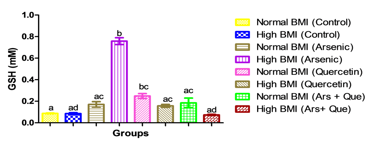
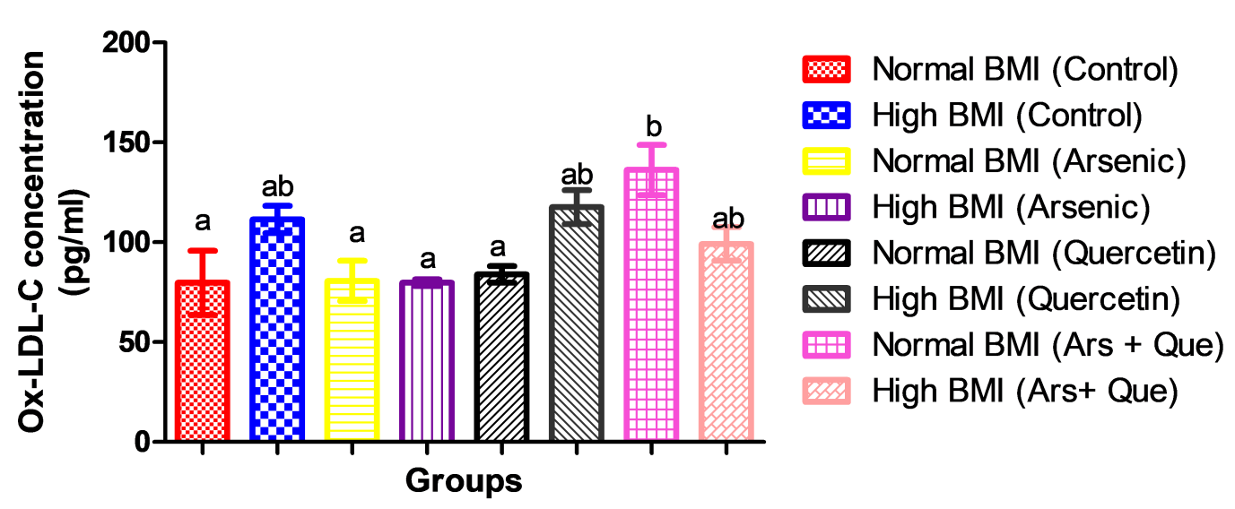
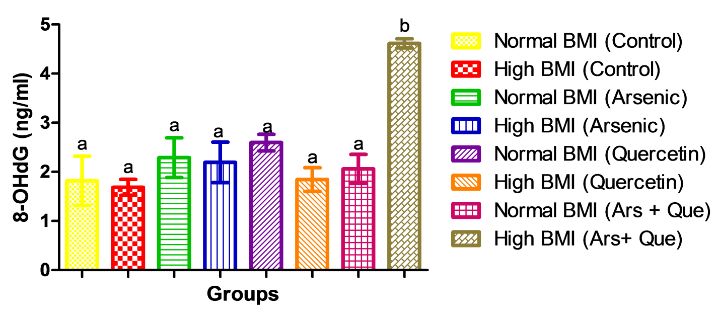
In Figure 1, there were significant increases (p<0.05) in the serum reduced glutathione concentrations in groups D and E rats. In the same vein, the concentration of oxidized-low density lipoprotein cholesterol in the serum was elevated significantly (p<0.05) in groups B, F, G and H (Figure 2). The serum concentrations of 8-hydroxydeoxyguanosine gave significant increases (p<0.05) in rats of group H only (Figure 3).


Effect of Arsenic and Quercetin on Immunological Proteins
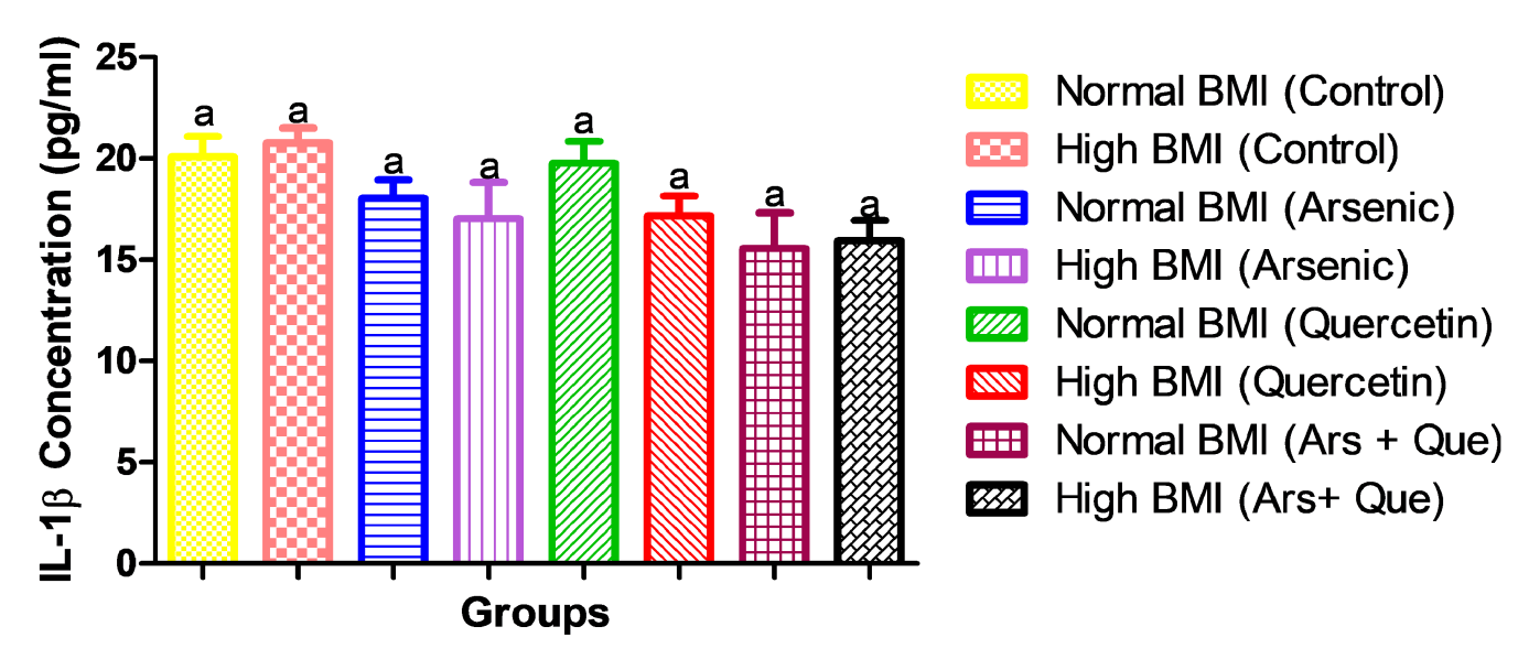
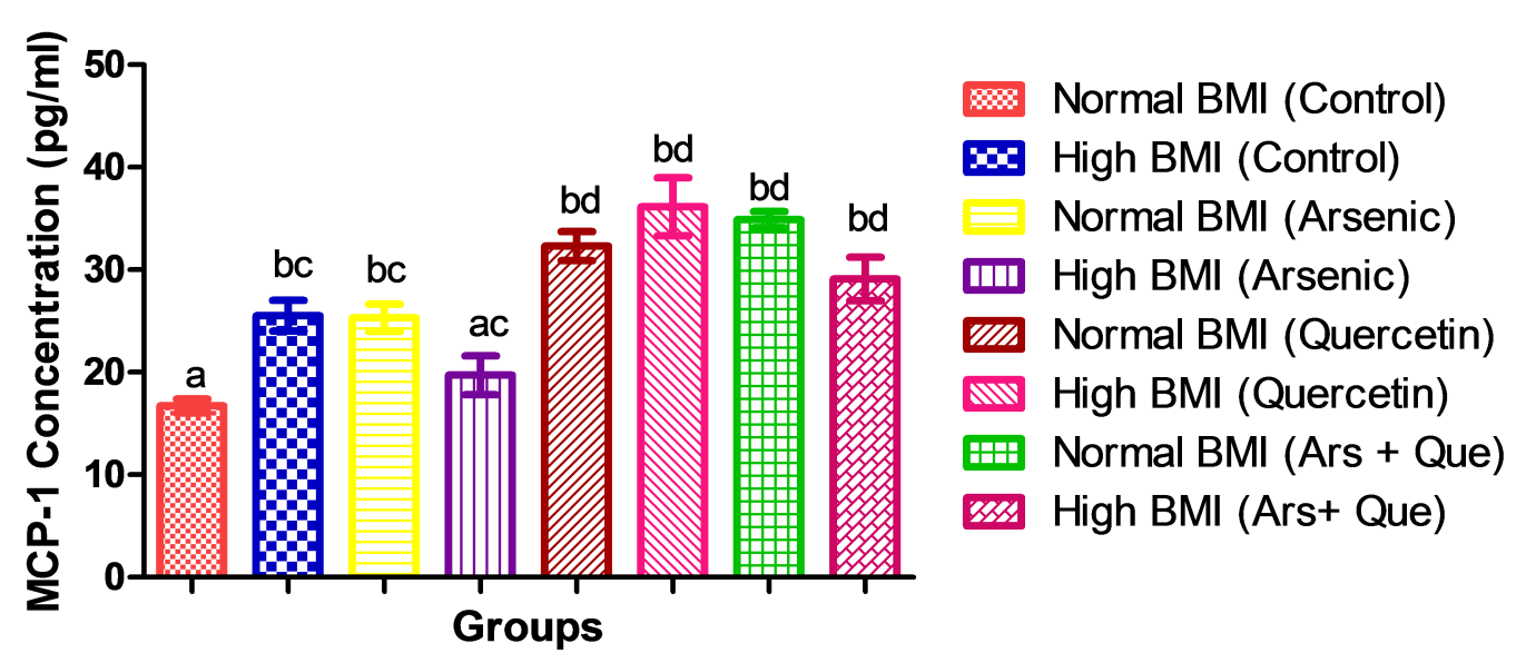
The results obtained in the serum interleukin-1β and vascular cell adhesion molecule-1 concentrations gave no significant alterations (p>0.05) across the groups (Figures 4 and 5). However, there was significant increases (p<0.05) in serum the concentrations of monocyte chemoattractant protein-1 in groups B, C, E, F, G and H, but insignificant increase (at p˃0.05) in group D (Figure 6).


Histological Assessment of the Brain, Liver and Heart Tissues
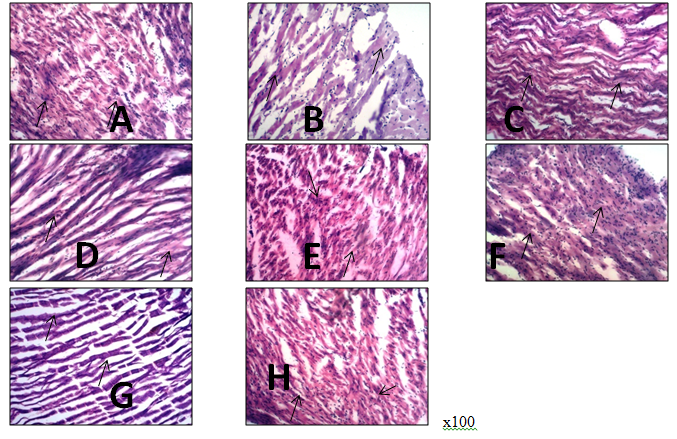
The photomicrographs of the prefrontal cortex of rats administered with arsenic and quercetin in plate 1 (A-H) showed the external granular layer with constituent granular neurons. The normal and intact neuronal cells are depicted with black arrow heads, while the presence of mild vacuoles perceived as histopathological alterations were depicted with red arrows in all the groups (A-H). In Plate 2, the photomicrographs of the heart in rats administered with arsenic and quercetin (A-H) depicted the cardiomyocytes containing the cardiomyocytes nuclei (black arrow). The cellular delineation, cellularity and morphological delineation of groups A,C and E appeared normal, while groups B, D, F, G, and H appeared atypical. The photomicrographs of the liver sections of rats administered arsenic and quercetin is presented in plate 3 (A-H). The central vein, sinusoidal space and hepatocytes are depicted by dotted circles, S, and H respectively. Mild-moderate sinusoidal space dilation and atrophy of the central veins were observed in all the groups (B, E, F, G and H).


DISCUSSION:

In experimental animals, arsenic exposure has been associated with increases in body weight, changes in fat metabolism and deposition, and other related outcomes but the evidence that arsenic is a cause of obesity is not clear [44], hence, the associations between exposure to inorganic arsenic and body mass index (BMI) have been inconsistent. For, instance, Aliyu et al., (2012) [45] showed that arsenic treatment reduced body weight in a concentration dependent manner. In contrast, the findings in this present investigation revealed that arsenic treatment had no effect on the organ (brain, heart and liver) to body weight ratio (Table 2) regardless of the body mass index (BMI) status of the rats or quercetin treatment. The observation in this study is in agreement with the findings of Singh et al., (2017) [46] in which arsenic treatment had no significant effect on body weight and liver to body weight ratio. In addition, certain rodent studies that evaluated lipid lowering effects of quercetin supplementation showed reduction in body weight, serum lipid levels, hepatic lipid accumulation, and/or white adipose tissue mass. However, these effects were not seen in all studies and were sometimes conflicting [47-53]. The trend in the organ (brain, heart and liver) to body weight ratio across the groups (Table 1 and 2) in this present study reflected that quercetin at the administered dose and duration had no effect on the organ (brain, heart and liver) to body weight ratio.


Furthermore, the primary mechanism of arsenic toxicity is not known, but has been associated with oxidative stress and inflammatory response [10, 15], while some other study have also suggested a pathological crosstalk between obesity, oxidative stress, and inflammatory process [54]. Oxidative stress occurs as a result of an imbalance between reactive oxygen species (ROS) and antioxidant defenses, and the inability of the biological system to eliminate free radicals which results in oxidative damage of lipids, proteins and deoxyribonucleic acid (DNA). Consequently, oxidative stress is associated with several pathological implications and might be a major mechanism underlying obesity-related complications [15, 55, 56].


In biological systems, various antioxidant defense systems, including enzymatic and nonenzymatic routes, act to regulate excessive levels of ROS [57]. Reduced glutathione (GSH) which is the most abundant nonprotein sulfhydryl (NPSH) in most cells, acts as a nucleophilic scavenger of free radicals and their metabolites through enzymatic and chemical mechanisms, and plays crucial roles in the protection against oxidative damage caused by ROS either directly as an antioxidant or indirectly by preserving other cellular antioxidants in a functional state [15].  Significant to oxidative stress is that arsenic blocks the generation of glutathione which protects the cell against oxidative damage [58]. Moreover, arsenic is capable of activating the antioxidant system and may increase the expression of antioxidant molecules such as superoxide dismutase, catalase and glutathione that are involved in removal of excess free radicals and peroxides [59]. However, when the level of oxidation overwhelms the capacity of the antioxidant defense system, the level of GSH and other antioxidant molecules will be reduced [15, 60]. Hence, in this present study, the elevation observed in the serum GSH level of rats with high BMI administered arsenic only (group D) (Figure 1) reflected an oxidative progression in which arsenic activated the antioxidant defense system and increased the expression of GSH but the degree of oxidation has not exceeded the antioxidant molecules (buffering capabilities). The oxidative progression might also be an effect of arsenic treatment coupled with the high BMI status of the rats since previous studies implicated elevated BMI in increased oxidative stress conditions [10].


Similarly, various pieces of evidence have revealed that exposure to arsenic increases the morbidity and mortality of cardiovascular diseases [61, 62]. The plasma level of oxidized-low density lipoprotein cholesterol (Ox-LDL-C) has been used as a sensitive marker for oxidative stress in vascular systems [63, 64] and has also been associated with obesity. In 2014, Albuali reported an increase in the level of Ox-LDL-C in obese children, and increased levels of Ox-LDL-C has been linked to increased oxidative stress with lowered antioxidant activities [65]. In this current study, we observed an increase in Ox-LDL-C levels in normal BMI rats administered arsenic and quercetin. This suggests an arsenic-induced oxidative stress progression and might lead to complications in the cardiovascular system. This observation is consistent with the findings of Karim et al., (2013) [66] where it was evident that Ox-LDL-C increased in individuals exposed to arsenic. This opinion is further strengthened by the pattern observed in the levels of 8-hydroxydeoxyguanosine (8-OHdG).


Earlier studies have shown that 8-OHdG is a marker that is often used for evaluation of oxidative deoxyribonucleic acid (DNA) damage and of total systemic oxidative stress in vivo [67]. 8-OHdG is believed to be involved in tissue cell injury through the induction of apoptotic cell death [68] and it is also regarded as a risk factor that can be assessed for pathological conditions like cancer, atherosclerosis, and diabetes [69]. A rise in 8-OHdG levels is considered to reflect an increase in the degree of oxidative stress affecting tissue function and integrity and therefore gives essential information on oxidative stress and disease progression [70]. In this present study, the increase in serum levels of 8-OHdG in High BMI rats administered arsenic and quercetin (group H) (Figure 3) suggests that arsenic exposure might lead to oxidative DNA damage and a systemic oxidative stress state which might be further implicated in various diseases progression. Paradoxically, establishing the relationship between 8-OHdG and BMI has been conflicting, while some studies showed a negative correlation existing between them [71], another showed the opposite [72]. However, in this present investigation, the increase recorded in High BMI rats administered arsenic and quercetin (Figure 3). This increase might be due to the role of arsenic coupled with the high BMI status of the rats. This back-up the reported changes in GSH levels of High BMI rats administered arsenic  (Group D) and is consistent with the findings of [73] who reported a positive correlation between 8-OHdG and BMI. It is also important to note that the oxidative injuries caused by arsenic are dependent on time and dose [60]. Therefore, this might explain the trend observed in High BMI rats administered with arsenic whose level of oxidative stress markers remained unchanged when compared with the control H-BMI rats (Figures 2 and 3).


As demonstrated in earlier studies, quercetin has been regarded as a powerful antioxidant and free radical scavenger [74, 75]. In various studies, the administration of quercetin to rodents resulted in increased antioxidant activity [76], decreased lipid peroxidation [77, 78] and inhibition of LDL-C oxidation [79]. Thus, the increase in GSH level in normal BMI rats administered quercetin only (group E) (Figure 1) suggests that quercetin increased antioxidant capacity, and this contradicts the observation of Boots et al.,. (2008) [80] who previously reported that GSH was unaffected by quercetin supplementation. There are controversies on quercetin’s potential antioxidant effects based on results obtained in a few small scale human quercetin supplementation studies. In one of the studies, Egert et al., (2009) [81] reported that 6 weeks of quercetin supplementation decreased Ox-LDL-C, but other human studies reported no effect of quercetin on a variety of measures of antioxidant capacity and oxidative stress [80-86]. In this present study, we recorded no change in levels of Ox-LDL-C as well as 8-OHdG in rats given quercetin only (group E and F) and in rats administered with arsenic and quercetin (group G and H) (Figure 2 and 3). This reflected that at the given dose and duration, quercetin administration did not affect lipid peroxidation and thus indicated no ameliorative effect on elevated BMI and arsenic induced oxidative stress. This opinion agreed with the finding of Shanley et al.,. (2010) [87] where quercetin supplementation in doses of 500 mg or 1000 mg/day did not improve antioxidant capacity or decrease oxidative stress in a large population of subjects ranging widely in age, BMI and disease state.


The overall trend recorded in the levels of GSH, Ox-LDL-C and 8-OHdG in this study implicated high BMI and arsenic in oxidative stress progression. Consequently, enhanced oxidative stress could alter the integrity of biological membranes and contribute to inflammation and increase the secretions of pro- inflammatory cytokines [15, 46, 87]. In humans and rodents, arsenic exposure is said to be involved in the activation of inflammatory cytokines involved in immune related disorders [88]. Arsenic toxicity results in secretion of cytokines including tumor necrosis factor-alpha (TNF-α), interleukin-1beta (IL-1β) and interleukin-6 (IL-6) and generates inflammatory responses [15, 46, 89,  90, 91].These pro-inflammatory mediators are involved in the various biological and cellular comebacks including tumor progression, growth factor, transcription factor and activation of proapoptotic proteins [92]. In addition, increasing epidemiologic evidence revealed that arsenic exposure, even at low concentrations, increased the risk of developing cardiovascular diseases such as atherosclerosis [93-95].Initiation of atherosclerosis involves endothelial cell activation by several stimuli, including cytokines, high levels of reactive oxygen species (ROS), and oxidized low density lipoprotein. Atherosclerosis is said to involve chronic inflammation because of the roles of several cytokine, chemokine and immune cells in its progression [96].


Arsenic has been described to participate in endothelial cell activation, and the first adhesion molecule expressed on endothelial cell activation is vascular cell adhesion molecule-1 (VCAM-1), which is virtually absent on the vasculature prior to activation [97]. VCAM-1 expression is uniquely up-regulated upon atherosclerotic stimuli [98] and exacerbation of cellular recruitment to VCAM-1 contributes to atherosclerosis [99]. Similarly, monocyte chemoattractant protein-1 (MCP-1), a monocyte recruiting chemokine is expressed in endothelial cells, foam cells and vascular smooth muscle cells of artheroslerotic lesions [100] and possess a strong chemotactic activity for immune cells [101]. Previous reports have also shown that obesity is linked with alterations in immunity attributed to elevated levels of these circulating proinflammatory cytokines [102] as well as over expression of MCP-1[103].


Exposure to arsenic in an earlier study significantly increased the levels of serum IL-1β in mice [104] and significant increases were observed in VCAM-1 levels in plasma of individuals exposed to arsenic [66]. However, arsenic is a potent immunotoxicant which modulates non-specific immune responses and alters the expression of cytokines in time and dose dependent manners [105]. This explained the insignificant effect of arsenic at the administered dose and duration on serum levels of IL-1β as well as VCAM-1, even in rats with high BMI in this study (Figure 4 and 5). Conversely, the reasons for the recorded increase in serum monocyte chemoattractant protein (MCP-1) concentrations in rats administered arsenic only (group C) and rats administered arsenic and quercetin (group G and H) (Figure 6). These were probably reflection of a possible progression of inflammatory events that might result in atherosclerosis if the duration of exposure to arsenic was longer, because significant increase of chemokines and cytokines were reported as atherosclerotic plaques started to form [106-108]. This is consistent with the findings of Wu et al., (2003) [109] who reported a positive correlation between arsenic exposure and expression of MCP-1 in human subjects. Similarly, in previous studies, MCP-1 increased in obese mice when compared with the lean control [110, 111]. Thus, the increase we observed in serum MCP-1 levels in groups B, F and G in this present study suggested that high BMI could enhance the risk of arsenic-induced inflammatory responses.


In several studies, quercetin has been recognized as a long lasting anti-inflammatory substance which has previously been shown to possess and exert its anti-inflammatory activities [112-114]. Stewart et al.,. (2008) [115] reported that quercetin was effective in reducing circulating markers of inflammation including IL-1β after 8 weeks of administration. However, the administration of quercetin did not have such an effect on the levels of IL-1β, MCP-1 and VCAM-1 across the groups administered quercetin (groups E, F, G, H) (Figure 4, 5 and 6) in this present study. This showed that quercetin, at the administered doses and duration of administration was not sufficient to exert any noticeable anti-inflammatory activity and did not regulate arsenic and high BMI induced inflammatory responses. Thus, we opined that the anti-inflammatory roles of quercetin might be significantly expressed at increased doses or if the duration of administration was extended.


Figure 1: Concentration of reduced glutathione serum of rats following administration of arsenic and quercetin
Values are means ± SEM, n=4 and mean values bearing different alphabets are significantly different (P<0.05).
BMI (body mass index)


Figure 2: Serum concentration of oxidized-density lipoprotein cholesterol in rats administered arsenic and quercetin. Values are means ± SEM, n=4 and mean values bearing different alphabets are significantly different (P<0.05). BMI (body mass index)


Figure 3: 8-hydroxydeoxyguanosine levels in serum of rats administered arsenic and quercetin
Values are means ± SEM, n=4 and mean values bearing different alphabets are significantly different (P<0.05). BMI (body mass index).


Figure 4: Concentration of Interleukin-1 β in serum of rats administered with arsenic and 
quercetin. Values are means ± SEM, n=4 and mean values bearing different alphabets are significantly 
different (P<0.05). BMI (body mass index).


Figure 5: Concentration of Vascular cell adhesion molecule-1 in serum of rats following administration with arsenic and quercetin. Values are means ± SEM, n=4 and mean values bearing different alphabets are significantly different (P<0.05). BMI (body mass index)


Figure 6: Monocyte chemoattractant protein-1 concentrations in serum of rats administered with arsenic and quercetin. Values are means ± SEM, n=4 and mean values bearing different alphabets are significantly different (P<0.05). BMI (body mass index)


Plate 1: Photomicrographs of the brain section of rats administered with arsenic and quercetin (H&E). Keys: A-NBMI, B HBMI, C- NBMI + Arsenic, D-HBMI + Arsenic, E-NBMI + Quercetin, F-HBMI + Quercetin, G-NBMI + Arsenic + Quercetin, H-HBMI + Arsenic + Quercetin. NBMI (normal body mass index) and HBMI (high body mass index)


Plate 2: Photomicrograph of the heart of rats administered with arsenic and quercetin (H&E).

Keys: A-NBMI, B HBMI, C- NBMI + Arsenic, D-HBMI + Arsenic, E-NBMI + Quercetin, F-HBMI + Quercetin, G-NBMI + Arsenic + Quercetin, H-HBMI + Arsenic + Quercetin. NBMI (normal body mass index) and HBMI (high body mass index)


Plate 3: Photomicrograph of the liver of rats administered with arsenic and quercetin (H&E,).

Keys: A-NBMI, B HBMI, C- NBMI + Arsenic, D-HBMI + Arsenic, E-NBMI + Quercetin, F-HBMI + Quercetin, G-NBMI + Arsenic + Quercetin, H-HBMI + Arsenic + Quercetin. NBMI (normal body mass index) and HBMI (high body mass index)


In addition, the trend observed in levels of the assessed oxidative stress markers were reflected in the histological examinations of the organs of interest (brain, heart and liver). Degenerations in various organs have been shown to be indicators of arsenic induced oxidative stress during exposure [116], thus, previous studies suggested that histological changes that occur as a result of arsenic mediated oxidative stress might be due to the association of chronic arsenic exposure with methyl insufficiency and loss of DNA methylation in animals [117, 118]. Samuel and Adewale (2019) [119] showed that arsenic exposure is toxic to various organs in animals including the brain and the heart in animals. The brain is known to contain a high level of polyunsaturated fatty acids and relatively low levels of antioxidant defenses, thereby making it vulnerable to arsenic mediated oxidative damage [120, 121]. Consequently, the unregulated production of reactive oxygen species in the brain and alteration in balance of the antioxidants are associated with several pathological changes in neurodegenerative diseases [122]. Histological examination of the prefrontal cortex of rat in this present study revealed the presence of mild vacuoles in all the normal BMI and high BMI rats administered arsenic with or without subsequent quercetin treatment (Groups C, D, G, H) (Plate 1). This suggested that arsenic is potentially toxic to the brain and might result in severe oxidative damage and neurodegenerative disorders if the duration of administration was extended. This observation was corroborated by the findings of Ghosh, (2011) [123] who implicated  chronic exposure to arsenic in oxidative stress induction,  Koehler et al., (2014) [124] who indicated the accumulation inorganic arsenicals in brain astrocytes and Noman et al., (2015) [116] that reported alterations such as edema, intracellular space, edematous changes in arsenic exposed brain tissue.


The liver has long been identified as a target organ of arsenic toxicity due to its unique metabolic functions and its association with the gastrointestinal tract [116]. Several histopathological changes such as mild sinusoidal dilation and atrophy of the central vein were observed across the groups (A-H) (Plate 3) in this present study and supported the hepatotoxic potential of arsenic. Our findings in this study were consistent with a previous study where mild to moderate sinusoidal dilation is usually indicated by several features such as widening of hepatic capillaries [125, 127]. In the same study, Al-forkan et al.,. (2016) [126] reported that the heart had lesser amount of arsenic and least histopathological injury, and explained that it might be due to the short half-life of arsenic in the blood which makes its chance of  accumulation in the heart low when compared with other organs [128]. However, in this present study, the cellular delineation, cellularity and morphological delineation of the heart in normal BMI rats (A, C and E) appeared normal, while that of high BMI rats administered arsenic only or with subsequently quercetin treatment (B, D, F, G, and H) appeared atypical (Plate 2) and reflected the possible effects of arsenic induced toxicity in heart tissues.


CONCLUSION:

The overall trend of the results in this study indicated alterations in the health indices that could result in dysfunctions in immune functions and health following arsenic exposure irrespective of the body mass index. However, a high body mass index increased the risk of arsenic-induced dysfunctions in the health indices and quercetin did not ameliorate the dysfunction nor improved the quality of life in high body mass index subjects.


Ethical Approval and Funding
This study was carried out in accordance with national ethical laws on animal handling. We did not receive any grant for the conduct of this study. The study was funded personally.


Authors’ Contributions
EBO conceived, designed and supervised the study, and drafted the manuscript. AGE did the statistical analyses and provided some materials. AAL and OBT provided some reagents and performed some of the experimental procedures. AJO and FJO provided some materials and reagents used in the study. All authors read and approved the final manuscript with the order of author’s names.


Acknowledgements
We acknowledge the immense efforts and laboratory assistance of Dr. R. A. Ajani (LAUTECH), the technical inputs of Biolab (Ogbomoso) and Messr Olabanji Debo of Bridge Scientifik Limited (Ilorin).


Consent for Publication and Competing Interests
All the authors gave their consents for the publication of the study and there were no competing interests among the authors.


Funding: No funding sources.

Conflict of interest: None declared.

Ethical approval: The study was approved by the Institutional Ethics Committee of Ladoke Akintola University of Technology, Ogbomoso.


REFERENCES:
  1. Mandal, Badal Kumar, and Kazuo T. Suzuki. "Arsenic round the world: a review." Talanta 58.1 (2002): 201-235. https://www.sciencedirect.com/science/article/pii/S0039914002002680 

  2. Martinez, Victor D., et al. "Arsenic exposure and the induction of human cancers." Journal of toxicology 2011 (2011). https://doi.org/10.1155/2011/431287 

  3. Dangleben, Nygerma L., Christine F. Skibola, and Martyn T. Smith. "Arsenic immunotoxicity: a review." Environmental Health 12 (2013): 1-15. https://link.springer.com/article/10.1186/1476-069X-12-73 

  4. Hughes, Michael F., et al. "Arsenic exposure and toxicology: a historical perspective." Toxicological sciences 123.2 (2011): 305-332.https://academic.oup.com/toxsci/article-abstract/123/2/305/1685876 

  5. World Health Organization. Guidelines for Drinking-Water Quality: Second Addendum. Vol. 1, Recommendations. (2008).

  6. Jomova, Klaudia, et al. "Arsenic: toxicity, oxidative stress and human disease." Journal of applied toxicology 31.2 (2011): 95-107.https://analyticalsciencejournals.onlinelibrary.wiley.com/doi/abs/10.1002/jat.1649 

  7. Yoshida, Takahiko, Hiroshi Yamauchi, and Gui Fan Sun. "Chronic health effects in people exposed to arsenic via the drinking water: dose–response relationships in review." Toxicology and applied pharmacology 198.3 (2004): 243-252.https://www.sciencedirect.com/science/article/pii/S0041008X04000948 

  8. Rahman, Mohammad Mahmudur, Jack C. Ng, and Ravi Naidu. "Chronic exposure of arsenic via drinking water and its adverse health impacts on humans." Environmental geochemistry and health 31 (2009): 189-200.https://link.springer.com/article/10.1007/s10653-008-9235-0 

  9. Naujokas, Marisa F., et al. "The broad scope of health effects from chronic arsenic exposure: update on a worldwide public health problem." Environmental health perspectives 121.3 (2013): 295-302. https://ehp.niehs.nih.gov/doi/abs/10.1289/ehp.1205875 

  10. Steinmaus, Craig, et al. "Obesity and excess weight in early adulthood and high risks of arsenic-related cancer in later life." Environmental research 142 (2015): 594-601. https://doi.org/10.1016/j.envres.2015.07.021 

  11. Coussens, Lisa M., and Zena Werb. "Inflammation and cancer." Nature 420.6917 (2002): 860-867. https://www.nature.com/articles/nature01322 

  12. Black, Paul H. "The inflammatory response is an integral part of the stress response: Implications for atherosclerosis, insulin resistance, type II diabetes and metabolic syndrome X." Brain, behavior, and immunity 17.5 (2003): 350-364. https://www.sciencedirect.com/science/article/pii/S0889159103000485 

  13. Leemans, Jaklien C., et al. "Pattern recognition receptors and the inflammasome in kidney disease." Nature Reviews Nephrology 10.7 (2014): 398-414.https://www.nature.com/articles/nrneph.2014.91 

  14. Selgrade, MaryJane K. "Immunotoxicity—The risk is real." Toxicological Sciences 100.2 (2007): 328-332. https://academic.oup.com/toxsci/article-abstract/100/2/328/1710476 

  15. Vahter, Marie. "Health effects of early life exposure to arsenic." Basic & clinical pharmacology & toxicology 102.2 (2008): 204-211.https://onlinelibrary.wiley.com/doi/abs/10.1111/j.1742-7843.2007.00168.x 

  16. Okagu, Innocent U., et al. "Erythropoietic and hepatocurative profile of Yoyo Bitters®-A pilot study." Medicine 5 (2020): 24. https://www.researchgate.net/profile/Innocent-Okagu/publication/342902656_Erythropoietic_and_hepatocurative_profile_of_Yoyo_BittersR_-_A_pilot_study/links/5f0cc20ba6fdcca32ae972c4/Erythropoietic-and-hepatocurative-profile-of-Yoyo-BittersR-A-pilot-study.pdf 

  17. Flora, Swaran JS. "Arsenic-induced oxidative stress and its reversibility." Free Radical Biology and Medicine 51.2 (2011): 257-281.https://doi.org/10.1016/j.freeradbiomed.2011.04.008 

  18. Morgan, Michael J., and Zheng-gang Liu. "Crosstalk of reactive oxygen species and NF-κB signaling." Cell research 21.1 (2011): 103-115. https://www.nature.com/articles/cr2010178 

  19. National Research Council. Critical Aspects of EPA's IRIS Assessment of Inorganic Arsenic: Interim Report. National Academies Press, (2014). Available at: www.nap.edu/catalog.php?record_id=18594. Accessed 02 June 2014.

  20. Gomez-Rubio, Paulina, et al. "Association between body mass index and arsenic methylation efficiency in adult women from southwest US and northwest Mexico." Toxicology and applied pharmacology 252.2 (2011): 176-182.https://doi.org/10.1016/j.taap.2011.02.007 

  21. Lindberg, Anna-Lena, et al. "Metabolism of low-dose inorganic arsenic in a central European population: influence of sex and genetic polymorphisms." Environmental health perspectives 115.7 (2007): 1081-1086.  https://ehp.niehs.nih.gov/doi/abs/10.1289/ehp.10026 

  22. De Pergola, Giovanni, and Franco Silvestris. "Obesity as a major risk factor for cancer." Journal of obesity 2013 (2013). https://doi.org/10.1155/2013/291546 

  23. Marseglia, Lucia, et al. "Oxidative stress in obesity: a critical component in human diseases." International journal of molecular sciences 16.1 (2014): 378-400. https://www.mdpi.com/1422-0067/16/1/378 

  24. Erdman Jr, John W., et al. "Flavonoids and Heart Health: Proceedings of the ILSI North America Flavonoids Workshop, May 31–June 1, 2005, Washington, DC1." The Journal of nutrition 137.3 (2007): 718S-737S. https://doi.org/10.1093/jn/137.3.718S 

  25. Davis, J. Mark, et al. "Quercetin increases brain and muscle mitochondrial biogenesis and exercise tolerance." American Journal of Physiology-Regulatory, Integrative and Comparative Physiology 296.4 (2009): R1071-R1077.https://journals.physiology.org/doi/abs/10.1152/ajpregu.90925.2008 

  26. Nutrient Data Laboratory (US), and Food Composition Laboratory (US). "USDA Database for the Flavonoid Content of Selected Foods." Beltsville, MD: US Department of Agriculture, Agricultural Research Service, Beltsville Human Nutrition Research Center, Nutrient Data Laboratory, (2007).

  27. Sampson, Laura, et al. "Flavonol and flavone intakes in US health professionals." Journal of the American Dietetic Association 102.10 (2002): 1414-1420. https://doi.org/10.1016/S0002-8223(02)90314-7 

  28. Egert, Sarah, et al. "Daily quercetin supplementation dose-dependently increases plasma quercetin concentrations in healthy humans." The Journal of nutrition 138.9 (2008): 1615-1621. https://doi.org/10.1093/jn/138.9.1615 

  29. Harwood, M., et al. "A critical review of the data related to the safety of quercetin and lack of evidence of in vivo toxicity, including lack of genotoxic/carcinogenic properties." Food and chemical toxicology 45.11 (2007): 2179-2205. https://www.sciencedirect.com/science/article/pii/S0278691507001780 

  30. Dias, Alexandre Simoes, et al. "Quercetin decreases oxidative stress, NF-κB activation, and iNOS overexpression in liver of streptozotocin-induced diabetic rats." The Journal of nutrition 135.10 (2005): 2299-2304. https://doi.org/10.1093/jn/135.10.2299 

  31. Číž, Milan, et al. "The influence of wine polyphenols on reactive oxygen and nitrogen species production by murine macrophages RAW 264.7." Physiological Research 57.3 (2008). https://www.biomed.cas.cz/physiolres/pdf/57/57_393.pdf 

  32. Comalada, Mònica, et al. "Inhibition of pro-inflammatory markers in primary bone marrow-derived mouse macrophages by naturally occurring flavonoids: analysis of the structure–activity relationship." Biochemical pharmacology 72.8 (2006): 1010-1021. https://doi.org/10.1016/j.bcp.2006.07.016 

  33. Comalada, Mònica, et al. "In vivo quercitrin anti‐inflammatory effect involves release of quercetin, which inhibits inflammation through down‐regulation of the NF‐κB pathway." European journal of immunology 35.2 (2005): 584-592.https://onlinelibrary.wiley.com/doi/abs/10.1002/eji.200425778 

  34. Nair, M. P. "Mahajan S, Reynolds JL, Aalinkeel R, Nair H, Schwartz SA, Kandaswami C." The flavonoid quercetin inhibits proinflammatory cytokine (tumor necrosis factor alpha) gene expression in normal peripheral blood mononuclear cells via modulation of the NF-κB system. Clin Vaccine Immunol 13 (2006): 319-328. https://doi.org/10.1016/S0074-7742(09)88008-2 

  35. Nair, Madhavan P., et al. "The flavonoid quercetin inhibits proinflammatory cytokine (tumor necrosis factor alpha) gene expression in normal peripheral blood mononuclear cells via modulation of the NF-κβ system." Clinical and vaccine immunology 13.3 (2006): 319-328. https://journals.asm.org/doi/abs/10.1128/cvi.13.3.319-328.2006 

  36. Vrijsen, Raf, Ludwig Everaert, and Albert Boeyé. "Antiviral activity of flavones and potentiation by ascorbate." Journal of General Virology 69.7 (1988): 1749-1751.https://www.microbiologyresearch.org/content/journal/jgv/10.1099/0022-1317-69-7-1749 

  37. Chiang, L. C., et al. "In vitro antiviral activities of Caesalpinia pulcherrima and its related flavonoids." Journal of Antimicrobial Chemotherapy 52.2 (2003): 194-198. https://academic.oup.com/jac/article-abstract/52/2/194/719677 

  38. Dimova, S., et al. "Safety-assessment of 3-methoxyquercetin as an antirhinoviral compound for nasal application: effect on ciliary beat frequency." International journal of pharmaceutics 263.1-2 (2003): 95-103. https://doi.org/10.1016/S0378-5173(03)00363-6 

  39. Chen, Lili, et al. "Binding interaction of quercetin-3-β-galactoside and its synthetic derivatives with SARS-CoV 3CLpro: Structure–activity relationship studies reveal salient pharmacophore features." Bioorganic & medicinal chemistry 14.24 (2006): 8295-8306. https://doi.org/10.1016/j.bmc.2006.09.014 

  40. Cushnie, TP Tim, and Andrew J. Lamb. "Antimicrobial activity of flavonoids." International journal of antimicrobial agents 26.5 (2005): 343-356. https://doi.org/10.1016/j.ijantimicag.2005.09.002 

  41. Neuhouser, Marian L. "Dietary flavonoids and cancer risk: evidence from human population studies." Nutrition and cancer 50.1 (2004): 1-7. https://doi.org/10.1207/s15327914nc5001_1 

  42. Davis, J. Mark, et al. "Quercetin reduces susceptibility to influenza infection following stressful exercise." American Journal of Physiology-Regulatory, Integrative and Comparative Physiology (2008). https://doi.org/10.1152/ajpregu.90319.2008 

  43. Askari, Gholamreza, et al. "The effect of quercetin supplementation on selected markers of inflammation and oxidative stress." Journal of research in medical sciences: the official journal of Isfahan University of Medical Sciences 17.7 (2012): 637. https://www.ncbi.nlm.nih.gov/pmc/articles/PMC3685779/ 

  44. Reagan‐Shaw, Shannon, Minakshi Nihal, and Nihal Ahmad. "Dose translation from animal to human studies revisited." The FASEB journal 22.3 (2008): 659-661.https://faseb.onlinelibrary.wiley.com/doi/abs/10.1096/fj.07-9574LSF 

  45. Avwioro, O. G. "Histochemistry and tissue pathology, principles and techniques." Nigeria: Claverianum (2010).

  46. Eick, Stephanie M., and Craig Steinmaus. "Arsenic and obesity: a review of causation and interaction." Current environmental health reports 7 (2020): 343-351. https://link.springer.com/article/10.1007/s40572-020-00288-z 

  47. Oyewo, E. B., et al. "Patterns of Some Immunological Proteins and Oxidative Indices in Quercetin Treated Normal and High Body Mass Index Arsenic Administered Male Wistar Rats." Himalayan Journal of Applied Medical Sciences and Research 2.5 (2021).

  48. Singh, Nrashant, et al. "Adverse health effects due to arsenic exposure: modification by dietary supplementation of jaggery in mice." Toxicology and applied pharmacology 242.3 (2010): 247-255. https://doi.org/10.1016/j.taap.2009.10.014v 

  49. De Boer, V. C. J., et al. "Chronic quercetin exposure affects fatty acid catabolism in rat lung." Cellular and molecular life sciences CMLS 63 (2006): 2847-2858. https://link.springer.com/article/10.1007/s00018-006-6316-z 

  50. Odbayar, Tseye-Oidov, et al. "Comparative studies of some phenolic compounds (quercetin, rutin, and ferulic acid) affecting hepatic fatty acid synthesis in mice." Journal of agricultural and food chemistry 54.21 (2006): 8261-8265.https://doi.org/10.1021/jf061135c 

  51. Stewart, L. K., et al. "Failure of dietary quercetin to alter the temporal progression of insulin resistance among tissues of C57BL/6J mice during the development of diet-induced obesity." Diabetologia 52 (2009): 514-523.https://link.springer.com/article/10.1007/s00125-008-1252-0 

  52. Wein, Silvia, et al. "Quercetin enhances adiponectin secretion by a PPAR-γ independent mechanism." European Journal of Pharmaceutical Sciences 41.1 (2010): 16-22. https://www.sciencedirect.com/science/article/pii/S092809871000182X 

  53. Hoek-Van Den Hil, Elise F., et al. "Quercetin induces hepatic lipid omega-oxidation and lowers serum lipid levels in mice." PloS one 8.1 (2013): e51588.https://journals.plos.org/plosone/article?id=10.1371/journal.pone.0051588 

  54. Jung, Chang Hwa, et al. "Quercetin reduces high‐fat diet‐induced fat accumulation in the liver by regulating lipid metabolism genes." Phytotherapy Research 27.1 (2013): 139-143. https://doi.org/10.1002/ptr.4687 

  55. Kobori, Masuko, et al. "Chronic dietary intake of quercetin alleviates hepatic fat accumulation associated with consumption of a Western‐style diet in C57/BL6J mice." Molecular nutrition & food research 55.4 (2011): 530-540.https://doi.org/10.1002/mnfr.201000392 

  56. Dludla, Phiwayinkosi V., et al. "Inflammation and oxidative stress in an obese state and the protective effects of gallic acid." Nutrients 11.1 (2018): 23. https://www.mdpi.com/2072-6643/11/1/23 

  57. Martín‐Gallán, P., et al. "Changes in oxidant‐antioxidant status in young diabetic patients from clinical onset onwards." Journal of Cellular and Molecular Medicine 11.6 (2007): 1352-1366. https://onlinelibrary.wiley.com/doi/abs/10.1111/j.1582-4934.2007.00068.x 

  58. Othman, Zaidatul Akmal, et al. "Anti-atherogenic effects of orlistat on obesity-induced vascular oxidative stress rat model." Antioxidants 10.2 (2021): 251. https://www.mdpi.com/2076-3921/10/2/251 

  59. Peng, Jun, Graham L. Jones, and Kenneth Watson. "Stress proteins as biomarkers of oxidative stress: effects of antioxidant supplements." Free radical biology and medicine 28.11 (2000): 1598-1606. https://doi.org/10.1016/S0891-5849(00)00276-8 

  60. Miller Jr, Wilson H., et al. "Mechanisms of action of arsenic trioxide." Cancer research 62.14 (2002): 3893-3903. https://aacrjournals.org/cancerres/article-abstract/62/14/3893/508952 

  61. Santra, Amal, et al. "Hepatic damage caused by chronic arsenic toxicity in experimental animals." Journal of Toxicology: Clinical Toxicology 38.4 (2000): 395-405. https://www.tandfonline.com/doi/abs/10.1081/CLT-100100949 

  62. Xu, Mengchuan, et al. "Oxidative damage induced by arsenic in mice or rats: a systematic review and meta-analysis." Biological trace element research 176 (2017): 154-175. https://link.springer.com/article/10.1007/s12011-016-0810-4 

  63. Cheng, Tain-Junn, Der-Shin Ke, and How-Ran Guo. "The association between arsenic exposure from drinking water and cerebrovascular disease mortality in Taiwan." Water research 44.19 (2010): 5770-5776. https://doi.org/10.1016/j.watres.2010.05.040 

  64. Chen, Yu, et al. "Arsenic exposure from drinking water and mortality from cardiovascular disease in Bangladesh: prospective cohort study." Bmj 342 (2011). https://www.bmj.com/content/342/bmj.d2431.short 

  65. Steinberg, Daniel. "Lewis A. Conner Memorial Lecture: oxidative modification of LDL and atherogenesis." Circulation 95.4 (1997): 1062-1071. https://doi.org/10.1161/01.CIR.95.4.1062 

  66. Heinecke, Jay W. "Oxidants and antioxidants in the pathogenesis of atherosclerosis: implications for the oxidized low density lipoprotein hypothesis." Atherosclerosis 141.1 (1998): 1-15. https://doi.org/10.1016/S0021-9150(98)00173-7 

  67. Kelly, Aaron S., et al. "Relation of circulating oxidized LDL to obesity and insulin resistance in children." Pediatric diabetes 11.8 (2010): 552-555.https://doi.org/10.1111/j.1399-5448.2009.00640.x 

  68. Karim, Md Rezaul, et al. "Increases in oxidized low-density lipoprotein and other inflammatory and adhesion molecules with a concomitant decrease in high-density lipoprotein in the individuals exposed to arsenic in Bangladesh." toxicological sciences 135.1 (2013): 17-25. https://academic.oup.com/toxsci/article-abstract/135/1/17/1659986 

  69. Kasai, Hiroshi. "Analysis of a form of oxidative DNA damage, 8-hydroxy-2′-deoxyguanosine, as a marker of cellular oxidative stress during carcinogenesis." Mutation Research/Reviews in Mutation Research 387.3 (1997): 147-163. https://doi.org/10.1016/S1383-5742(97)00035-5 

  70. Tsuruya, Kazuhiko, et al. "Accumulation of 8-oxoguanine in the cellular DNA and the alteration of the OGG1 expression during ischemia-reperfusion injury in the rat kidney." DNA repair 2.2 (2003): 211-229. https://doi.org/10.1016/S1568-7864(02)00214-8 

  71. Wu, Lily L., et al. "Urinary 8-OHdG: a marker of oxidative stress to DNA and a risk factor for cancer, atherosclerosis and diabetics." Clinica chimica acta 339.1-2 (2004): 1-9. https://doi.org/10.1016/j.cccn.2003.09.010 

  72. Choi, Seongwon, et al. "Anti-inflammatory effects of 8-hydroxy-2′-deoxyguanosine on lipopolysaccharide-induced inflammation via Rac suppression in Balb/c mice." Free Radical Biology and Medicine 43.12 (2007): 1594-1603.https://www.sciencedirect.com/science/article/pii/S0891584907005588 

  73. Irie, Masahiro, et al. "Occupational and lifestyle factors and urinary 8‐hydroxydeoxyguanosine." Cancer science 96.9 (2005): 600-606. https://onlinelibrary.wiley.com/doi/abs/10.1111/j.1349-7006.2005.00083.x 

  74. Tondel, Martin, et al. "Urinary 8-hydroxydeoxyguanosine in Belarussian children relates to urban living rather than radiation dose after the Chernobyl accident: a pilot study." Archives of environmental contamination and toxicology 48 (2005): 515-519. https://link.springer.com/article/10.1007/s00244-004-0079-z 

  75. Al-Aubaidy, Hayder A., and Herbert F. Jelinek. "8-Hydroxy-2-deoxy-guanosine identifies oxidative DNA damage in a rural prediabetes cohort." Redox Report 15.4 (2010): 155-160. https://www.tandfonline.com/doi/abs/10.1179/174329210X12650506623681 

  76. Hou, Lifen, et al. "Inhibition of human low density lipoprotein oxidation by flavonols and their glycosides." Chemistry and physics of lipids 129.2 (2004): 209-219.https://doi.org/10.1016/j.chemphyslip.2004.02.001 

  77. Loke, Wai Mun, et al. "Quercetin and its in vivo metabolites inhibit neutrophil-mediated low-density lipoprotein oxidation." Journal of agricultural and food chemistry 56.10 (2008): 3609-3615. https://doi.org/10.1021/jf8003042 

  78. Justino, Gonçalo C., et al. "Plasma quercetin metabolites: structure–antioxidant activity relationships." Archives of biochemistry and biophysics 432.1 (2004): 109-121. https://www.sciencedirect.com/science/article/pii/S0003986104005193 

  79. Coskun, Omer, et al. "Quercetin, a flavonoid antioxidant, prevents and protects streptozotocin-induced oxidative stress and β-cell damage in rat pancreas." Pharmacological research 51.2 (2005): 117-123.https://doi.org/10.1016/j.phrs.2004.06.002 

  80. Gong, Maokai, et al. "Quercetin up-regulates paraoxonase 1 gene expression with concomitant protection against LDL oxidation." Biochemical and Biophysical Research Communications 379.4 (2009): 1001-1004. https://www.sciencedirect.com/science/article/pii/S0006291X09000102 

  81. Moon, Jae-Hak, et al. "Identification of quercetin 3-O-β-D-glucuronide as an antioxidative metabolite in rat plasma after oral administration of quercetin." Free Radical Biology and Medicine 30.11 (2001): 1274-1285.https://doi.org/10.1016/S0891-5849(01)00522-6 

  82. Boots, Agnes W., et al. "In vitro and ex vivo anti-inflammatory activity of quercetin in healthy volunteers." Nutrition 24.7-8 (2008): 703-710. https://doi.org/10.1016/j.nut.2008.03.023 

  83. Egert, Sarah, et al. "Quercetin reduces systolic blood pressure and plasma oxidised low-density lipoprotein concentrations in overweight subjects with a high-cardiovascular disease risk phenotype: a double-blinded, placebo-controlled cross-over study." British journal of nutrition 102.7 (2009): 1065-1074. https://www.cambridge.org/core/journals/british-journal-of-nutrition/article/quercetin-reduces-systolic-blood-pressure-and-plasma-oxidised-lowdensity-lipoprotein-concentrations-in-overweight-subjects-with-a-highcardiovascular-disease-risk-phenotype-a-doubleblinded-placebocontrolled-crossover-study/8DB47B7FB4C09E5D3995A2F3F577D473 

  84. Beatty, Emily R., et al. "Effect of dietary quercetin on oxidative DNA damage in healthy human subjects." British journal of nutrition 84.6 (2000): 919-925.https://www.cambridge.org/core/journals/british-journal-of-nutrition/article/effect-of-dietary-quercetin-on-oxidative-dna-damage-in-healthy-human-subjects/4D6A7BA5BB4ACF1B64211C5777F69EC9 

  85. Boyle, S. P., et al. "Bioavailability and efficiency of rutin as an antioxidant: a human supplementation study." European Journal of Clinical Nutrition 54.10 (2000): 774-782. https://www.nature.com/articles/1601090 

  86. Quindry, John C., et al. "Oral quercetin supplementation and blood oxidative capacity in response to ultramarathon competition." International journal of sport nutrition and exercise metabolism 18.6 (2008): 601-616.https://journals.humankinetics.com/view/journals/ijsnem/18/6/article-p601.xml 

  87. Edwards, Randi L., et al. "Quercetin Reduces Blood Pressure in Hypertensive Subjects1." The Journal of nutrition 137.11 (2007): 2405-2411.https://doi.org/10.1093/jn/137.11.2405 

  88. Shanely, R. Andrew, et al. "Quercetin supplementation does not alter antioxidant status in humans." Free radical research 44.2 (2010): 224-231.https://www.tandfonline.com/doi/abs/10.3109/10715760903407293 

  89. Fry, Rebecca C., et al. "Activation of inflammation/NF-κB signaling in infants born to arsenic-exposed mothers." PLoS genetics 3.11 (2007): e207. https://journals.plos.org/plosgenetics/article?id=10.1371/journal.pgen.0030207 

  90. Duramad, Paurene, Ira B. Tager, and Nina T. Holland. "Cytokines and other immunological biomarkers in children's environmental health studies." Toxicology letters 172.1-2 (2007): 48-59. https://doi.org/10.1016/j.toxlet.2007.05.017 

  91. Das, Nandana, et al. "Arsenic exposure through drinking water increases the risk of liver and cardiovascular diseases in the population of West Bengal, India." BMC public health 12 (2012): 1-9. https://link.springer.com/article/10.1186/1471-2458-12-639 

  92. Afolabi, O., et al. "Assessment Of Lipid Peroxidation Markers And Roinflammatory Cytokines In Arsenite-Exposed Rats." Assessment 2 (2013): 8-18.https://www.researchgate.net/profile/Olusegun-Afolabi-5/publication/246544806_ASSESSMENT_OF_LIPID_PEROXIDATION_MARKERS_AND_ROINFLAMMATORY_CYTOKINES_IN_ARSENITE-EXPOSED_RATS/links/0deec51d9691995122000000/ASSESSMENT-OF-LIPID-PEROXIDATION-MARKERS-AND-ROINFLAMMATORY-CYTOKINES-IN-ARSENITE-EXPOSED-RATS.pdf 

  93. Manna, Sunil K., Asok Mukhopadhyay, and Bharat B. Aggarwal. "Resveratrol suppresses TNF-induced activation of nuclear transcription factors NF-κB, activator protein-1, and apoptosis: potential role of reactive oxygen intermediates and lipid peroxidation." The Journal of Immunology 164.12 (2000): 6509-6519. https://journals.aai.org/jimmunol/article/164/12/6509/33049 

  94. Engel, Robert R., and Allan H. Smith. "Arsenic in drinking water and mortality from vascular disease: an ecologic analysis in 30 counties in the United States." Archives of Environmental Health: An International Journal 49.5 (1994): 418-427. https://doi.org/10.1080/00039896.1994.9954996 

  95. Medrano, Ma José, et al. "Arsenic in public water supplies and cardiovascular mortality in Spain." Environmental Research 110.5 (2010): 448-454.https://doi.org/10.1016/j.envres.2009.10.002 

  96. Moon, Katherine A., et al. "Association between exposure to low to moderate arsenic levels and incident cardiovascular disease: a prospective cohort study." Annals of internal medicine 159.10 (2013): 649-659.https://doi.org/10.7326/0003-4819-159-10-201311190-00719 

  97. Libby, Peter, Paul M. Ridker, and Attilio Maseri. "Inflammation and atherosclerosis." Circulation 105.9 (2002): 1135-1143. https://www.ahajournals.org/doi/abs/10.1161/hc0902.104353 

  98. Iiyama, Kaeko, et al. "Patterns of vascular cell adhesion molecule-1 and intercellular adhesion molecule-1 expression in rabbit and mouse atherosclerotic lesions and at sites predisposed to lesion formation." Circulation research 85.2 (1999): 199-207.https://www.ahajournals.org/doi/abs/10.1161/01.res.85.2.199 

  99. Galkina, Elena, and Klaus Ley. "Vascular adhesion molecules in atherosclerosis." Arteriosclerosis, thrombosis, and vascular biology 27.11 (2007): 2292-2301. https://www.ahajournals.org/doi/abs/10.1161/ATVBAHA.107.149179 

  100. Lessner, Susan M., et al. "Atherosclerotic lesions grow through recruitment and proliferation of circulating monocytes in a murine model." The American journal of pathology 160.6 (2002): 2145-2155. https://doi.org/10.1016/S0002-9440(10)61163-7 

  101. Nelken, N. A., et al. "Monocyte chemoattractant protein-1 in human atheromatous plaques." The Journal of clinical investigation 88.4 (1991): 1121-1127.https://www.jci.org/articles/view/115411 

  102. Rollins, Barrett J. "Chemokines." Blood, The Journal of the American Society of Hematology 90.3 (1997): 909-928. https://ashpublications.org/blood/article-abstract/90/3/909/237107 

  103. Lee, Hansongyi, In Seok Lee, and Ryowon Choue. "Obesity, inflammation and diet." Pediatric gastroenterology, hepatology & nutrition 16.3 (2013): 143. https://doi.org/10.5223%2Fpghn.2013.16.3.143 

  104. Krzysztoszek, Jana, Ewelina Wierzejska, and Alicja Zielińska. "Systematic review obesity. An analysis of epidemiological and prognostic research." Archives of Medical Science 11.1 (2015): 24-33. https://www.termedia.pl/Journal/-19/pdf-21343-10?filename=Obesity.pdf 

  105. Singh, Manish K., et al. "Immunomodulatory role of Emblica officinalis in arsenic induced oxidative damage and apoptosis in thymocytes of mice." BMC Complementary and Alternative Medicine 13 (2013): 1-13. https://link.springer.com/article/10.1186/1472-6882-13-193 

  106. Das, Subhashree, et al. "Sodium arsenite mediated immuno-disruption through alteration of transcription profile of cytokines in chicken splenocytes under in vitro system." Molecular biology reports 38 (2011): 171-176.https://link.springer.com/article/10.1007/s11033-010-0091-5 

  107. Li, Hongmei, et al. "An atherogenic diet rapidly induces VCAM-1, a cytokine-regulatable mononuclear leukocyte adhesion molecule, in rabbit aortic endothelium." Arteriosclerosis and thrombosis: a journal of vascular biology 13.2 (1993): 197-204.Libby, P. (2002). Inflammation in atherosclerosis. Nature420(6917), 868-874. https://www.ahajournals.org/doi/abs/10.1161/01.atv.13.2.197 

  108. Braunwald, Eugene, et al. "Braunwald's heart disease: a textbook of cardiovascular medicine." Braunwald's heart disease: A textbook of cardiovascular medicine. 2015. 1028-1028. https://pesquisa.bvsalud.org/portal/resource/pt/dan-4227 

  109. Wu, Meei-Maan, et al. "Gene expression of inflammatory molecules in circulating lymphocytes from arsenic-exposed human subjects." Environmental health perspectives 111.11 (2003): 1429-1438. https://ehp.niehs.nih.gov/doi/abs/10.1289/ehp.6396 

  110. Sartipy, Peter, and David J. Loskutoff. "Monocyte chemoattractant protein 1 in obesity and insulin resistance." Proceedings of the National Academy of Sciences 100.12 (2003): 7265-7270. https://www.pnas.org/doi/abs/10.1073/pnas.1133870100 

  111. Higa, Jason K., et al. "Supplement of bamboo extract lowers serum monocyte chemoattractant protein-1 concentration in mice fed a diet containing a high level of saturated fat." British journal of nutrition 106.12 (2011): 1810-1813.https://www.cambridge.org/core/journals/british-journal-of-nutrition/article/supplement-of-bamboo-extract-lowers-serum-monocyte-chemoattractant-protein1-concentration-in-mice-fed-a-diet-containing-a-high-level-of-saturated-fat/43931C93553E286678B83DF382DEE6C3 

  112. Read, Margaret A. "Flavonoids: naturally occurring anti-inflammatory agents." The American journal of pathology 147.2 (1995): 235.https://www.ncbi.nlm.nih.gov/pmc/articles/PMC1869815/ 

  113. Lee, Kyung Mi, et al. "Protective effect of quercetin against arsenite-induced COX-2 expression by targeting PI3K in rat liver epithelial cells." Journal of agricultural and food chemistry 58.9 (2010): 5815-5820. https://doi.org/10.1021/jf903698s 

  114. Abdelkarem, Hala M., and Lila H. Fadda. "Flaxseed and quercetin improve anti-inflammatory cytokine level and insulin sensitivity in animal model of metabolic syndrome, the fructose-fed rats." Arabian Journal of Chemistry 10 (2017): S3015-S3020. http:/dx.doi.org/10.1016/j.arabjc.2013.11. 042.

  115. Stewart, Laura K., et al. "Quercetin transiently increases energy expenditure but persistently decreases circulating markers of inflammation in C57BL/6J mice fed a high-fat diet." Metabolism 57 (2008): S39-S46. https://doi.org/10.1016/j.metabol.2008.03.003 

  116. Noman, Abu Shadat Mohammod, et al. "Arsenic-induced histological alterations in various organs of mice." Journal of cytology & histology 6.3 (2015).https://doi.org/10.4172%2F2157-7099.1000323 

  117. Chen, Hua, et al. "Chronic inorganic arsenic exposure induces hepatic global and individual gene hypomethylation: implications for arsenic hepatocarcinogenesis." Carcinogenesis 25.9 (2004): 1779-1786. https://academic.oup.com/carcin/article-abstract/25/9/1779/2475973 

  118. Reichard, John F., Michael Schnekenburger, and Alvaro Puga. "Long term low-dose arsenic exposure induces loss of DNA methylation." Biochemical and biophysical research communications 352.1 (2007): 188-192.https://doi.org/10.1016/j.bbrc.2006.11.001 

  119. Oyewo, E. B., et al. "Patterns of Some Immunological Proteins and Oxidative Indices in Quercetin Treated Normal and High Body Mass Index Arsenic Administered Male Wistar Rats." Himalayan Journal of Applied Medical Sciences and Research 2.5 (2021).

  120. Coyle, Joseph T., and Pamela Puttfarcken. "Oxidative stress, glutamate, and neurodegenerative disorders." Science 262.5134 (1993): 689-695. https://doi.org/10.1126/science.7901908 

  121. Butterfield, D. Allan, et al. "Evidence that amyloid beta-peptide-induced lipid peroxidation and its sequelae in Alzheimer’s disease brain contribute to neuronal death." Neurobiology of aging 23.5 (2002): 655-664.https://www.sciencedirect.com/science/article/pii/S0197458001003402 

  122. Halliwell, Barry. "Oxidative stress and neurodegeneration: where are we now?." Journal of neurochemistry 97.6 (2006): 1634-1658.https://doi.org/10.1111/j.1471-4159.2006.03907.x 

  123. Ghosh, Aparajita, et al. "Hepatoprotective and neuroprotective activity of liposomal quercetin in combating chronic arsenic induced oxidative damage in liver and brain of rats." Drug Delivery 18.6 (2011): 451-459.https://www.tandfonline.com/doi/abs/10.3109/10717544.2011.577110 

  124. Koehler, Yvonne, et al. "Uptake and toxicity of arsenite and arsenate in cultured brain astrocytes." Journal of Trace Elements in Medicine and Biology 28.3 (2014): 328-337. https://doi.org/10.1016/j.jtemb.2014.04.007 

  125. Bruguera, Miguel, et al. "Incidence and clinical significance of sinusoidal dilatation in liver biopsies." Gastroenterology 75.3 (1978): 474-478. https://doi.org/10.1016/0016-5085(78)90853-3

  126. Al-Forkan, M., et al. "A sub-chronic exposure study of arsenic on hematological parameters, liver enzyme activities, histological studies and accumulation pattern of arsenic in organs of Wistar albino rats." J Cytol Histol S 5.2 (2016): 1-7. http://dx.doi.org/10.4172/2157-7099.S5-006.

  127. Hall, Marni, et al. "Determinants of arsenic metabolism: blood arsenic metabolites, plasma folate, cobalamin, and homocysteine concentrations in maternal–newborn pairs." Environmental health perspectives 115.10 (2007): 1503-1509.https://ehp.niehs.nih.gov/doi/abs/10.1289/ehp.9906 

  128. Im Chang, Soo, et al. "Arsenic-induced toxicity and the protective role of ascorbic acid in mouse testis." Toxicology and applied pharmacology 218.2 (2007): 196-203. https://doi.org/10.1016/j.taap.2006.11.009 

Advertisement
Recommended Articles
Original Article
Role of Audits in Helathcare associated Infections
Published: 20/06/2024
Download PDF
Research Article
Role of Hydroxychloroquine Therapy in COVID 19 Patients. A Systematic Review and Meta Analysis
...
Published: 30/05/2022
Download PDF
Research Article
Thyroid Health in Shimla: Recognizing and Addressing Common Disorders
...
Published: 29/05/2024
Download PDF
Review Article
Hospital-Acquired Infections: Comprehensive Insights into Epidemiology, Pathophysiology, Risk Factors, Diagnosis, and Treatment
Published: 30/06/2024
Download PDF
Chat on WhatsApp
Flowbite Logo
Najmal Complex,
Opposite Farwaniya,
Kuwait.
Email: kuwait@iarcon.org

Editorial Office:
J.L Bhavan, Near Radison Blu Hotel,
Jalukbari, Guwahati-India
Useful Links
Order Hard Copy
Privacy policy
Terms and Conditions
Refund Policy
Others
About Us
Contact Us
Online Payments
Join as Editor
Join as Reviewer
Subscribe to our Newsletter
Follow us
MOST SEARCHED KEYWORDS
scientific journal
 | 
business journal
 | 
medical journals
 | 
Scientific Journals
 | 
Academic Publisher
 | 
Peer-reviewed Journals
 | 
Open Access Journals
 | 
Impact Factor
 | 
Indexing Services
 | 
Journal Citation Reports
 | 
Publication Process
 | 
Impact factor of journals
 | 
Finding reputable journals for publication
 | 
Submitting a manuscript for publication
 | 
Copyright and licensing of published papers
 | 
Writing an abstract for a research paper
 | 
Manuscript formatting guidelines
 | 
Promoting published research
 | 
Publication in high-impact journals
Copyright © iARCON Internaltional LLP . All Rights Reserved.