Medicinal plants have emerged as important sources of natural antibacterial chemicals and other activities that could help heal many complex ailments worldwide. Hyptis capitata, which belongs to the Lamiaceae family and is also known as knobweed, is used by most of the Indigenous people of the Philippines as one of their traditional treatments for treating various ailments. The plant’s phytochemical constituents were screened in a qualitative test. The biological activities were evaluated, such as antioxidants through the DPPH assay, and antibacterial activity through the disc diffusion test. The results showed that Hyptis capitata leaf extract contains alkaloids, flavonoids, saponins, steroids, and tannins. The antioxidant activity showed more radical scavenging activity in 1,1-diphenyl-2-picrylhydrazyl (IC50 of 57ppm) and higher phenolic content (22 mg/GAE). The plant extract's 30 mg/ml concentration had the highest ZOI of 9.11 mm against Bacillus cereus and 6 mm against Proteus vulgaris. The phytochemical constituents of Hyptis capitata and its antioxidant and antibacterial activity may validate the use of the plant in traditional medicine. Thus, further studies should be conducted to evaluate the plant’s potential for drug discovery and development.
Nature has produced medical chemicals for thousands of years, and several modern pharmaceuticals have been obtained from natural sources. Many medicinal plants have emerged as key sources of natural antibacterial compounds and other activities that may aid in treating a variety of complex ailments[1]. According to the World Health Organization, more than 80% of the global population uses traditional medicines for primary health care. In addition to their curative effects on primary and secondary metabolites, medicinal plants exhibit a wide range of bioactive compounds that may be used as pesticides, molting hormones, repellents, juvenile hormone mimics, and growth inhibitors[2].
The Anak Dalam Tribe, indigenous to Indonesia Jambi Province, used the leaves of Hyptis capitata as herbal medicine to treat exterior and internal wounds by crushing or squeezing the leaves before applying them to cure the wound[3]. Locals in Kalimantan have long utilized this herb to heal various ailments, especially the Bentian people of East Kalimantan, Indonesia, who use the leaves to prevent external wound infection and the roots as a food poisoning remedy[4]. The Guyanese utilized the leaf extract as a sedative and calmative and lower heart rate, and an infusion of the leaves was used to cure black diarrhea[5]. The kadazan/Dusun ethnic group of east Malaysia used the roots of H. capitata for treating fevers and colds[6]. In Bangladesh, tribal people in the Chittagong hill tracts, including Chakma, Marma, and Tripura, used the whole plant of H. capitata by making it a juice to cure malaria and leaf paste for wounds prevent infections[7]. In an enclave of central Sulawesi's Lore-Lindu national park, Indonesia, the Kaili ethnic group, the local community used the leaves, flowers, and roots of H. capitata for renal stones, liver disease, wounds, cough, shortness of breath, and diabetes[8].
Many plant species have long been used by the indigenous people of Mindanao in the Philippines as traditional remedies; one of them is Hyptis capitata Jacq., which belongs to the family of Lamiaceae, also known as knobweed[9]. Mindanao is mostly inhabited, accounting for 61% of the total number of indigenous peoples, and has distinct identities, languages, and cultural traditions[10]. In previous ethnomedicinal studies, H. capitata was frequently used by the locality and tribes in Mindanao like Obo Manobo, Maranao, Ati tribes, T’boli tribes, and local people of Agusan Del Sur to treat skin infections such as boils and to treat diarrhea, vomiting, and wounds[11-18].
Medicinal plants from the Lamiaceae family are also featured. Many antibacterial antioxidant, anti-inflammatory, and insecticidal properties have been documented for compounds derived from this family of plants[19]. Several studies have found that Lamiaceae plants are a source of phytochemical substances with health benefits depending on the amount and type of phenolic compounds found[20]. The Hyptis genus has a diverse range of chemical components, including flavonoids. In a study by Kusuma et al.[4], phytochemical screening of H. capitata demonstrated that alkaloids, flavonoids, carbohydrates, and coumarins were discovered in the sample. Furthermore, the samples did not detect phytoconstituents like terpenoids, carotenoids, and steroids. The ethanolic extracts of Hyptis spp., H. dilatata, and H. conferta displayed the strongest antioxidant capabilities, according to García et al.[18]. Their Total Antioxidant Activity was the highest, which was connected to their highest Total Phenolic Content and lowest EC50 values.
Hyptis is often used to treat gastrointestinal and skin infections and pain, headaches, cramps, stomach discomfort, menstrual pain, and fever [21, 22]. Most people trust and rely on medicinal plants to treat various ailments based on traditional knowledge passed down from their ancestors. This traditional medicinal plant is based on folk knowledge and has not been scientifically proven here in the Philippines. As a prospective medicinal plant, it is important to investigate the biological activity of Hyptis capitata the potential of leaves as a source of bioactive compounds.
Plant Collection and Authentication
The leaves of Hyptis capitata Jacq. (Lamiaceae) were collected from Tilapass, Poblacion, Linamon, Lanao Del Norte, Philippines, and authenticated by the botany faculty of the Department of Biological Sciences, Mindanao State University-Iligan Institute of Technology (MSU-IIT). After collecting, the leaves were thoroughly rinsed under running water to remove dust and then washed with distilled water. They were air-dried in the shade at room temperature for two weeks. A voucher specimen was deposited at the MSU-IIT Museum of Botany for future reference.
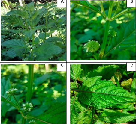
Figure 1. Photograph of Hyptis capitata Jacq., showing its (A) habit of growth, (B) stem, (C) flower, and (D) leaf
Preparation of Plant Materials and Extraction
The air-dried leaves were coarsely powdered using an electric blender. A total of 250 g of the powdered samples were extracted with 2.23 L of 90% methanol. The mixture was soaked for 48 hours with intermittent shaking and then filtered through Whatman No. 1 filter paper. The filtrates were concentrated by evaporating the solvent to dryness at 40°C using a rotary evaporator. The resulting extracts were wrapped in aluminum foil to protect them from light and stored in a dark room at room temperature.
Phytochemical Analysis
Preliminary phytochemical screening was performed qualitatively to identify the presence of phytoconstituents, including phenolics, alkaloids, flavonoids, steroids, terpenoids, tannins, saponins, and anthraquinones, following the methods outlined by Harborne[23].
Determination of Antioxidant Activity Using the 2,2-Diphenyl-1- picrylhydrazyl (DPPH) Radical Scavenging Method
The DPPH assay was conducted by the Department of Chemistry at Mindanao State University-Iligan Institute of Technology. As described by Alam et al.[24], Pisoschi and Negulescu[25], and Oyeyinka and Afolayan[26], 1,1-diphenyl-2-picrylhydrazyl (DPPH) is a stable free radical due to the delocalization of its spare electron, which imparts a deep violet color with an absorption band centered at 517 nm in ethanol. When DPPH is mixed with a substrate capable of donating a hydrogen atom, the DPPH radical is reduced, causing a loss of the violet color.
To assess the antioxidant potential of the methanolic leaf extract of Hyptis capitata, changes in the optical density of the DPPH radicals were monitored. A 0.2 mL sample extract was diluted with methanol and mixed with 2 mL of DPPH solution (0.5 mM). After 30 minutes, the absorbance was measured at 517 nm. The percentage inhibition of DPPH radicals was calculated using the following formula:
Where Abr represents the absorbance before the reaction, and Aar represents the absorbance after the reaction.
Determination of Total Phenolic Content
The total phenolic content of the extract was determined by the Department of Chemistry at Mindanao State University-Iligan Institute of Technology (MSU-IIT) using the Folin–Ciocalteu method[27] 200 μL of crude extract (1 mg/mL) was diluted to 3 mL with distilled water. Then, 0.5 mL of Folin–Ciocalteu reagent was added and mixed thoroughly for 3 minutes. Afterward, 2 mL of 20% sodium carbonate was added to the mixture, which was allowed to stand in the dark for 60 minutes. The absorbance was then measured at 765 nm. The total phenolic content was calculated from a standard calibration curve and expressed as milligrams of gallic acid equivalents (mg GAE) per gram of extract
Antibacterial Analysis
Preparation of Materials
The culture medium was prepared by dissolving 15.4 grams of Muller-Hinton agar in 400 mL of distilled water. A Normal Saline Solution (NSS) was also prepared for the inoculum. All materials were sterilized using an autoclave at 121 °C for 15 minutes. Additionally, Petri dishes were sterilized in a dry oven at 160 °C for 2 hours.
Preparation of Inoculum
Cultures of Bacillus cereus and Proteus vulgaris were prepared over 24 hours. The microorganisms were inoculated into a sterile saline solution using an inoculating loop until the turbidity was comparable to the 0.5 McFarland standard. The bacterial suspension was prepared using aseptic techniques inside a biological safety cabinet.
Preparation of Different Concentrations
A total of 1 mL of extract concentrations of 20 mg/mL, 30 mg/mL, 40 mg/mL, and 1000 mg/mL were prepared by weighing the desired amount in milligrams and dissolving it in dimethyl sulfoxide (DMSO) as the diluting solvent.
Disc Diffusion Method
The antibacterial activity of Hyptis capitata plant extracts was assessed following the Disc diffusion method by Biemer[28] with modification.
Sterile Muller-Hinton agar was cooled to 50-55 °C before being poured into individual sterile Petri dishes. An inoculum of test bacteria, prepared to be comparable to the 0.5 McFarland standard, was swabbed uniformly onto the solidified sterile Muller-Hinton agar plates using sterile cotton swabs. Ten microliters of each prepared extract concentration were then added to sterile discs (6 mm diameter, Whatman No. 1) using a micropipette. The discs were placed aseptically on the swabbed plates using sterile forceps, which were sterilized by soaking in 80% ethanol and passing through a flame. The inoculated plates were incubated upright at 37 °C for 24 hours. After incubation, the zones of inhibition formed around the discs were measured using a microcaliper. Known antibiotics, such as streptomycin discs, were introduced to the plates following the initial procedure. The diameters of the inhibition zones created by the extract and the known antibiotic were compared. All procedures were performed in triplicate across three trials at the Applied Microbiology Laboratory, University of San Carlos, Talamban, Cebu City, Philippines.
Statistical Analysis
One-way analysis of variance (ANOVA) was employed to analyze the data on the zones of inhibition (in mm) to assess whether the different concentrations, including the control, produced significantly different effects.
Phytochemicals are bioactive substances that exist naturally in plants that work with nutrients and dietary fiber to prevent illnesses[29]. A preliminary phytochemical investigation of Hyptis capitata leaf extract showed phytochemical components (Table 1).
The results showed a medium intensity reaction (++) to alkaloids and tannins. Flavonoids, saponins, and steroids have a strong (+++) intensity reaction, but cyanogenic glycosides and anthraquinones were not detected in the sample (-). According to Anyanwu and Dawet[30], phytochemical constituents found in plants such as alkaloids, flavonoids, saponins, steroids, and tannins are recognized to have antiprotozoal, antibacterial activities. Alkaloids are used as medicines, stimulants, narcotics, and poisons due to their powerful biological activity[31]. It is also known as tender nature drugs[32]. Several experimental studies have proven various biological and pharmacological flavonoids in recent years, particularly their antimicrobial effects, anti-inflammatory, antioxidant, and anti-tumor effects, all related to free radical scavenging[33].
Table 1. The different tests and their corresponding indication in the presence of phytochemical constituents of methanolic leaf extract of Hyptis capitata
Phytochemical Constituents | Tests | Hyptis capitata |
Alkaloids | Mayers test | ++ |
Flavonoids | Sodium hydroxide test | +++ |
Saponins | Frothing test | +++ |
Steroids | Libermann-Buccard test | +++ |
Tannins | Ferric chloride test | ++ |
Cyanogenic glycosides | Ferriferrocyanide test | - |
Anthraquinones | Bontragers test | - |
(+++): Strong intensity reaction; (++): Medium intensity reaction; (-): Nondetected
Saponins are highly poisonous, as they cause hemolysis of blood and cause cattle poisoning[34]. Hemolytic factor, anti-inflammatory, antibacterial, antifungal, antiviral, insecticidal, anti-cancer, and molluscicidal actions are among its biological and therapeutic properties[35]. Phytochemical constituents such as steroids, also known as cardiac glycosides, are among the most discovered plant phytoconstituents with therapeutic applications such as cardiac medicines; they also have anti-diarrheal properties, boosting intestinal absorption of Na+ water[36]. Tannins have anti-diarrheal properties and may cause proteins to precipitate on enterocytes, limiting bowel movements and intestinal discharge. Cyanogenic glycosides, which inhibit the formation of autocoids and prostaglandins, also have anti-diarrheal properties[36]. It also has antibacterial and anthelminthic activities[37]. Anti-cancer and anti- inflammatory characteristics are among the many bioactivities of anthraquinones[38]. Even though anthraquinones were not discovered in the leaf extract, this phytochemical offers a lot of potential for clinical usage in various disorders.
Several ethnic tribes in the Philippines used Hyptis capitata as a primary treatment for illnesses as well as diseases, and phytochemical analysis showed that no cyanogenic glycosides were present. Therefore, there is no risk of cyanide poisoning due to how traditional recipes are prepared. These results may aid in understanding its application in traditional medicine in the management of various diseases.
Determination of Antioxidant Activity Using the 2,2-Diphenyl-1-picrylhydrazyl (DPPH) Radical Scavenging Method
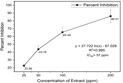
The investigated free radical activity of Hyptis capitata methanolic leaf extract was measured by DPPH assay. The leaf extract of H. capitata was analyzed in the blank due to its heavy color because it might affect the absorbance readings. As a result of its absorbance reading and % inhibition, there is evidence of antioxidant capabilities. Antioxidant molecules can quench DPPH radicals (by providing a hydrogen atom or electron donation) and convert them into colorless products. The lower IC50 value indicates a stronger ability of the extract to act as a DPPH scavenger. In contrast, the higher IC50 values indicate a lower scavenging activity of the scavengers as more scavengers were required to achieve a 50 % scavenging reaction[39]. The IC50 value of the methanolic leaf extract of H. capitata was determined to be 57ppm by using the calibration curves and regression equation (Figure 2).
Figure 2. DPPH radical scavenging result: concentration of extract (ppm) vs. Percent inhibition and its IC50 value.
Based on IC50 values of the methanolic leaf extracts of H. capitata, it shows potent and active antioxidant activity. The results also show novel aspects of traditional medicine therapeutic value that different Indigenous People of the Philippines used. Antioxidant and anti-free radical activities are established as important for cardioprotective prevention. Thus, the positive effects of long-term usage of H. capitata preparations could be attributed to the plants found significant antioxidant characteristics.
Determination of Total Phenolic
The Total Phenolic Content (TPC) of Hyptis capitata methanolic leaf extract was measured spectrophotometrically and expressed in milligrams of gallic acid equivalents per extract (GAE/g) as the standard. The total phenolic content of the extracts was determined using the calibration curves regression equation (y= 0125x+0.0452; R2= 0.9995) shown in (Figure 3). As a result, the methanolic leaf extract of H. capitata contains a considerable amount total of phenolic content (22 mg gallic acid per gram extract). The result suggested that the phenolic content contributed significantly to the antioxidant activities of the medicinal herb. Plant substances containing phenolic compounds exert their antiproliferative effects through increased oxidant stress in cancer cells by inhibiting ROS-scavenging systems, inactivating pro-survival signals, activating apoptosis-related signals, and inducing DNA damage and inhibiting favorable signaling pathways to cancer cell growth[40]. It is also considered why this plant is utilized in various traditional herbal medicines because of these qualities.
Figure 3. Total Phenolic Content result: Absorbance Reading at 765nm Vs. Standard Deviation of Concentration of gallic acid, µg/ml
Determination of Antibacterial Activity Using the Disc Diffusion Method
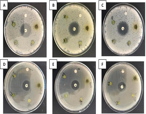
The disc diffusion method was used to assess the antibacterial activity of Hyptis capitata leaf extract against gram-positive and gram-negative bacteria (Bacillus cereus and Proteus vulgaris), respectively. Bacillus cereus is a foodborne pathogen that can produce toxins, causing two types of gastrointestinal illness: vomiting and diarrheal syndrome[41]. Proteus vulgaris are frequently recovered from infected wounds as they contaminate wounds and thus cause infections[42]. The efficacy of antibacterial substances was investigated by measuring the diameter of the zone of inhibition (ZOI), which contained paper discs with a diameter of 6mm. The test organisms were tested by a methanolic leaf extract of H. capitata. (Figure 4). DMSO serves as negative control and streptomycin as a positive control.
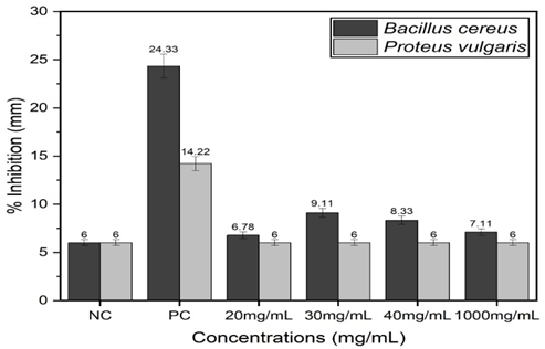
Methanolic leaf extracts show inhibitory zones from gram-positive bacteria (Table 4). In the concentration of 20 mg/mL gained, a mean ZOI of (6.78 mm), 30 mg/mL (9.1 mm), 40 mg/mL (8.33mm), and 1000mg/mL gained mean ZOI of (7.11mm). In contrast, gram-negative bacteria show resistance to the methanolic leaf extract of H. capitata. These findings can also be seen in the graph representation in Figure 5.
Figure 4. Antibacterial inhibition of H. capitata extract on Gram-positive bacteria Bacillus cereus (A-C) and Gram-negative bacteria Proteus vulgaris (D- F), with D - DMSO, S - Streptomycin and different extract concentrations (20mg/mL, 30mg/mL, 40mg/mL, and 1000mg/mL).
Table 4. Mean Zone of Inhibition in 3 trials from Gram-positive and Gram- negative bacteria on Hyptis capitata methanolic leaf extract.
Mean Zone of Inhibition (mm) | ||||||
Bacteria | DMSO | Streptomycin | 20 mg/ml | 30 mg/ml | 40 mg/ml | 1000 mg/ml |
Bacillus cereus |
6 |
24.33±0.57 |
6.78±0.19 |
9.11±0.50 |
8.33±0.88 |
7.11±0.50 |
Proteus vulgaris | ||||||
6 | 14.22±0.84 | 6 | 6 | 6 | 6 |
Note: The size of the disc is 6 mm.
According to [43], antimicrobial activity was assessed using the following rating system: (< 10 mm zone of inhibition maybe expressed as weak), (10-13 mm zone of inhibition is partially active); (14-19 mm zone of inhibition is active), and (> 19 mm zone of inhibition is very active). The antibacterial activity of H. capitata is rated as "weak" based on the rating system and the calculated mean of ZOI. Therefore, the methanolic leaf extract of H. capitata is not an excellent antibacterial agent.
Figure 5. Mean ZOI formed between gram-positive and gram-negative bacteria using different concentrations (mg/ml) of Hyptis capitata methanolic leaf extracts with negative and positive controls.
In this experiment, the highest inhibition occurred at 30 mg/mL concentrations. The antibacterial effect of H. capitata is attributed to the phytochemical constituents present in it. The leaves of H. capitata are rich in phytonutrients such as alkaloids, flavonoids, steroids, saponins, and tannins. These secondary metabolites exert an antimicrobial effect through different mechanisms on different microorganisms[30]. Saponin is one of the secondary metabolites with a potent antimicrobial property[44]. Saponins have a lytic action on erythrocyte membranes, a property that has been used for their detection. According to Natta and Orapan[45], the cell walls of gram- positive bacteria are more susceptible to antibiotics and even many natural products.
However, gram-negative bacteria show resistance to the methanolic leaf extract of H. capitata. According to [46], gram-negative bacteria tend to be more resistant to lipophilic and amphiphilic inhibitors than gram-positive bacteria. Recent research has revealed that many drug efflux pumps, many with exceptionally broad specificities, play an important role in Gram-negative bacteria intrinsic resistance[47]. The antibacterial drugs capable of bypassing or blocking efflux pumps and re-establishing the antibacterial effectiveness of other antibiotics must be discovered and developed[48].
Phytochemical analysis revealed the presence of Alkaloids, flavonoids, saponins, steroids, and tannins. In relation to the phytochemical constituents present in the leaf of Hyptis capitata, there is an active antioxidant property (IC50 57ppm). The disc diffusion antibacterial assay demonstrated a minor inhibition in gram-positive bacteria. However, it was weak compared to standard antibacterial activity criteria. Nonetheless, the phytochemical constituents present in the leaf extract contribute to antibacterial activities. Gram-negative bacteria showed resistance to the sample.
In conclusion, the findings demonstrate the antioxidant properties of plant extracts due to their phytochemical constituents. It was also discovered that the methanol extracts of Hyptis capitata contain phenolic content, which is beneficially attributed to antioxidant properties. These qualities of leaf extract could help explain why these plants are so valuable in traditional medicine. The methanolic leaf extract of this plant has potential antibacterial activities against the gram-positive bacteria (Bacillus cereus) and thus supports the IP claims that this plant can treat diarrhea, but gram-negative bacteria show no potential antibacterial activities in treating wound infections.
Acknowledgment
This research would not be possible without the support of the DOST-ASTHRDP for the research funding and financial assistance.
The authors declare that they have no conflict of interest
No funding sources
The study was approved by the Mindanao State University – Iligan Institute of Technology, Iligan City, 9200 Philippines
Iwu, M. W., A. R. Duncan, and C. O. Okunji. "New Antimicrobials of Plant Origin." ASHS Press, 1999, pp. 457-462.
Govindarajulu, B., et al. "Mosquito Larvicidal Efficacy of the Leaf Extracts of Annona reticulata Against Aedes aegypti." International Journal of Current Microbiology and Applied Sciences, vol. 4, 2015, pp. 132-140.
Rupa, D., Y. C. Sulistyaningsih, D. Dorly, and D. Ratnadewi. "Identification of Secretory Structure, Histochemistry and Phytochemical Compounds of Medicinal Plant Hyptis capitata Jacq." Biotropia, vol. 24, no. 2, 2017, pp. 94-103. doi: 10.11598/btb.2017.24.2.499.
Kusuma, I. W., et al. "Biological Activities and Phytochemicals of Hyptis capitata Grown in East Kalimantan, Indonesia." Journal of Applied Biology & Biotechnology, vol. 8, no. 2, 2020, pp. 58-64. doi: 10.7324/jabb.2020.80210.
DeFilipps, R. A., S. L. Maina, and J. Crepin. Medicinal Plants of the Guianas (Guyana, Surinam, French Guiana). Smithsonian Institution Press, 2003.
Ahmad, F. B., and D. K. Holdsworth. "Medicinal Plants of Sabah, East Malaysia – Part I." Pharmaceutical Biology, vol. 41, no. 5, 2008, pp. 340-346. doi: 10.1076/phbi.41.5.340.15940.
Biswas, A., M. A. B. Miah, M. Roy, and S. Bhadra. "Inherited Folk Pharmaceutical Knowledge of Tribal People in the Chittagong Hill Tracts Bangladesh." Indian Journal of Traditional Knowledge, vol. 9, 2010, pp. 77-89.
Gailea, R., A. A. Bratawinata, R. Pitopang, and I. W. Kusuma. "The Use of Various Plant Types as Medicines by Local Community in the Enclave of the Lore-Lindu National Park of Central Sulawesi, Indonesia." Global Journal of Research on Medicinal Plants & Indigenous Medicine, vol. 5, no. 1, 2016, pp. 29-40.
Dapar, M. L. G. Ethnobotany of the Mountain Regions of Southeast Asia. Springer, 2020.
"Labor Market Intelligence Report." Technical Education and Skills Development Authority, issue 3, 2020.
Dapar, M. L. G., et al. "Ethnomedicinal Plants Used for the Treatment of Cuts and Wounds by the Agusan Manobo of Sibagat, Agusan del Sur, Philippines." Ethnobotany Research and Applications, vol. 19, 2020. doi: 10.32859/era.19.31.1-18.
Lañojan, R., et al. "Ethnomedicinal Resources of the Indigenous People’s (IP) Groups in the SOCSARGEN Region." Journal of Health Research and Society, vol. 1, 2018. doi: 10.34002/jhrs.v1i0.8.
Olowa, L., and C. Demayo. "Ethnobotanical Uses of Medicinal Plants Among the Muslim Maranaos in Iligan City, Mindanao, Philippines." Advances in Environmental Biology, vol. 9, no. 27, 2015, pp. 204-215.
Cordero, C., and G. J. D. Alejandro. "Medicinal Plants Used by the Indigenous Ati Tribe in Tobias Fornier, Antique, Philippines." Biodiversitas: Journal of Biological Diversity, vol. 22, no. 2, 2021. doi: 10.13057/biodiv/d220203.
Tantengco, O. A. G., et al. "Ethnobotanical Survey of Medicinal Plants Used by Ayta Communities in Dinalupihan, Bataan, Philippines." Pharmacognosy Journal, vol. 10, no. 5, 2018, pp. 859-870. doi: 10.5530/pj.2018.5.145.
Arquion, R. D., et al. "Ethnobotanical Study of Indigenous Plants Used by Local People of Agusan del Sur, Philippines." Asia Pacific Higher Education Research Journal, vol. 2, no. 2, 2015. doi: 10.56278/apherj.v2i2.102.
Campilan, J. R., et al. "Quantitative Ethnobotanical Study, Phytochemical Screening and Antibacterial Assay of Ethnomedicinal Plants of T’boli in Lemsnolon, Tboli, South Cotabato." International Journal of Pharmacology, Phytochemistry and Ethnomedicine, vol. 13, 2019, pp. 45-61. doi: 10.18052/www.scipress.com/IJPPE.13.45.
Tafurt García, G., L. Jiménez Vidal, and A. Calvo Salamanca. "Antioxidant Capacity and Total Phenol Content of Hyptis spp., P. heptaphyllum, T. panamensis, T. rhoifolia, and Ocotea sp." Revista Colombiana de Química, vol. 44, no. 2, 2016, pp. 28-33. doi: 10.15446/rev.colomb.quim.v44n2.55217.
Falcão, D. Q., et al. "Triterpenos de Hyptis fasciculata Benth." Revista Brasileira de Farmacognosia, vol. 13, 2003, pp. 81-83. doi: 10.1590/s0102-695x2003000300030.
Shan, B., et al. "Antioxidant Capacity of 26 Spice Extracts and Characterization of Their Phenolic Constituents." Journal of Agricultural and Food Chemistry, vol. 53, no. 20, 2005, pp. 7749-7759. doi: 10.1021/jf051513y.
McNeil, M., P. Facey, and R. Porter. "Essential Oils from the Hyptis Genus- A Review (1909-2009)." Natural Product Communications, vol. 6, no. 11, 2011. doi: 10.1177/1934578x1100601149.
Bezerra, J. W. A., et al. "Chemical Composition and Toxicological Evaluation of Hyptis suaveolens (L.) Poiteau (Lamiaceae) in Drosophila melanogaster and Artemia salina." South African Journal of Botany, vol. 113, 2017, pp. 437-442. doi: 10.1016/j.sajb.2017.10.003.
Harborne, J. B. Phytochemical Methods, 3rd ed., Chapman and Hall, 1973.
Alam, M. N., N. J. Bristi, and M. Rafiquzzaman. "Review on in Vivo and in Vitro Methods Evaluation of Antioxidant Activity." Saudi Pharmaceutical Journal, vol. 21, no. 2, 2013, pp. 143-152. doi: 10.1016/j.jsps.2012.05.002.
Pisoschi, A. M., and G. P. Negulescu. "Methods for Total Antioxidant Activity Determination: A Review." Biochemistry & Analytical Biochemistry, vol. 01, no. 01, 2012. doi: 10.4172/2161-1009.1000106.
Oyeyinka, B. O., and A. J. Afolayan. "Comparative and Correlational Evaluation of the Phytochemical Constituents and Antioxidant Activity of Musa sinensis L. and Musa paradisiaca L. Fruit Compartments (Musaceae)." The Scientific World Journal, vol. 2020, 2020, pp. 1-12. doi: 10.1155/2020/4503824.
Waterhouse, A. L. "Determination of Total Phenolics." Current Protocols in Food Analytical Chemistry, 2003. doi: https://doi.org/10.1002/0471142913.fai0101s06.
Biemer, J. "Antimicrobial Susceptibility Testing by the Kirby-Bauer Disc Diffusion Method." Annals of Clinical Laboratory Science, vol. 3, no. 2, 1973.
M. C., D. "Proximate, Phytochemical and Nutrient Compositions of Some Fruits, Seeds and Leaves of Some Plant Species at Umudike, Nigeria." ARPN Journal of Agricultural and Biological Science, vol. 5, no. 1, 2010.
Anyanwu, C. U., and G. C. Nwosu. "Assessment of the Antimicrobial Activity of Aqueous and Ethanolic Extracts of Piper guineense Leaves." Journal of Medicinal Plants Research, vol. 8, no. 10, 2014, pp. 436-440. doi: 10.5897/jmpr12.976.
Heinrich, M., J. Mah, and V. Amirkia. "Alkaloids Used as Medicines: Structural Phytochemistry Meets Biodiversity—An Update and Forward Look." Molecules, vol. 26, no. 7, 2021, p. 1836. https://doi.org/10.3390/molecules26071836.
Hannan, J. M. A., et al. "Evaluation of Phytochemical Screening and Antimicrobial Activities of Ethanolic Extracts of Leaves and Bark of Ardisia Colorata." International Journal of Pharmaceutical and Life Sciences, vol. 2, no. 4, 2013, pp. 158-164. https://doi.org/10.3329/ijpls.v2i4.17115.
Auwal, M. S., et al. "Preliminary Phytochemical and Elemental Analysis of Aqueous and Fractionated Pod Extracts of Acacia Nilotica (Thorn Mimosa)." Veterinary Research Forum, vol. 5, no. 2, 2014, pp. 95-100.
Kar, A. Pharmacognosy and Pharmacobiotechnology. 2nd ed., New Age International, 2007, p. 898.
Moghimipour, E., and S. Handali. "Saponin: Properties, Methods of Evaluation and Applications." Annual Research & Review in Biology, vol. 5, no. 3, 2015, pp. 207-220. https://doi.org/10.9734/arrb/2015/1167
Tiwari, P., et al. "Phytochemical Screening and Extraction: A Review." Internationale Pharmaceutica Scientia, vol. 1, no. 1, 2011.
Su, Y., et al. "Antioxidant Activity of Flavonoids Isolated from Scutellaria Rehderiana." Journal of the American Oil Chemists' Society, vol. 77, no. 8, 2000, pp. 807-812. https://doi.org/10.1007/s11746-000-0129-y.
Chien, S. C., et al. "Naturally Occurring Anthraquinones: Chemistry and Therapeutic Potential in Autoimmune Diabetes." Evid Based Complement Alternat Med., vol. 2015, p. 357357, 2015. https://doi.org/10.1155/2015/357357.
Ghasemi Pirbalouti, A., et al. "Antioxidant Activity, Total Phenolic and Flavonoid Contents of Some Medicinal and Aromatic Plants Used as Herbal Teas and Condiments in Iran." Journal of Medicinal Food, vol. 17, no. 10, Oct. 2014, pp. 1151-1157. https://doi.org/10.1089/jmf.2013.0057.
Chikara, S., et al. "Oxidative Stress and Dietary Phytochemicals: Role in Cancer Chemoprevention and Treatment." Cancer Letters, vol. 413, 28 Jan. 2018, pp. 122-134. https://doi.org/10.1016/j.canlet.2017.11.002.
Fazeli-Nasab, B., et al. "Investigation of Antimicrobial Activity of Medicinal Plant Extracts on Bacillus Cereus Isolated from Soil." Gene, Cell and Tissue, vol. 9, no. 1, 2021. https://doi.org/10.5812/gct.115133.
Samy, R. P., and S. Ignacimuthu. "Antibacterial Activity of Some Folklore Medicinal Plants Used by Tribals in Western Ghats of India." Journal of Ethnopharmacology, vol. 69, no. 1, Jan. 2000, pp. 63-71. https://doi.org/10.1016/s0378-8741(98)00156-1.
Gutierrez, R. M., et al. "Antibacterial Potential of Some Medicinal Plants of the Cordillera Region, Philippines." Indian Journal of Traditional Knowledge, vol. 12, no. 4, 2013, pp. 630-637.
Ameri, A., et al. "Medicinal Plants Contain Mucilage Used in Traditional Persian Medicine (TPM)." Pharmaceutical Biology, vol. 53, no. 4, 2014, pp. 615-623. https://doi.org/10.3109/13880209.2014.928330.
Norajit, K., et al. "Antibacterial Effect of Five Zingiberaceae Essential Oils." Molecules, vol. 12, no. 8, 23 Aug. 2007, pp. 2047-2060. https://doi.org/10.3390/12082047.
Vaara, M. "Antibiotic-Supersusceptible Mutants of Escherichia Coli and Salmonella Typhimurium." Antimicrobial Agents and Chemotherapy, vol. 37, no. 11, Nov. 1993, pp. 2255-2260. https://doi.org/10.1128/AAC.37.11.2255.
Nikaido, H. "Multidrug Efflux Pumps of Gram-Negative Bacteria." Journal of Bacteriology, vol. 178, no. 20, Oct. 1996, pp. 5853-5859. https://doi.org/10.1128/jb.178.20.5853-5859.1996.
Piddock, L. J. "Clinically Relevant Chromosomally Encoded Multidrug Resistance Efflux Pumps in Bacteria." Clinical Microbiology Reviews, vol. 19, no. 2, Apr. 2006, pp. 382-402. https://doi.org/10.1128/CMR.19.2.382-402.2006.