he author describes a case of mesial temporal lobe epilepsy who has unsual early age presentation. Otherwise normally born child and developmentally achieving milestones as per age had recent onset seizure episodes. The child presented in emergency of our hospital with status epilepticus. His seizures were aborted with medication and on subsequent work-up he was diagnosed as a case of mesial temporal lobe epilepsy.
Key findings: The key findings of the abstract include the unusual early age presentation of mesial temporal lobe epilepsy in an otherwise normally developing child. Despite achieving developmental milestones appropriately for age, the child experienced recent-onset seizure episodes, culminating in status epilepticus. Subsequent evaluation confirmed the diagnosis of mesial temporal lobe epilepsy. What is known and what is new? Rectal cancer management typically involves multimodal treatment. This study contrasts preoperative hypofractionated radiotherapy with concurrent chemoradiotherapy for locally advanced cases, assessing their efficacy and sphincter preservation outcomes. Findings reveal comparable tumor response rates, highlighting potential treatment options for clinicians managing rectal carcinoma. What is the implication, and what should change now? |
The study underscores the importance of evaluating alternative treatment modalities, such as preoperative hypofractionated radiotherapy, in managing locally advanced rectal carcinoma. Clinicians should consider these options to tailor treatment based on patient and resource considerations, potentially optimizing outcomes and preserving sphincter function in rectal cancer patients.
Epilepsy is one of the most common neurologic conditions in the world and is of a major public health concern. Epileptic seizures of the temporal lobe constitute the most frequent presentation of drug-resistant epilepsy which satisfactorily responds to surgical treatment [1]. In children with temporal lobe epilepsy, mesial temporal sclerosis (MTS) is considered an infrequent etiology [2, 3]. MTS was thought to be a disease of adults, but with the advances in neuroimaging, it started to be diagnosed at an increasingly younger age [4]. Magnetic Resonance Imaging (MRI) of people with epilepsy helps in diagnosis and further management of such patients. Here the author presents a case of a 1.5 year old, male child, who had recent onset tonic- clonic seizures and on investigations diagnosed as a case of mesial temporal lobe epilepsy.
A 1.5 year old, male child was brought to the emergency of a regional hospital with ongoing active seizure with semiology as uprolling of eyes, tonic posturing of all limbs, and impaired consciousness. He had no bladder and bowel incontinence during seizure. This episode started 15 to 20 minutes back while the child was on the way to the same hospital to consult the Pediatrician for seizure episodes the child was having for the past 12 hours. Child was managed as a case of status epilepticus in the emergency room with intravenous midazolam to abort the seizure. He was loaded with intravenous levetiracetam. Seizures were managed adequately.
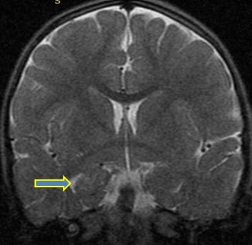
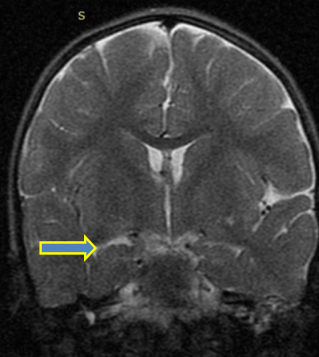
He was apparently well until one day back when he had similar four episodes of tonic-clonic movements in the last 12 hours. Each episode lasted for 3-5 minutes and used to abort itself. There was no history of fever, excessive crying, poor feeding and lethargy. He was the first born child to a non-consanguineously married couple with an uneventful antenatal history. He was born through normal vaginal delivery at term, with an average birth weight and smooth perinatal transition. He was developmentally gaining milestones age appropriate. There was no family history of developmental delay and seizure disorder. On examination the child was drowsy post seizure with stable vitals. Neurological examination was unremarkable. Clinical diagnosis of the first episode unprovoked a generalized seizure with status epilepticus was kept and neuro-imaging was advised. Contrast enhanced MRI (CT MRI) brain showed increased T2-weighted signal on magnetic resonance imaging (Figure 1 and 2) suggestive of right mesial temporal sclerosis with partially empty sella-turcica. Diagnosis of mesial temporal lobe epilepsy was kept and the child was continued on maintenance dose of levetiracetam. He was observed for seizure and encephalopathy for next 48 hours in which there was no seizure recurrence. EEG was planned in subsequent
follow up visits and had no seizure after a 3 week follow up.
Picture 1 and 2: Contrast enhanced MRI brain of the child. The increased T2-weighted are suggestive of right mesial temporal sclerosis.
Epilepsy is one of the most common neurologic conditions in the world and is of a major public health concern. The Medial Temporal Lobe (also known as the Mesial Temporal Lobe) structures are most commonly involved in Temporal Lobe Epilepsy. Mesial temporal sclerosis is the most common pathologic entity associated with refractory temporal lobe epilepsy (TLE); it is seen in as many as 60% to 80% of cases [5]. The etiology of mesial temporal sclerosis is unknown but studies suggest an increased incidence among family members and an association with precipitating insults during the first four to five years of life [6]. Mesial temporal sclerosis has a poor prognosis, considering its tendency of worsening epileptic seizures and a risk of becoming refractory epilepsy. Antiepileptic drugs such as carbamazepine, oxcarbazepine, levetiracetam, lamotrigine, and topiramate are used as medical management of mesial temporal lobe epilepsy. In refractory epilepsy, surgical treatment may be indicated. Studies have shown a better long-term outcome in patients with MTS receiving surgical therapy in comparison with medical therapy [7]. Currently, this patient is responding to medical treatment alone but if seizures are not controlled satisfactorily in the future, temporal lobectomy may be an option to improve quality of life by controlling symptoms.
No funding sources
None declared
The study was approved by the Institutional Ethics Committee of Regional Hospital of Himachal Pradesh.