Background: The present study was done for Quantitative Analysis of 68Ga-PSMA PET/CT in Prostate Cancer Patients. Material and Methods: Forty four patients with suspected PCa on clinical assessment and/or raised serum PSA levels (>4ng/ml) were prospectively recruited. Patient who underwent prostatic biopsy prior to 68Ga-PSMA PET/CT and mpMRI, deranged renal function tests or coagulogram and refused consent were excluded. Forty four patients underwent 68Ga-PSMA PET/CT and 33 patients underwent biopsy. Any focal tracer uptake on 68Ga-PSMA PET/CT was regarded as pathological. Histopathology was taken as gold standard. SUVmaxof the lesions were measured for quantization. Results: Mean age of patients was 66.61±9.3 years and median PSA level was 13.4 ng/ml (IQR 13.9). Malignancy was detected in 16/33 (48.5%) patients who underwent biopsy. The median SUVmax for cases with confirmed malignancy (n=16) was found out to be 17.6 (IQR 18.9). Median SUVmax for cases with no evidence of malignancy was 6.3 (IQR 2.80). The difference between the two groups was found to be statistically significant (Mann Whitney U test, P =0.002). Conclusions: The present study concluded that SUVmax (PSMA expression) was significantly higher in Prostate cancer cases as compared to benign cases.
Keywords
Quantitative Analysis
SUVmax
68GA PSMA PET/CT
Prostate Cancer Patients
INTRODUCTION
68Ga PSMA PET/CT is a relatively new hybrid functional imaging modality which has been extensively used in PCa in the last decade. The basis of 68Ga PSMA PET/CT is targeting of PSMA receptors which are over expressed (100-1000x) in PCa as compared to normal prostate cells. Initial studies on bio distribution of 68Ga PSMA PET/CT reported tracer uptake (PSMA expression) in normal prostate as measured by SUVmax. [1-4]
The reported SUVmax values in a normal prostate ranged from 2.5-8.8. 68Ga PSMA PET/CT has shown encouraging results in staging, restaging, response assessment and evaluation of biochemical recurrence in PCa.[5-7]
Aims & Objectives
The present study was done with the objective of Quantitative Analysis of 68Ga-PSMA PET/CT in Prostate Cancer Patients
MATERIAL AND METHODS
Study Design: Prospective pilot study
Place of Study: This study have been conducted at the Department of Nuclear Medicine, PGIMER Chandigarh in collaboration with the Department Urology, Radiodiagnosis and Histopathology, PGIMER, Chandigarh
Period of study: The study was done during the period between January, 2018 and June, 2019.
Inclusion Criteria
Patients having suspicion of carcinoma prostate on clinical assessment and/or raised serum PSA levels(>4ng/ml).
Patient not suffering from bleeding diathesis or renalfailure.
Patient consent to participate in thestudy.
Exclusion Criteria
Patient with prior diagnosed PCa or who underwent prostatic biopsy priorto68Ga-PSMA PET/CT and mpMRI
Patients with deranged renal function tests orcoagulogram
Patient refused to participate in thestudy
Study Population: A total of 44patients suspected for PCa on the basis of clinical assessment and/or raised PSA were recruited for the study.
Figure-1: Study Methodology
Patients suspected for prostate adenocarcinoma and/or serum PSA levels >4ng/ml and willing to participate in the study were recruited. Study was approved by Institutional Ethics Committee, PGIMER Chandigarh. Written informed consent was obtained. After clinical assessment, mpMRI and68Ga-PSMA PET/CT were done. Biopsy was done in cases with high clinical suspicion of PCa.
Statistical Analysis
Qualitative variables are described using number and percentages. Normally distributed continuous quantitative data is described using mean and standard deviation. Skewed quantitative data is described using median and interquartile range. The normality of data will be checked by measures of Kolmogorov-Smirnov tests of normality. The sensitivity, specificity, NPV and PPV of 68Ga-PSMA PET/CT was estimated using a 2x2 table. The statistical analysis was conducted using the IBM SPSS STATISTICS (version 23.0)
RESULTS
In this prospective study, a total of 44 patients suspected to have PCa were recruited on the basis of clinical assessment and serum PSA levels. All patients were subjected to 68Ga-PSMA PET/CT acquisition. Out of the total 44 patients, 33 underwent biopsy. The remaining 11 patients did not undergo biopsy after revised clinical assessment based on negative. Therefore, 33 patients underwent 68Ga-PSMA PET/CT and biopsy.
In this study, malignancy was detected in 16/33 (48.5%) patients on biopsy. In 17/33 (51.5%), no evidence of malignancy was noted.
The mean and SD of the injected 68Ga PSMA radioactivity was 2.79 ±0.89 mCi (103.2 ± 32.9MBq). The median time between the injection and scan acquisition was 65 min (IQR 32.5 min). SUVmaxof the lesions were measured for quantitation and the results were compared with histopathological reference standard.
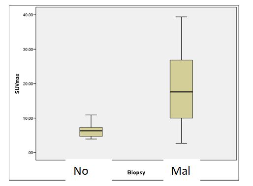
The median SUVmax for cases with confirmed malignancy (n=16) was found out to be 17.6 (IQR 18.9). Median SUVmax for cases with no evidence of malignancy was 6.3 (IQR 2.80). The difference between the two groups was found to be statistically significant (Mann Whitney U test, P =0.002). Fig. 2 shows box plot comparing SUVmax between the 2 subgroups.
Figure -2: Box Plot showing SUVmax of PSMA in malignant and non malignant lesions
DISCUSSION
In the present study, PSMA expression measured by SUVmax on 68Ga PSMA PET/CT was found to be significantly higher in PCa as compared to benign cases (p <0.002). In our study, median and range of SUVmax of patients with PCa was found to be 17.6 and 2.7 - 39.4. In a study [6] of biopsy proven PCA cases, they found median and range of SUVmax was 11.5 and 2.7 – 66.4.
CONCLUSION
The present study concluded that SUVmax (PSMA expression) was significantly higher in Prostate cancer cases as compared to benign cases .
Conflict of Interest:
The authors declare that they have no conflict of interest
Funding:
No funding sources
Ethical approval:
The study was approved by the PGIMER, Chandigarh
REFERENCES
Von Eyben, F. E., et al. "68Ga-Labeled Prostate-Specific Membrane Antigen Ligand Positron Emission Tomography/Computed Tomography for Prostate Cancer: A Systematic Review and Meta-Analysis." Eur Urol Focus 4. (2018), pp. 686-93.
Zhang, J., et al. "Diagnostic Performance of 68 Ga-PSMA PET/CT in the Detection of Prostate Cancer Prior to Initial Biopsy: Comparison with Cancer-Predicting Nomograms." Eur J Nucl Med Mol Imaging 46. (2019), pp. 908-20.
Bray, F., et al. "Global Cancer Statistics 2018: GLOBOCAN Estimates of Incidence and Mortality Worldwide for 36 Cancers in 185 Countries." CA: A Cancer Journal for Clinicians 68. (2018), pp. 394-424.
Alizadeh, M., and S. Alizadeh. "Survey of Clinical and Pathological Characteristics and Outcomes of Patients With Prostate Cancer." Glob J Health Sci 6. (2014), pp. 49-57.
Bancroft, E. K., et al. "Targeted Prostate Cancer Screening in BRCA1 and BRCA2 Mutation Carriers: Results from the Initial Screening Round of the IMPACT Study." Eur Urol 66. (2014), pp. 489-99.
Uprimny, C., et al. "68Ga-PSMA-11 PET/CT in Primary Staging of Prostate Cancer: PSA and Gleason Score Predict the Intensity of Tracer Accumulation in the Primary Tumour." Eur J Nucl Med Mol Imaging 44. (2017), pp. 941-49.
Demirci, E., et al. "Normal Distribution Pattern and Physiological Variants of 68Ga-PSMA-11 PET/CT Imaging." Nuclear Medicine Communications 37. (2016), pp. 1169-79.
License
Creative Commons Attribution-NonCommercial-NoDerivatives 4.0 International License
All papers should be submitted electronically. All submitted manuscripts must be original work that is not under submission at another journal or under consideration for publication in another form, such as a monograph or chapter of a book. Authors of submitted papers are obligated not to submit their paper for publication elsewhere until an editorial decision is rendered on their submission. Further, authors of accepted papers are prohibited from publishing the results in other publications that appear before the paper is published in the Journal unless they receive approval for doing so from the Editor-In-Chief.
Himalayan Journal of Applied Medical Sciences and Research open access articles are licensed under a Creative Commons Attribution-Share A like 4.0 International License. This license lets the audience to give appropriate credit, provide a link to the license, and indicate if changes were made and if they remix, transform, or build upon the material, they must distribute contributions under the same license as the original.
Advertisement
Recommended Articles
Research Article
Clinical and Functional Outcomes of Interlocking and Küntscher Nailing in the Management of Femoral Shaft Fractures: A Prospective Study
Ahmed Abdalzahra Mohaisen ,
...
Mohammed Saad Abdulzahra
Published: 10/01/2026
Download PDF
Cite
x
APA
None, A. A. M., None, H. A. A. & None, M. S. A. (2026). Clinical and Functional Outcomes of Interlocking and Küntscher Nailing in the Management of Femoral Shaft Fractures: A Prospective Study. Himalayan Journal of Applied Medical Sciences and Research, 7(1), 1-7.
MLA
None, Ahmed Abdalzahra Mohaisen, Hassan Ali Abid and Mohammed Saad Abdulzahra . "Clinical and Functional Outcomes of Interlocking and Küntscher Nailing in the Management of Femoral Shaft Fractures: A Prospective Study." Himalayan Journal of Applied Medical Sciences and Research 7.1 (2026): 1-7.
Chicago
None, Ahmed Abdalzahra Mohaisen, Hassan Ali Abid and Mohammed Saad Abdulzahra . "Clinical and Functional Outcomes of Interlocking and Küntscher Nailing in the Management of Femoral Shaft Fractures: A Prospective Study." Himalayan Journal of Applied Medical Sciences and Research 7, no. 1 (2026): 1-7.
Harvard
None, A. A. M., None, H. A. A. and None, M. S. A. (2026) 'Clinical and Functional Outcomes of Interlocking and Küntscher Nailing in the Management of Femoral Shaft Fractures: A Prospective Study' Himalayan Journal of Applied Medical Sciences and Research 7(1), pp. 1-7.
Vancouver
Ahmed Abdalzahra Mohaisen AAM, Hassan Ali Abid HAA, Mohammed Saad Abdulzahra MSA. Clinical and Functional Outcomes of Interlocking and Küntscher Nailing in the Management of Femoral Shaft Fractures: A Prospective Study. Himalayan Journal of Applied Medical Sciences and Research. 2026 Jan;7(1):1-7.
Download PDF
Research Article
Bones in Balance: Awareness of Rickets and Nutritional Deficiencies Among the Hamirpur Community
Vishal Dhatwalia,
Swati Chandel
Published: 10/12/2024
Download PDF
Cite
x
APA
Dhatwalia, V. & Chandel, S. (2024). Bones in Balance: Awareness of Rickets and Nutritional Deficiencies Among the Hamirpur Community. Himalayan Journal of Applied Medical Sciences and Research, 5(2), 1-5.
MLA
Dhatwalia, Vishal and Swati Chandel. "Bones in Balance: Awareness of Rickets and Nutritional Deficiencies Among the Hamirpur Community." Himalayan Journal of Applied Medical Sciences and Research 5.2 (2024): 1-5.
Chicago
Dhatwalia, Vishal and Swati Chandel. "Bones in Balance: Awareness of Rickets and Nutritional Deficiencies Among the Hamirpur Community." Himalayan Journal of Applied Medical Sciences and Research 5, no. 2 (2024): 1-5.
Harvard
Dhatwalia, V. and Chandel, S. (2024) 'Bones in Balance: Awareness of Rickets and Nutritional Deficiencies Among the Hamirpur Community' Himalayan Journal of Applied Medical Sciences and Research 5(2), pp. 1-5.
Vancouver
Dhatwalia V, Chandel S. Bones in Balance: Awareness of Rickets and Nutritional Deficiencies Among the Hamirpur Community. Himalayan Journal of Applied Medical Sciences and Research. 2024 Jul;5(2):1-5.
Download PDF
Research Article
The Role of Elevated CRP in Assessing the Severity and as a Prognostic Marker in Covid19 Patients
V. S. Sheshan,
...
Shekhar N. Chandra
Published: 04/07/2021
Download PDF
Cite
x
APA
S. Sheshan, V., Mulla Bahuddeen, M., None, M. & N. Chandra, S. (2021). The Role of Elevated CRP in Assessing the Severity and as a Prognostic Marker in Covid19 Patients. Himalayan Journal of Applied Medical Sciences and Research, 2(2), 1-4.
MLA
S. Sheshan, V., et al. "The Role of Elevated CRP in Assessing the Severity and as a Prognostic Marker in Covid19 Patients." Himalayan Journal of Applied Medical Sciences and Research 2.2 (2021): 1-4.
Chicago
S. Sheshan, V., M. Mulla Bahuddeen, Mahendra and Shekhar N. Chandra. "The Role of Elevated CRP in Assessing the Severity and as a Prognostic Marker in Covid19 Patients." Himalayan Journal of Applied Medical Sciences and Research 2, no. 2 (2021): 1-4.
Harvard
S. Sheshan, V., Mulla Bahuddeen, M., None, M. and N. Chandra, S. (2021) 'The Role of Elevated CRP in Assessing the Severity and as a Prognostic Marker in Covid19 Patients' Himalayan Journal of Applied Medical Sciences and Research 2(2), pp. 1-4.
Vancouver
S. Sheshan V, Mulla Bahuddeen M, Mahendra M, N. Chandra S. The Role of Elevated CRP in Assessing the Severity and as a Prognostic Marker in Covid19 Patients. Himalayan Journal of Applied Medical Sciences and Research. 2021 Jul;2(2):1-4.
Download PDF
Research Article
A Critical Analysis of Spinal Block Anesthesia under Ultrasonographic Guided Technique: Is Levobupivacaine Alone Effective or on Adjuvant is Warranted?
Verma AK
Published: 20/09/2021
Download PDF
Cite
x
APA
AK, V. (2021). A Critical Analysis of Spinal Block Anesthesia under Ultrasonographic Guided Technique: Is Levobupivacaine Alone Effective or on Adjuvant is Warranted?. Himalayan Journal of Applied Medical Sciences and Research, 2(2), 1-4.
MLA
AK, Verma. "A Critical Analysis of Spinal Block Anesthesia under Ultrasonographic Guided Technique: Is Levobupivacaine Alone Effective or on Adjuvant is Warranted?." Himalayan Journal of Applied Medical Sciences and Research 2.2 (2021): 1-4.
Chicago
AK, Verma. "A Critical Analysis of Spinal Block Anesthesia under Ultrasonographic Guided Technique: Is Levobupivacaine Alone Effective or on Adjuvant is Warranted?." Himalayan Journal of Applied Medical Sciences and Research 2, no. 2 (2021): 1-4.
Harvard
AK, V. (2021) 'A Critical Analysis of Spinal Block Anesthesia under Ultrasonographic Guided Technique: Is Levobupivacaine Alone Effective or on Adjuvant is Warranted?' Himalayan Journal of Applied Medical Sciences and Research 2(2), pp. 1-4.
Vancouver
AK V. A Critical Analysis of Spinal Block Anesthesia under Ultrasonographic Guided Technique: Is Levobupivacaine Alone Effective or on Adjuvant is Warranted?. Himalayan Journal of Applied Medical Sciences and Research. 2021 Jul;2(2):1-4.
Sharma, V., Kumar, S. & Chadha, R. (2022). Quantitative Analysis of 68GA PSMA PET/CT in Prostate Cancer Patients at a Tertiary Care Hospital. Himalayan Journal of Applied Medical Sciences and Research, 3(2), 1-3.
MLA
Sharma, Viper, Sunil Kumar and Rishabh Chadha. "Quantitative Analysis of 68GA PSMA PET/CT in Prostate Cancer Patients at a Tertiary Care Hospital." Himalayan Journal of Applied Medical Sciences and Research 3.2 (2022): 1-3.
Chicago
Sharma, Viper, Sunil Kumar and Rishabh Chadha. "Quantitative Analysis of 68GA PSMA PET/CT in Prostate Cancer Patients at a Tertiary Care Hospital." Himalayan Journal of Applied Medical Sciences and Research 3, no. 2 (2022): 1-3.
Harvard
Sharma, V., Kumar, S. and Chadha, R. (2022) 'Quantitative Analysis of 68GA PSMA PET/CT in Prostate Cancer Patients at a Tertiary Care Hospital' Himalayan Journal of Applied Medical Sciences and Research 3(2), pp. 1-3.
Vancouver
Sharma V, Kumar S, Chadha R. Quantitative Analysis of 68GA PSMA PET/CT in Prostate Cancer Patients at a Tertiary Care Hospital. Himalayan Journal of Applied Medical Sciences and Research. 2022 Jul;3(2):1-3.