Research Article | Volume 3 Issue 2 (July-Dec, 2022) | Pages 1 - 9
Bacteria Isolate from Different Soil Types Contaminated with Crude Oil in Birnin Kebbi Local Government Area, Kebbi State, Nigeria
 ,
1
Kebbi State University of Science and Technology Aliero, Birnin Kebbi, Kebbi State
2
Department of General Studies, School of Nursing and Midwifery Gusau, Zamfara State
Under a Creative Commons license
Open Access
Received
July 22, 2022
Revised
Aug. 12, 2022
Accepted
Sept. 17, 2022
Published
Oct. 20, 2022
Abstract

This paper was conducted to enumerate, isolate, and identify bacteria using cultural, morphological, and biochemical characteristics of different soil types (sandy, Clay, and Loamy) contaminated with crude oil in Birnin Kebbi Local Government Area, Kebbi State, Nigeria. The study was conducted between August and December 2020. 12 artificially polluted soil samples were collected and tested using appropriate methods (Grams staining, coagulase test, catalase test, oxidase test, urease test, citrate test, nitrate reduction test, etc.) in the study. The result of bacterial populations estimated in each original and saturated sample showed that the control (oil free) soil was found to be higher than the crude oil treated soil at different concentration (2.3 x 10¹⁰ CFU/g, 1.8 x 10¹⁰ CFU/g and 1.9 x 10⁹ CFU/g for Loamy Clay and Sandy soil respectively). In loamy treated soil, the organisms show growth even at 15% crude oil (3.0 x 10⁸ CFU/g, 1.8 x 10⁷ CFU/g and 1.1 x 10⁶ CFU/g for 5%, 10% and 15% crude oil respectively), whereas in clay soil the organisms were recovered at 5 – 10% crude oil (1.4 x 10⁷ CFU/g and 1.0 x 10⁶ CFU/g at 5% and 10% respectively). In sandy soil, the growth was only observed at concentration of 5% crude oil which was 1.3 x 10⁸ CFU/g. 21 hydrocarbon-degrading bacterial species were isolated and identified, the highest potential to utilize crude oil as a source of carbon and energy are as follows; Pseudomonas aerogenusa (A09 andC04), Acinetobacter (A04, B03), Bacillus cereus (A02, B05, and C01), Bacillus polymyxa (A07) Micrococcus luteus (B02, C07), Enterobacter (A06), Providentia spp (A03). In conclusion, this finding supports the fact that crude oil-degrading bacteria is widely distributed in the environment and therefore can be "easily" collectible from sites with no apparent history of crude oil pollution as it was in our case study.

Keywords
INTRODUCTION

Many microorganisms such as bacteria, fungi, and microalgae can degrade petroleum hydrocarbons and utilize them as the sole source of energy in their metabolism. Microorganisms, more especially bacteria, had opened up a new era of discussing oil pollution[1]. In addition, numerous genera of bacteria are known as good hydrocarbon degraders. Most of them belong to Pseudomonas, RhodococcusAeromonas, Alcaligenes, Acinetobacter, Ewingella Americana, Arthobacter, Bacillus, Brevibacterium, Flavobacterium, Geobacillus, Micrococcus, Mycobacterium, Ochrobactrum, Sphingomonas, Thermus,Xanthomonas species, Bacillus sphaericus and Bacillus azotoformans[2]

[3]reported bacterial degradation of petroleum hydrocarbons in a polluted tropical stream in Lagos, Nigeria. The investigator isolated nine bacterial strains, namely, Pseudomonasfluorescens, Pseudomonas aeruginosa, Bacillus subtilis,Bacillus species., Alcaligenes species, Acinetobacter lwoffi,Flavobacterium species, Micrococcus roseus, and Corynebacterium species were all found to degrade crude oil. [4]used Micrococcus sp. GS2-22, Corynebacterium sp. GS5-66, Flavobacterium sp. DS6-86 and Pseudomaonas sp. DS10-129 individually as well as their co-culture for crude oil biodegradation studies. They reported that bacterial co-culture degraded 78% of 1% crude oil, which was higher than the percentage degradation by individual cultures. [5]also reported two novel crude oil-degrading bacteria (Microbacterium oleivorans and Microbacterium hydrocarbonoxydans).

 In addition, tested the capacity of three bacterial strains, (Bacillus sp. SV9, Acinetobacter sp. SV4, and Pseudomonas sp., SV17) from contaminated soil in Ankleshwar (21°60' N 73°00' E), India for their ability to degrade the complex mixture of crude oil hydrocarbon (alkanes, aromatics, resins, and asphaltenes), sediments, heavy metals, and oily sludge. [6]isolated a psychrotrophic Rhodococcus species from oil-contaminated groundwater, which could utilize various petroleum hydrocarbons such as crude oil, diesel oil, and gasoline by almost 90% within 20 days. [7]observed two strains (R. wratislaviensis and R. aetherivorans) that could degrade more than 15 petroleum compounds from a mixture of benzene, toluene, cyclohexane, cyclohexanol, naphthalene ethylbenzene, m-xylene, p-xylene, o-xylene, octane, hexadecane, 2,2,4-trimethylpentane, methyl tert-butyl ether, ethyl tert-butyl ether, tert-butyl alcohol, and 2-ethylhexyl nitrate. The co-culture degraded 13 compounds completely. Interestingly, these strains had broad degradation capacities towards the more recalcitrant compounds such as isooctane, cyclohexane. Similarly, [8]studied the hydrocarbon degrading potential of Proteus vulgaris strain isolated from fish samples. However, [9]studied the utilization of engine oil, diesel oil, jet fuel, and kerosene as carbon sources by autotrophic nitrifying bacteria, Nitrobacter and Nitrosomonas species and reported that mixed culture of the isolates degraded 52% of crude oil followed by 40% by Nitrosomonas sp. and 20% by Nitrobacter species.

Furthermore, [10]reported qualitative analysis which confirmed that B. subtilis was one of the best crude oil degrading bacteria isolated from produced formation water of an onshore oil field in Sudan. It completely degraded short-chain n-alkanes while medium-chain n-alkanes were partially degraded considerably. In Norwegian crude oil, B. subtilis degraded n-alkanes with chain lengths C7, C8 and C9 completely but 31% of C10 n-alkanes were not degraded[10]. The metabolic pathways that hydrocarbon-degrading heterotrophs use can be classified as either aerobic or anaerobic. The aerobic degradation usually proceeds more rapidly and is considered to be more effective than the anaerobic one[11].

The major problem associated with crude oil exploration is the pollution/contamination of the environment. Release of hydrocarbons into the environment as a result of the activities taking place in the oil-producing states in Nigeria and Nigerian National Petroleum Corporation (NNPC) whether accidentally or due to human activities is the main cause of soil and water pollution. According to a report, an estimate of nine to thirteen million (1.5 million tons) of oil has been spilled into the Niger Delta ecosystem over the past 50 years and this was estimated 50 times the volume spilled in the Exxon Valdez oil spill in Alaska 1989[12]. 

Soil contamination with hydrocarbons causes extensive damage to the local system since the accumulation of pollutants in animals and plant tissues may cause death or mutations. Over the years, attempt to remediate hydrocarbon polluted sites using physiochemical methods had been in existence, but due to other implications, their applications were discouraged. The technology commonly used for soil remediation includes mechanical, burying, evaporation, dispersion, and washing. However, these technologies are expensive and can lead to incomplete decomposition of the contaminants[13].

In addition, bioremediation using microorganisms capable of degrading hydrocarbon is now believed to be more promising, effective, and environmentally friendly. Although many bacteria can metabolize organic pollutants, a single bacterium does not possess the enzymatic capability to degrade all or even most of the organic compounds in polluted soil. Mixed microbial communities have the most powerful biodegradative potential because the genetic information of more than one organism is necessary to degrade the complex mixtures of organic compounds present in contaminated areas[13].

Moreover, another major problem associated with bioremediation studies is the contaminants concentrations. These directly influence microbial activity. When concentrations are too high, the contaminants may have toxic effects on the resident microbes. In contrast, low contaminants concentration may prevent the induction of bacterial degradation enzymes[14]. 

There is a paucity of data on the isolation and characterizations of bacteria from different soil types contaminated with crude oil in the Birnin Kebbi Local Government Area. Thus, 

This research is aimed at identifying the competent indigenous bacterial species for bioremediation of crude oil contaminated soil which would help in knowing specific approach for a particular soil type in tackling soil environmental problems associated with crude oil pollution.

MATERIAL AND METHOD

The research study was conducted in Kebbi State, and the different soil type samples were collected from Birnin Kebbi Local Government Area, the State Capital of Kebbi State of the Northwestern part of Nigeria. The State has a total area of 36,800km2; it lies between latitude 12.450N in the north and 4.20E to the east. The state has three seasons mainly, dry, rainy, and harmattan seasons. The temperature of the State usually ranges from a maximum of 42.70C to a minimum of 24.90C. The major types of soil in Kebbi State just like many other regions are sandy, clay, and loamy soil.[15].


2.1 Collection of Samples (Collection of crude oil/ Soil Samples)

Nigerian crude oil (Bonny light crude oil) was collected from Kaduna Refinery Petrochemical Company (KRPC), Kaduna Nigeria. About 500-600 grams of different soil types sandy, loamy, and clay soil were collected within Birnin Kebbi Local Government Area of Kebbi State. Different concentrations of sterilized crude oil (5ml, 10ml, and 15ml) were used to artificially contaminate a fixed amount of Sandy soil collected using the probe into three conical flasks which were labeled A1, A2, and A3 for 5ml, 10ml, and 15ml crude oil respectively. One conical flask for control containing Sandy soil was left uncontaminated and regarded as control, the flask was labeled AC. The same procedures were performed for other Clay and Loamy soils.

The samples were placed in a clean plastic container and labeled A, B, and C for sandy, clay, and loamy respectively. The samples were then transported to the microbiology laboratory, biological science department of Kebbi State University of Science and Technology, Aliero, for processing.


2.2 Selection of hydrocarbon tolerant strains

To select the hydrocarbon utilizing or hydrocarbon tolerating bacteria, different concentrations of sterilized crude oil (5ml, 10ml, and 15ml) were used to artificially contaminate a fixed amount of Sandy soil in three conical flasks which were labeled A1, A2, and A3 for 5ml, 10ml, and 15ml crude oil respectively. One conical flask for control containing Sandy soil was left uncontaminated and regarded as control, the flask was labeled AC. The same procedures were performed for other Clay and Loamy soils.

The labeling followed the order given below:

  1. A1 A2 A3 AC - Sandy Soil 

  2. B1 B2 B3 BC - Clay Soil

  3. C1 C2 C3 CC - Loamy Soil 

All contaminated and control flasks were incubated at room temperature for seven days to select the crude oil tolerant strains.


2.3 Media Preparation

All media used in this research work was prepared according to the manufacturer’s instructions.  


2.4 Bacteriological Analysis

2.5 Isolation, Enumeration, and characterization of Crude oil Degrading Bacteria from different soil type samples contaminated with crude oil.

After the period of incubation, Ringer’s solution was prepared, 9ml was aliquot into test tubes, the Ringer’s solution was then autoclaved and allowed to cooled, 1g of each soil treatment was serially diluted into the cooled ringer solution, dilution factor 6 7 and 8 were inoculated on a crude oil base media and then incubate at 37 0C for 24hrs.After the incubation period colonies were counted, sub cultured on the same media, and incubated at the same temperature and period for further characterization and identification. The cultural and morphological characterization of the isolates was carried out using the standard microbiological method.


2.5.1Gram staining 

A drop of normal saline was placed on a clean slide; a sterile wire loop was used to pick a colony from the Petri-dish which was emulsified on the slide to make a smear. The smear was heat fixed by passing over the flame. It was then covered with crystal violet stain for 1 minute and washed off with distilled water. This was followed by covering the smear with Lugol's iodine for 1 minute; it was washed off with distilled water and decolorized rapidly with acetone-alcohol for 30 seconds; then washed immediately with distilled water. The smear was flowed with safranin and left for 1 minute after which it was washed with distilled water and allowed to dry[16]. An oil immersion was placed on the surface of the smear and the slide was viewed with a microscope using x 100 objectives.


2.6 Biochemical Characterization of Bacterial Isolates 

  1. Coagulase test

Two drops of physiological saline were placed on a clean, grease-free slide. A colony of the test organism (previously checked by Gram’s Staining) was emulsified on the drop of the physiological saline. Loopful of plasma was added to the suspension and mixed gently. Clumping of organisms within 10 seconds indicated a positive coagulase test while the absence of clumping within 10 seconds indicated a negative coagulase test.

  1. Catalase Test

A catalase test was done by placing a drop of 3% hydrogen peroxide on a clean, grease-free slide. With sterile inoculating wire, a colony of the organism was picked and placed in the 3% hydrogen peroxide. The presence of bubbles indicated catalase positive while the absence of bubbles indicated a catalase-negative result[16].

  1. Oxidase Test 

A piece of filter paper was placed in a clean Petri dish and 3 drops of freshly prepared oxidase reagent were added. Using a glass rod, a colony of the test organism was removed and smeared on the filter paper containing the oxidase reagent. The development of a blue-purple color within 10 seconds indicated a positive result while the absence of blue-purple color within 10 seconds indicated a negative result[16].

  1. Urease Activity

This test is used to detect the organism's ability to produce enzyme urease that hydrolyses urea into ammonia and carbon dioxide. When the strain is urease-producing, the enzyme will break down the urea by hydrolysis to give ammonia and carbon dioxide. With the release of ammonia, the pale yellow of urea changes to pink-red which signifies positive for urease. Colony from the stock culture was subcultured into nutrient agar to obtain a fresh culture. Heavy inoculums were fetched from the nutrient agar using a sterile wire loop and streaked on the slant surface of the urea medium. It was incubated for 24hrs at 370c. The development of a pink/red signifies urease positive; if color remains unchanged (yellow/orange) it signifies negative[16].

  1. Citrate Test 

This test is one of the several techniques used to assist in the identification of some groups of bacteria. The test is based on the ability of an organism to use citrate as its source of carbon. Simon citrate agar was inoculated with the isolate and incubation was done at 370C for 48hrs. The presence of a bright blue color indicates citrate positive i.e. the organism utilizes citrate as its source of carbon while when there is no change in the color of the medium, it indicates citrate negative[16].

  1. Nitrate Reduction Test

About 0.5 mL of the sterile nitrate broth was inoculated with the test organism and incubated at 370C for 4hours. 1 drop of sulphanilic acid reagent and one drop of alpha-naphthylamine reagent were added. The test tube was shaken and observed for the presence of red color, which indicates a positive result. The absence of red color indicates a negative result [16].

  1. Indole Test 

This test has used the determination of the ability of bacteria to produce Indole from tryptophan. Indole production is detected by Kovac's reagent which contains 4–dimethylaminobenzaldehyde. The reaction of the reagent with indole produces a red-colored compound. The isolate was grown for 48hrs in a test tube containing 5ml peptone water; 0.5ml of Kovac's reagent was added and shaken gently. The presence of a red or pink layer signifies the presence of indole which also indicates positive for indole; for indole negative, the color remains yellow[16].

  1. VIII. Methyl Red-Voges Proskauer (MRVP) Test

This test is used in differentiating bacteria that ferment glucose with the production of acetyl methyl carbonyl (acetone). The media contains peptone salt and glucose. Five (5) milliliters of methyl red- Voges-Proskauer broth was inoculated with the test organism and incubated at 35°C for 48h. After the period of incubation, one (1) milliliter of the broth was transferred into a test tube and five (5) drops of methyl red reagent were added. The red color on the addition of the indicator signified a positive methyl red reaction while the yellow color signified a negative test[17].

  1. Triple Sugar Iron Agar Test 

This medium contains three sugar namely glucose, sucrose, and lactose. Some organism can ferment all three sugars present and produces acid which changes the color of the indicator from red to yellow. The inoculum was streaked on the surface of the TSI agar slant and stabbed using a sterile wire loop and incubated at 37°C for 24h. After the incubation period, the tubes were observed for sugar fermentation, hydrogen sulfide, and gas production. The gas formation was determined by the appearance of one or several bubbles in the bottom. The formation of hydrogen sulfide was determined by the blackening of the whole bottom or a streak or ring of blackening at the slant bottom junction. The bottom becoming yellow indicated glucose fermentation. If no other sugar was fermented, the slant was red while the bottom was yellow[17].

  1.  Mannitol Fermentation Test.

The purpose of the test was to see if the isolates can ferment mannitol as a carbon source. Inoculums from a pure culture were transferred aseptically using a sterile wire loop to a sterile tube of phenol red mannitol broth. The inoculated tubes were then incubated at 35 – 37 oC for 24 hours and the results were determined. The positive test consists of a color change from red to yellow, indicating a pH change to acidic, while the negative test was indicated by hot pink color.

  1. XI. Motility Test

The stabbing technique was used to perform this test. Test tubes containing sterilized sulfide, indole, and motility (SIM) agar were prepared. The sterilized inoculating needle was used to pick up isolates from their pure cultures. Each test tube was stabbed with the needle rubbed with each isolate in the middle. The test tubes were then incubated at 37°C for 24h after which the tubes were observed for the motility. A motile organism mostly grew away from the point where the medium was stabbed[17].


  1. XII. Spore Staining

A smear of the bacterial colony was prepared using a sterile wire loop. It was heat fixed by passing it over a Bunsen flame. A drop of 5% malachite green was added. It was then washed off with distilled water and 0.5% safranin was added to the smear for 30 seconds, washed, and air-dried. The slide was viewed using the oil immersion objective (x100). The spore stained green under the microscope [17] while the vegetative cell appeared pink.


2.7 STATISTICAL ANALYSIS

Data obtained from the study were analyzed using IBM SPSS version 23 to determine the counts. Descriptive statistical tests were used to describe the data.

RESULT

3.1 Bacterial Colony Count of three polluted soil

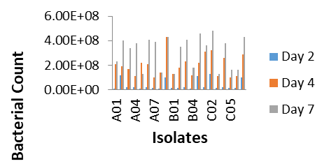
Before the screening of crude oil-degrading microorganisms, the bacterial populations were estimated in each original and saturated sample. In each case, the count of the control (oil-free) soil was found to be higher than the crude oil treated soil at different concentrations (2.3 x 10¹⁰ CFU/g, 1.8 x 10¹⁰ CFU/g, and 1.9 x 10⁹ CFU/g for Loamy Clay and Sandy soil respectively). In loamy treated soil, the organisms show growth even at 15% crude oil (3.0 x 10⁸ CFU/g, 1.8 x 10⁷ CFU/g and 1.1 x 10⁶ CFU/g for 5%, 10% and 15% crude oil respectively), whereas in clay soil the organisms were recovered at 5 – 10% crude oil (1.4 x 10⁷ CFU/g and 1.0 x 10⁶ CFU/g at 5% and 10% respectively). In sandy soil, the growth was only observed at a concentration of 5% crude oil which was 1.3 x 10⁸ CFU/g (Figure 1).

Figure 1: Crude Oil-Degrading Bacterial Counts (Cfu/ml) in of individual isolates

3.2 Cultural and Morphological Characterization of the Isolate from three different soils.

The morphology and type of bacterial colonies were also investigated in cell count experiments. A summary of the results is presented in Table 1. A total of 21 hydrocarbon-degrading bacteria species were segregated from three crude oil contaminated soil samples (namely sandy and clay and loamy respectively). From Sandy soil nine different isolates were obtained namely, A01 to A09. From Clay contaminated soil isolates B01, B02, B03, B04, and B05 were obtained. From Loamy contaminated soil, seven (7) bacteria were isolated and coined as C01, C02, C03, C04, C05, C06, and C07. The strains are characterized based on morphological features (Table.4.2). 

One of the objectives of this study was to isolate as many culturable strains as possible to determine their hydrocarbon biodegradation potential in standardized culture conditions. For this reason, the first screening of strain was done and followed by Gram staining and microscopic examination for bacteria to eliminate similar strains.


 Table 1: Cultural and Morphological Characteristics of Bacteria Isolated from three artificially Contaminated Soils.

I. Code

Cultural characteristics

Gram stain

Morphology

 Shape

Arrangement

A01

Smooth raised, glistering white colonies with circular or entire margin

G+ve

Cocci

pairs and irregular clusters
A02

Large Grey- white, circular colonies with undulate margins, 

G+ ve

Straight rods with rounded ends

Single / Chains
A03

Relatively large, dull grey, opaque colonies with convex

G- ve

Straight Rods 

Singly evenly spread
A04

Small, Smooth Pale yellow colonies , convex with entire margins

G- ve

Coccobacilli

Pairs
A05

Smooth grey colonies, convex, shiny surface with entire margins

G- ve

Long rods shaped

Single / Pairs
A06

Large grayish to white colonies , circular and convex

G- ve

Rods shaped with round ends

Singly evenly spread
A07

Slightly large white lobate colonies with irregular margins 

G+ ve

Coccobacilli

Single / Chains
A08

Pale white colonies with swarming growth and entire margins

G- ve

Straight Rods

Singles
A09

gray white, large low convex and wrinkle colonies

G- ve

Straight and slightly curved Rods

Singles
B01

Small, shiny grey, translucent with raised and irregular margins

G- ve

slightly curved Rods

Single / Pairs
B02

small, Circular,  convex and creamy yellow pigmented colonies with entire margins

G+ ve

Cocci

Pairs/ tetrad.
B03

Small, Smooth Pale yellow colonies , convex with entire margins

G- ve

Coccobacilli

Pairs/ small clusters
B04

Circular, and translucent yellow to cream-colored colonies 

G- ve

Rods shaped

Single / Pairs
B05

Grey- white, granular colonies with undulate margins

G+ ve

Straight rods with rounded ends

Single / Chains
C01

Grey- white, granular colonies with undulate margin   

G+ ve

Straight rods with rounded ends

Single / Chains
C02

Filamentous Mycelium, Aerial pale yellow and substrate ash with slimy colonies. 

G+ ve

Rods Shaped

Singles
C03

Smooth red and round colonies, slightly raised   convex surfaces, with entire margins

G- ve

Straight short Rods

Pairs/ Short chains
C04

gray white, large, low convex with irregular margins

G- ve

Straight and slightly curved Rods

Singles
C05

gray white, large, low convex and entire margins

G+ ve

slightly short curved Rods

Single / Pairs
C06

White moist colonies and Convex with irregular margins

G- ve

Coccobacillary  

Singles
C07

small, Circular, entire, convex with regular edges and creamy yellow pigmented colonies 

G+ ve

Cocci

Tetrads/ pairs

Key: A (isolated from sandy soil), B (isolated from Clay soil), C (isolated Loay soil), G+ ve (Gram positive), G- ve (Gram negative), I. Code (Isolate code)

Table 2 Biochemical Characterization of Crude Oil-Degrading Bacteria isolated from three Different Soil types. 

TestAcinetobacterProvidentia sppB. cereusB. polymyxaEnterobacter sppMicrococcus luteusP. aerogenusaActinomyces spp
Grm +-++-+-+
Spo--++----
Cat+++++++-
Oxi-----++NA
Cit+++++-+-
Ure---+-++-
Coa---_----
Nit-+-++-++
Ind-+------
Mr-+-+---+
Vp--+++---
Mot-++++-+-
Glu++++++++
Suc-DD++--+
Lac---++--+
TSIK/KK/AK/AK/AK/AK/KK/NK/A
Gas-+--+--+
H2S-------+
Man-+NA++-+-


Key: d = dubious, NA= not applicable; - = Negative, + = Positive. Gram’s stain (Grm), Spore stain (Spo), Catalase (Ca), Oxidase test (Ox), Citrate (Cit), Urea (Ure), Coagulase (Coa), Nitrate reduction test (Nit),  Indole (Ind), Methyl red test (Mr), Voges proskauer test (VP), Motility (Mot), Glucose fermentation (Glu), Sucrose (Suc), Lactose (Lac). Hydrogen sulphide (H2S), Mannitol (Man), Alkaline slant / Acid butt (A/A), Alkaline slant / Alkaline butt (K/K). Alkaline slant / Neutral butt (K/N), Acid slant / alkaline butt (A/K

DISCUSSION

Before isolation of biodegrading bacteria from the three soil sample types, the soil was artificially polluted with different concentrations of crude oil and incubated for seven days so as to select only the indigenous strain capable of utilizing or tolerating the hydrocarbons. One gram of each sample treatment was taken and serially diluted, plated on crude oil media for treated soil and Nutrient agar for control soil. Crude oil media were known to support the growth of only bacterial strains capable of tolerating or utilizing crude oil components as a sole carbon source. Many bacteria have been reported to exhibit hydrocarbon tolerance. A qualitative assay of growth in the presence of petroleum indicated that bacterial isolates in loamy soil were able to tolerate 15% of crude petroleum oil which is in contrast to that of sandy which grows only at 5% crude oil. The total and relative plate counts of the isolate obtained were decreased in comparison to oil-free (control) soil. Based on the colony morphology observed and stability in sub culturing, a total of 53 bacterial strains were segregated from both control and artificially polluted soil. 13, 10, and 9 bacterial isolates were recovered from the unpolluted (control) sandy, clay, and loamy soil sample respectively. When compared to the polluted treatment, the number of isolates reduced to 9, 5, and 7 corresponding to sandy, clay, and loamy treated soil respectively. This account for 21 hydrocarbon tolerant isolates. The replacement in isolate number points out that some of the isolates originally present in the unpolluted sample were not able to survive crude oil stress, hence only strains with a metabolic activity that would enable them to withstand the effects of crude oil hydrocarbons will be selected. The screening for potential degraders was further conducted even though the initial artificial pollution selected only those indigenous microorganisms that have especially acclimated to degrade hydrocarbons, it was necessary to characterize the biodegradation potential for an individual isolate. For this reason, each isolated microbial strain was further investigated in an MSN medium containing 1% crude oil for a details investigation of hydrocarbons utilization. The growth of the microorganisms was monitored regularly and estimated by visual observations of turbidity followed by the plate count technique. An increase in bacterial population was observed over time, reaching a maximum of 4.8 x 10⁸ for isolate C02 (at day 7), 4.6 x 10⁸ for isolate B05, 4.1 x 10⁸ for isolate B03, 4.0 x 10⁸ for isolate A02, 4.3 x 10⁸ for isolate C07, A09 (at day 7) etc. This study allows the selection of thirteen bacterial strains as potential candidates for crude oil biodegradation processes. Thirteen bacterial strains (A02, A03, A04, A06, A07, A09, B02, B03, B05, C01, C02, C04, and C07) have exhibited similar growth patterns as observed by turbidity and plate count. Therefore, they were considered highly oil-tolerant strains. Their increasing growth dynamics during degradation can be attributed to the constitutive expression of hydrocarbon assimilating capabilities or adaptation of the strains[18], whereas A01, B04, C05, and C06 displayed a little growth in comparison to the growth observed in the above isolate. The remaining strains showed very low tolerance.This could probably be due to the presence of some toxic volatile hydrocarbons affecting its growth such aspreviously reported [19]who reported that certain volatile hydrocarbon compounds in petroleum could affect microbial growth. Cultural and morphological studies of the selected isolate were also carried out, followed by physiological and biochemical analysis to identify the species involved.The identity of the bacterial isolates was confirmed by comparing their characteristics with thoseof known taxa as outlined in Bergey’s Manual of Systematic Bacteriology[20]. These isolates were identified as: pseudomonas aerogenusa (A09 andC04), Acinetobacter (A04, B03), Bacillus cereus (A02, B05, and C01), Bacillus polymyxa (A07) Micrococcus luteus (B02, C07), Enterobacter (A06), Providentia spp (A03), and Streptomyces (C02) respectively. According to many authors, bacteria have been described as being more efficient hydrocarbon degraders than other microorganisms, or at least bacteria are more commonly used as tested microorganisms[21]. Several strains of the genera Pseudomonas and Bacillus isolated from hydrocarbons-contaminated environments can grow and degrade aliphatic and aromatic hydrocarbons[22]. Pseudomonas aeruginosa has been isolated from hydrocarbon-contaminated soil. Studies regarding the ability of Bacillus megaterium and Bacillus cereus strains to degrade hydrocarbons and produce biosurfactants have also been reported[23]. However, few reports were found in the literature regarding hydrocarbon degradation capacity by Providentia. [18]also reported that hydrocarbon in spent lubricating oil was degraded by Micrococcus spp, Enterobacter spp, Acinetobacter spp, Bacillus spp, Pseudomonas, etc. 

CONCLUSION

The bacteria that can grow on bony light crude oil as a carbon and energy source were isolated from Sandy, Clay, and Loamy soil. The ability of the isolate to degrade hydrocarbons in crude oil was further assessed in liquid medium; this allowed the selection of potential hydrocarbon degraders. Out of the 21 bacterial colonies isolated from Sandy, Clay and Loamy treated soils, 13 were selected as the most promising degraders after the potential screening. Little growth was observed for any of the rest of the isolates. Four of these isolates turn out to be Bacillus spp, two turn out to be Pseudomanos sp., due to Acinetobacter spp, due to Microccocus spp, Actinomyces spp and Eenterobacter and Providentia spp. The discovery of Pseudomonas, Acinetobacter, Bacillus, Microccocus, Actinomyces was not surprising on their frequency in the soil as well as their frequent biodegradable ability. Nevertheless, isolation and good growth on hydrocarbon substrates were surprising for providentia. Thirteen isolates were tested for their ability to degrade crude oil in three different soil types. From three different crude oil contaminated soil samples, the highest degradability was detected in loamy polluted soil because it has highest colony count and thus, the organisms show grew at higher rate to degrade crude oil. it is Recommended that more screening of crude oil-degrading bacteria is required from different locations and on different soil that is not considered highly contaminated by crude oil and petroleum products for effective bioremediation of Bonny light crude oil. 

Conflict of Interest:

The authors declare that they have no conflict of interest

Funding:

No funding sources

Ethical approval:

The study was approved by the Birnin Kebbi, Kebbi State.

REFERENCE
  1. Van Beilen, J. B., and E. G. Funhoff. "Alkane Hydroxylases Involved in Microbial Alkane Degradation." Applied Microbiology and Biotechnology 74.1 (2007): 13-21. https://doi.org/10.1007/s00253-006-0796-2.

  2. Płaza, G. A., et al. "Estimation of the Environmental Risk Posed by Landfills Using Chemical, Microbiological, and Ecotoxicological Testing of Leachates." Environmental Science and Pollution Research 15.2 (2008): 1-12. https://doi.org/10.1007/s11356-008-0055-5.

  3. Adebusoye, S. A., et al. "Microbial Degradation of Petroleum Hydrocarbons in a Polluted Tropical Stream." World Journal of Microbiology and Biotechnology 23.7 (2007): 1149-1159. https://doi.org/10.1007/s11274-007-9374-4.

  4. Rahman, K. S., et al. "Bioremediation of Gasoline Contaminated Soil by a Bacterial Consortium Amended with Poultry Litter, Coir Pith, and Rhamnolipid Biosurfactant." Bioresource Technology 81.1 (2002): 25-32. https://doi.org/10.1016/S0960-8524(01)00157-4.

  5. Schippers, A., et al. "Microbacterium Oleivorans Sp. Nov. and Microbacterium Hydrocarbonoxydans Sp. Nov., Novel Crude Oil Degrading Gram-Positive Bacteria." International Journal of Systematic and Evolutionary Microbiology 55.3 (2005): 655-660. https://doi.org/10.1099/ijs.0.63403-0.

  6. Wook, K., et al. "Inhibition of Acrolein-Stimulated MUC5AC Production by Fucoidan in Human Bronchial Epithelial Cells." Die Pharmazie - An International Journal of Pharmaceutical Sciences 63.10 (2008): 757-759. https://doi.org/10.1691/ph.2008.7465.

  7. Auffret, M., et al. "Degradation of a Mixture of Hydrocarbons, Gasoline, and Diesel Oil Additives by Rhodococcus Aetherivorans and Rhodococcus Wratislaviensis." Applied and Environmental Microbiology 75.24 (2009): 7774-7782. https://doi.org/10.1128/AEM.01878-09.

  8. Olajide, P. O., and L. B. Ogbeifun. "Hydrocarbon Biodegrading Potentials of a Proteus vulgaris Strain Isolated from Fish Samples." American Journal of Applied Sciences 7.7 (2010): 922-928. https://doi.org/10.3844/ajassp.2010.922.928.

  9. John, R. C., and G. C. Okpokwasili. "Crude Oil-Degradation and Plasmid Profile of Nitrifying Bacteria Isolated from Oil-Impacted Mangrove Sediment in the Niger Delta of Nigeria." Bulletin of Environmental Contamination and Toxicology 88.6 (2012): 1020-1026. https://doi.org/10.1007/s00128-012-0621-0.

  10. Abdel-Rahman, M. A., et al. "Efficient Homofermentative L-(+)-Lactic Acid Production from Xylose by a Novel Lactic Acid Bacterium, Enterococcus mundtii QU 25." Applied and Environmental Microbiology 77.5 (2011): 1892-1895. https://doi.org/10.1128/AEM.02755-10.

  11. Watkinson, R. J., et al. "Automated Image Analysis Method to Determine Fungal Biomass in Soils and on Solid Matrices." Soil Biology and Biochemistry 23.7 (1990): 609-616. https://doi.org/10.1016/0038-0717(91)90030-W.

  12. Ayuba, O. O. A., et al. "Review on the Fate of Contaminants in the Niger Delta Environment." 2011.

  13. Nilanjana, D., and C. Preethy. "Role of Sophorolipid Biosurfactant in Degradation of Diesel Oil by Candida tropicalis." Bioremediation Journal 16.1 (2010): 19-30. https://doi.org/10.1080/10889860903468959.

  14. Goadleds, Adams O., et al. "Bioremediation, Biostimulation, and Bioaugmentation: A Review." International Journal of Environmental Bioremediation & Biodegradation 3.1 (2015): 28-39. https://doi.org/10.12691/ijebb-3-1-6.

  15. Usman, U. B., et al. "Molecular Characterization and Phylogenetic Analysis of Listeria monocytogenes Isolated from Milk and Milk Products in Kaduna, Nigeria." Canadian Journal of Infectious Diseases and Medical Microbiology 2016. https://doi.org/10.1155/2016/6065323.

  16. Cheeseborough, M. District Laboratory Practice in Tropical Countries, Part 2. Cambridge University Press, 2000. pp. 18-23.

  17. Oyeleke, S. B., and S. B. Manga. Essentials of Laboratory Practical. 1st ed., Best Publisher, 2008. pp. 36-63.

  18. Ekanem, J. O., and A. A. Ogunjobi. "Hydrocarbon Degradation Potentials of Bacteria Isolated from Spent Lubricating Oil Contaminated Soil." Journal of Applied Sciences and Environmental Management 21.5 (2017): 973-979. https://dx.doi.org/10.4314/jasem.v21i5.26.

  19. Leahy, J. G., and R. R. Colwell. "Microbial Degradation of Hydrocarbons in the Environment." Microbiological Reviews 54.3 (1990): 305-315. https://doi.org/10.1128/MMBR.54.3.305-315.1990.

  20. Ijah, U. J. J., and S. P. Antai. "Removal of Nigerian Light Crude Oil in Soil over 12 Months." International Biodeterioration & Biodegradation 51.2 (2003): 93-99. https://doi.org/10.1016/S0964-8305(02)00118-6.

  21. Cappello, S., et al. "Predominant Growth of Alcanivorax During Experiments on 'Oil Spill Bioremediation' in Mesocosms." Microbiological Research 162.3 (2007): 185-190. https://doi.org/10.1016/j.micres.2007.04.001.

  22. Assih, E. A., et al. "Stenotrophomonas Acidaminiphila Sp. Nov., a Strictly Aerobic Bacterium Isolated from an Upflow Anaerobic Sludge Blanket (UASB) Reactor." International Journal of Systematic and Evolutionary Microbiology 52.3 (2002): 559-568. https://doi.org/10.1099/ijs.0.01844-0.

  23. Thavasi, R., et al. "Effect of Biosurfactant and Fertilizer on Biodegradation of Crude Oil by Marine Isolates of Bacillus megaterium, Corynebacterium kutscheri, and Pseudomonas aeruginosa." Bioresource Technology 102.2 (2011): 772-778. https://doi.org/10.1016/j.biortech.2010.07.089.

Advertisement
Recommended Articles
Case Report
Mycosis Fungoides – A Case Report
...
Published: 20/10/2021
Download PDF
Research Article
Prevalence of Sacroiliac Joints Involvement in Iraqi Patients with Seropositive Rheumatoid Arthritis
Published: 30/12/2024
Download PDF
Research Article
A Critical Analysis of Spinal Block Anesthesia under Ultrasonographic Guided Technique: Is Levobupivacaine Alone Effective or on Adjuvant is Warranted?
Published: 20/09/2021
Download PDF
Research Article
The Role of Elevated CRP in Assessing the Severity and as a Prognostic Marker in Covid19 Patients
...
Published: 04/07/2021
Download PDF
Chat on WhatsApp
Flowbite Logo
Najmal Complex,
Opposite Farwaniya,
Kuwait.
Email: kuwait@iarcon.org

Editorial Office:
J.L Bhavan, Near Radison Blu Hotel,
Jalukbari, Guwahati-India
Useful Links
Order Hard Copy
Privacy policy
Terms and Conditions
Refund Policy
Others
About Us
Contact Us
Online Payments
Join as Editor
Join as Reviewer
Subscribe to our Newsletter
Follow us
MOST SEARCHED KEYWORDS
scientific journal
 | 
business journal
 | 
medical journals
 | 
Scientific Journals
 | 
Academic Publisher
 | 
Peer-reviewed Journals
 | 
Open Access Journals
 | 
Impact Factor
 | 
Indexing Services
 | 
Journal Citation Reports
 | 
Publication Process
 | 
Impact factor of journals
 | 
Finding reputable journals for publication
 | 
Submitting a manuscript for publication
 | 
Copyright and licensing of published papers
 | 
Writing an abstract for a research paper
 | 
Manuscript formatting guidelines
 | 
Promoting published research
 | 
Publication in high-impact journals
Copyright © iARCON Internaltional LLP . All Rights Reserved.