Hepatoid adenocarcinoma of stomach is a rare and clinically aggressive tumor. Most of these cases are associated with increased production of alpha fetoprotein, but not all AFP producing tumors have hepatoid features histologically. We report a case of 64 years male, with elevated Alpha Fetoprotein levels. The patient underwent gastrectomy. HPE suggested a diagnosis of hepatoid adenocarcinoma. PAS staining also revealed PAS positive hyaline globules.
The term Hepatoid Adenocarcinoma (HAC) was first proposed by Ishikura et al. in the year 1986. HAC is a malignant cancer manifesting outside liver, having morphological features identical to hepatocellular carcinoma. HAC is a rare tumor with an incidence varying from 0.38 to 0.73%. Stomach is the most common location of HAC. Some of the other sites for HAC include esophagus, papilla of vater, colon, lung, gall bladder, adrenal gland, kidney, urinary bladder, ovary uterus, vagina and testis.
About 64 year old male patient presented to the department of surgery with features of upper abdominal pain since 2 months and weight loss of more than 4 kilograms in the past 1 month. Patient had Hemoglobin 8gm% and raised Alpha Fetoprotein levels (AFP- 350ng/mL), Upper GI endoscopy revealed an ulcerated growth around the gastroesophageal junction. Biopsy was taken, which on histopathological findings revealed features suggestive of adenocarcinoma. Later, the patient underwent total gastrectomy left antrolateral thoracotomy with esophagojejumal anastomosis with jejunojejunostomy and feeding jejunostomy.
In the department of pathology, we received gastrectomy specimen measuring 17×6×3 cms with part of esophagus measuring 2 cms in length. On cut section, the gastroesophageal junction showed a ulcero-infilterative growth measuring 5×2.5×1 cms. There was flattening of surrounding rugal folds. The growth was gray white to gray brown in colour. Serosal surface and the resection margins appeared free from growth grossly.
Also received were left gastric and common hepatic lymph nodes.
Microscopic examination of sections from growth revealed nests, cords and trabeculae of polygonal tumor cells. The cells had mild to moderate nuclear cellular pleomorphism, round to oval nuclei, vesicular chromatin, small to inconspicuous nucleoli and abundant granular eosinophilic cytoplasm. Few cells revealed intracytoplasmic juxtanuclear vacuoles (pale bodies). Occasional signet ring cells also seen. Extracellular mucin, necrosis, frequent atypical mitotic Figures (1-5) also seen. There was evidence of peri neural and lymphovascular invasion.
Periodic Acid Schiff (PAS) with diastase resistance was done which showed PAS positive hyaline globules.
Figure 1: H&E Sections 40x Shows Gastric Lining Epithelium. Subepithelium Shows Neoplastic Cellsforming Nests, Cords and Trabeculae
Figure 2: H&E Sections 100x Shows Cords and Trabeculae of Polygonal Tumor Cells, there is Peritumoral Inflammatory Cell Infiltrate
Figure 3: H&E Sections 400x Reveal Cords and Trabeculae of Polygonal Tumor Cells. The Cells had Mild to Moderate Nuclear Cellular Pleomorphism, Round to Oval Nuclei, Vesicular Chromatin, Small to Inconspicuous Nucleoli and Abundant Granular Eosinophilic Cytoplasm
Figure 4: H&E Sections 100x Revealing Evidence of Lymphovascular Invasion by Tumor Cells (Arrowhead)
Figure 5: Periodic Acid Schiff (PAS) with Diastase Resistance 400x Showing PAS Positive Hyaline Globules (Arrowhead)
Esophageal and distal resection margins along with omentum were free from tumor invasion. Left gastric and common hepatic lymph nodes did not show any evidence of tumor metastasis.
Based on the above histopathological examination diagnosis of Hepatoid Adenocarcinoma, Stomach (HAS) was given. Immunohistochemistry for alpha fetoprotein, carcinoembryonic antigen for confirmation was suggested.
Hepatoid Adenocarcinoma (HAC) is a malignant tumor which resembles microscopic features of hepatcellular carcinoma, but occurs outside the liver. Hepatoid Adenocarcinoma of Stomach (HAS) is a rare tumor associated with poor prognosis [1]. According to Lauren’s classification HAS belongs to the indeterminate type of gastric carcinoma [2]. The pathological diagnosis of this tumor, is based on microscopic characteristics irrespective of serum Alpha Fetoprotein levels (AFP) or tissue AFP staining by Immunohistochemistry (IHC) [3,4].
Stomach is the most common location of HAC. The incidence of HAS is <1% [5]. Other sites for HAC include colon, lung, gall bladder, uterus, urinary bladder, etc. [6].
In the year 1985, Ishikura et al. first proposed this entity. Nagai et al suggested that HAC should be diagnosed on its morphological features and not on production of AFP levels [7,8].
HAS has a male to female ratio of 2.3:1. Mean age at the time of presentation is 63 years [5]. HAS is not associated with specific clinical features. Most symptoms include general fatigue, decreased appetite, epigastric pain, anemia, melena, etc. however, sometimes HAS, may present with gastric perforation [5]. Our patient presented with epigastric pain and weight loss. Wanget et al. reported that majority of HAS were located in gastric antrum (57%), followed by gastroesophageal junction and gastric body [9].
Most HAC are associated with production of AFP, but not all AFP producing carcinomas histologically show hepatoid features, Classically the tumors with elevated AFP levels are heptocellular carcinoma, hepatoblastoma, germ cell tumors (particularly Yolk sac tumor). Apart from them gastric cancer, lung cancer, renal cell carcinoma, nephroblastoma, pancreatic cancer, colorectal carcinoma, ovarian carcinoma and rhabdomyoblaastoma may also produce increased amount of AFP levels [10].
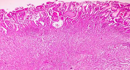
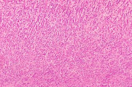
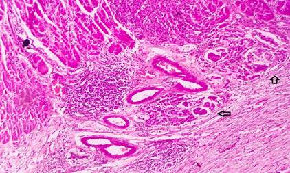
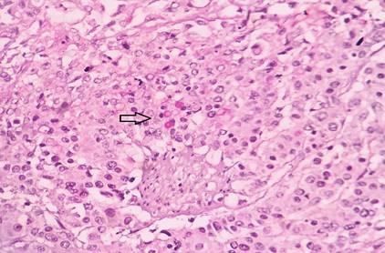
Kodama et al. described two histological types of HAS. The first was medullary type, characterized by solid nests ans sheets of polygonal cells. Along with scattered large pleomorphic or multinucleate giant cells. The second, being well differentiated papillary or tubular type with clear cytoplasm. Both the types can sometimes coexist in the same tumor [6]. The tumor cells, invade the surrounding tissue and are accompanied by significant venous infilteration [5].
Therefore HAC, should be diagnosed by characteristic histological features of a gastric tumor resembling hepatocellular carcinoma, distinction between HAS and ALP producing gastric cancer is necessary as HAS carry a dismal prognosis [4].
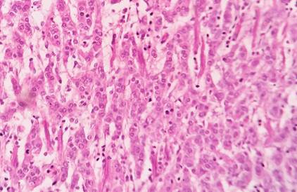
Only a minority of a HAC, show more definitive features of hepatoid differentiation like bile production, intracytoplasmic pale bodies and albumin m RNA production [10]. Presence of PAS positive diastase resistant intracytoplasmic eosinophilic granules is a non-specific finding but can be supportive of diagnosis [10]. About 9.7% cases can show hyaline globules [11].
Our case had characteristic tumor cell morphology, along with increased production of AFP levels, presence of pale bodies and PAS positive diastase resistant hyaline globules.
Most common sites of metastasis of HAS include lymph node (77%) and liver (42%), followed by lung, peritoneum, pancreas, spleen and brain [11].
On IHC, this tumor shows reactivity for AFP, Alpha antitrypsin, Alpha Chymotrypsin, CarcinoEmbryonic Antigen [12].
HAS carries a poor prognosis as compared to gastric adenocarcinoma with no hepatoid features regardless of AFP production. About 5 year survival rate is about 9% [12].
Hepatoid adenocarcinoma, is a rare yet important extra hepatic adenocarcinoma. Majority of HAC, arise in the stomach. The diagnosis does not depend on AFP levels or IHC, but not characteristics microscopic features. It is rapidly progressive and has a high incidence of lymph node and liver metastasis. Early diagnosis and radical surgery remain the gold standard treatment.
Inagawa, S. et al. "Hepatoid adenocarcinoma of the stomach." Gastric Cancer, vol. 4, no. 1, 2001, pp. 43–52. DOI: 10.1007/s10120010001.
Hu, B. et al. "Gastric cancer: Classification, histology and application of molecular pathology." Journal of Gastrointestinal Oncology, vol. 3, no. 3, 2012, pp. 251–261. DOI: 10.3978/j.issn.2078-6891.2012.021.
Xie, Y. et al. "Hepatoid adenocarcinoma of the stomach is a special and easily misdiagnosed or missed diagnosed subtype of gastric cancer with poor prognosis but curative for patients of pN0/1: the experience of a single center." International Journal of Clinical and Experimental Medicine, vol. 8, no. 5, 2015, pp. 6762–6772. PMID: 26221214; PMCID: PMC4509159.
Søreide, J.A. "Therapeutic approaches to gastric hepatoid adenocarcinoma: current perspectives." Therapeutics and Clinical Risk Management, vol. 15, 2019, pp. 1469–1477. DOI: 10.2147/TCRM.S204303. PMID: 31920320; PMCID: PMC6934111.
Yoshizawa, A. et al. "Hepatoid adenocarcinoma of the stomach with enteroblastic differentiation: an aggressive variant of gastric cancer." BMC Cancer, vol. 17, 2017, p. 368. DOI: 10.1186/s12885-017-3357-7.
Su, J.S. et al. "Clinicopathological characteristics in the differential diagnosis of hepatoid adenocarcinoma: a literature review." World Journal of Gastroenterology, vol. 19, no. 3, 2013, pp. 321–327. DOI: 10.3748/wjg.v19.i3.321. PMID: 23372352; PMCID: PMC3554814.
Ishikura, H. et al. "An AFP‐producing gastric carcinoma with features of hepatic differentiation: A case report." Cancer, vol. 56, no. 4, 1985, pp. 840–848. DOI: 10.1002/1097-0142(19850815)56:4<840::aid-cncr2820560423>3.0.co;2-e. PMID: 2410093.
Nagai, E. et al. "Hepatoid adenocarcinoma of the stomach: a clinicopathologic and immunohistochemical analysis." Cancer, vol. 72, no. 6, 1993, pp. 1827–1835. DOI: 10.1002/1097-0142(19930915)72:6<1827::aid-cncr2820720606>3.0.co;2-8. PMID: 7689918.
Wang, Y. et al. "Hepatoid adenocarcinoma of the stomach: a unique subgroup with distinct clinicopathological and molecular features." Gastric Cancer, vol. 22, no. 6, 2019, pp. 1183–1192. DOI: 10.1007/s10120-019-00965-5. PMID: 30989433; PMCID: PMC6811386.
Roberts, C.C., Colby, T.V. and Batts, K.P. "Carcinoma of the stomach with hepatocyte differentiation (hepatoid adenocarcinoma)." Mayo Clinic Proceedings, vol. 72, no. 12, 1997, pp. 1154–1160.
Yang, J. et al. "Clinicopathological and prognostic characteristics of hepatoid adenocarcinoma of the stomach." Gastroenterology Research and Practice, vol. 2014, 2014, Article ID 140587. DOI: 10.1155/2014/140587. PMID: 24669215; PMCID: PMC3942340.
Fakhruddin, N. et al. "Hepatoid adenocarcinoma of the stomach: A challenging diagnostic and therapeutic disease through a case report and review of the literature." Frontiers in Medicine, vol. 4, 2017, p. 164. DOI: 10.3389/fmed.2017.00164. PMID: 29034239; PMCID: PMC5627014.