Despite extraordinary advances, Cancer poses as a yet unmet threat to global public health with the number of cases surging drastically each year. At a time when conventional diagnostic and therapeutic modalities are challenged by the lack of target specificity, multi drug resistance, tradeoff between safety and efficacy, Cancer nanotechnology is an emerging branch that links nanotechnology and clinical research overcoming some of the most challenging impediments. Nanomaterials are characterized by a size of 1- 100nm and nanotechnology is the micromanufacturing by manipulating shapes and size at Nano length. Materials at nanoscale acquire high surface area, dominance of quantum effects and various electric, magnetic, thermal properties, while at the same time by the virtue of molecular scale, interact with biological systems at cellular level. Tailor made nanoparticles including carbon based, metallic, polymeric, surface functionalized, have shown target specificity, sensitivity and efficacy reaching otherwise unapproachable cancer targets. Quantum dots, nanowires, gold, magnetic nanoparticles have proven successful in enhanced imaging, detecting low concentration biomarkers providing prospects for early and accurate cancer detection. Nanotherapeutics, having improved half-life in blood, increased drug loading capacities, controlled release and multidrug delivery capacities has shown higher efficacy and better safety profiles. By integrating diagnostic and therapeutic functions into a single Nanoparticle formulation, theranostic nanomedicine offers a promising strategy providing prospects for future personalized medicine. However, there are barriers impeding clinical translation. Therefore, while addressing these limitations, this review aims to summaries developments in nanotechnology for cancer diagnosis and therapy for better patient outcomes in the future.
Keywords
Nanotechnology
Cancer
Diagnosis
Therapy
Theranostics.
INTRODUCTION
Cancer is a major cause of mortality worldwide [51,63]. Ranking as the first or second leading cause of death before the age of 70 years in 112 countries, it is a huge barrier to increasing life expectancy [51]. Furthermore, marking a 47% rise from the statistics in 2020, the global cancer burden is expected to be 28.4 million cases in 2040 [51] prompting researchers to redouble their research efforts.
Cancer isn’t actually a singular disease, but a heterogeneous group of disorders [3]. Characterized by uncontrolled cell division and absence of cell death, ultimately produces an abnormal cell mass. Initially starting as a localized disease, it is prone to spread to distance sites making it incurable Cree [12]. Host of rare alterations and high penetrant mutations are found in unique combinations in each patient and there is even more genetic variation between any two types of cancer [42]. The development of cancer, occurring through a multistep carcinogenesis process entailing numerous cellular and physiological systems adds to its complexity making it difficult to comprehend Misra et al. [36].
Conventional diagnostic techniques comprising of cytology, histopathology and imaging techniques are inadequate for early detection of cancer [65]. Frequent challenges encountered by conventional therapies such as surgery, chemotherapy and radiotherapy include nonspecific systemic distribution of antitumor agents, inadequate drug concentrations reaching the tumor, toxicity outside the tumor, multidrug resistance and the limited ability to monitor therapeutic responses [13,14]. Taking cues from the recent knowledge on molecular pathology of cancer, with the promise of specifically targeting malfunctioning molecules and pathways, Nano oncology has emerged as a field with great potential to address the intrinsic limits of conventional approaches, hence this review aims to provide an insight into various nanomaterials, Nano enabled diagnostic, therapeutic and theranostic approaches for the future potential for more effective and safe cancer diagnosis and treatment Mukherjee et al. [38].
Nanomaterials
Nanomaterials are characterized by a small size of 1- 100nm and nanotechnology is defined by the production and application of structures and devices by controlling the shape and size on the nanometer scale Kopeckova et al. [29]. Nanoparticles are prepared by using top-down approach where solid materials are broken down or bottom-up approaches where atoms, molecules or smaller particles are assembled to form the nanostructures. Three main types of medically important nanoparticles are organic nanoparticles including polymer nanoparticles, liposomes, dendrimers, Inorganic nanoparticles including metals and metal oxides and composite nanoparticles Yu et al. [63]. These nanomaterials by the virtue of the quantum effects acquire unique physical and chemical properties not present at the molecular level while at the same time they are able to interact with biological systems at cellular level [41]. In context, nanoscale devices smaller than 50nm can easily enter most cells while ones smaller than 20nm can easily exit blood vessels during circulation. Nanoscale materials possess ability to passively accumilate at the tumor site, actively target cancer cells and ability to cross biological barriers in the body such as Blood-Brian barrier [47,63]. Considering the fact that cancer is also a phenomenon occurring at nanoscale, nanotechnology is able to address cancer in ways unimagined before. Nano oncology refers to the application of Nano medical technologies to the diagnosis and treatment of cancer Riggio et al. [41]. Therefore, utilizing these unique properties of nanomaterials, integrating with anticancer agents and imaging agents scientists have been able to expand capacities of tumor diagnosis and therapeutics with enhanced imaging and targeted therapy to improve patient outcomes.
Nanotechnology in Cancer Diagnosis
Early and accurate diagnosis which plays an important role in treatment decision is one of the key aspects in the battle against cancer [49,56]. Ultrasound, Endoscopy, Computed Tomography (CT), Magnetic Resonance Imaging (MRI), Positron Emission Tomography (PET) are some of the imaging technologies that are currently employed, but are not so versatile to provide complete clinical information about various types and stages of tumor [10,53]. They can only detect cancer when there is a visible change, which is probably when the cancer has proliferated and metastasized. As a result of numerous research efforts over the past decade on combating limitations of prevailing technologies, nanotechnology has open doors to provide highly sensitive, specific, early detection of cancer, with multiplexed measurement capacity providing new hope for cancer management [18,63].
Utilizing nanoscale substances such as gold nanoparticles, quantum dots, iron oxide nanocrystals, biodegradable micelles and other nanomaterials that provides clear diagnostic clues of cancer Choi et al. [29], Nanotechnology have been investigated for the detection of cancer biomarkers, cancer cells as well as in vivo imaging. nanoparticles are being applied to capture cancer biomarkers, such as cancer-associated proteins, circulating tumor DNA, circulating tumor cells and exosomes [6,35,65].
Nanotechnology Assisted Detection of Cancer Biomarkers
Cancer biomarkers are biological features whose expression could be objectively measured and indicate the presence or state of a tumor Ye et al. [61]. Tumor biomarkers can be proteins, protein fragments, DNA, micro RNA, Oncofetal antigens, carbohydrates found in tissue or biological fluids such as blood, urine, saliva, cerebrospinal fluid etc. secreted by cancer cells themselves or by the body in response to cancer [9, 65]. As this unique molecular signature allows possible prediction of the risk of cancer development, screening and early diagnosis of patients experiencing cancer symptoms, provide a prognosis and even could be used in therapeutic monitoring and recurrence prediction, great effort is exerted in sensitivity enhancement and reliable detection of cancer biomarkers. The use of biomarkers had been limited due to heterogeneity and lower concentrations. But nanoparticles due to their unique physicochemical properties are highly versatile candidates for biomarker detection. Therefore, different types of Nano Particles (NP) including metal oxides, metals, lipids, semiconductor, hybrid nanoparticles are being explored in Nano particle-based assays, arrays and sensors for biomarker detection Choi et al. [9]. The basic principle behind nanoparticle-based detection of cancer biomarkers, is the engineering of the nanoparticle with a suitable ligand that can bind with high affinity to the cancer biomarker. Upon interaction with the specific biomarker, NP-conjugated ligand undergoes measurable changes, thus enabling the detection of biomarker Ye et al. [61]. Antibodies, peptides and aptamers are some of the examples of ligands used and click chemistry, linker chemistry, physical interaction or hybridization are conjugation strategies used in incorporation of functional moieties to the selected nanoparticle [62]. Nanoparticles are suitably surface modified and the ligands are required to maintain their functionality even after conjugation with the NP. Also, nanoparticles are employed as sensors in mass spectrometry, optical and electrical methods to improve the sensitivity and selectivity of biomarker detection [6].
Nanotechnology in Detecting Circulating Tumor Cells
Circulating Tumor Cells (CTC) is a hallmark of metastasis in cancer which are of high prognostic significance [35]. CTCs are known to accumulate during the transport of tumor cells that are shed from the primary tumor site throughout the bloodstream Bhana et al. [3]. The low occurrence of CTC’s in blood makes them difficult to enumerate and analyze. Research has shown that nanotechnology has been able to enhance CTC isolation and detection by enrichment of scares CTC’s. Carbon nanotubes for sensing applications, nanomaterials functionalized with antibodies specific to CTC’s and magnetic nanoparticles have shown prospects for future improvement in circulating tumor cell detection [28,35].
Nanotechnology Assisted Imaging
Contrast agents which can improve the visibility of features that would otherwise be difficult to detect are an emerging practice in medical imaging Smith et al. [47]. Nanoparticles have optical, magnetic, acoustic properties that can enhance imaging Yu et al. [63]. Biocompatibility and reduced toxicity are added advantages of nanoparticles as contrast agents. Common nanoparticle platforms in imaging are AuNP, dendrimers, liposomes, porous silica nanoparticles etc. Toy et al. [53] Nanotechnology has enabled novel imaging modalities such as Photoacoustic Tomography (PAT), Raman spectroscopic imaging, multimodal imaging which contrast agents specific to several imaging modalities simultaneously. Multimodal nanoparticles capable of delineating the margins of brain tumors preoperatively and intraoperatively have been developed Moritz et al. [37]. These MRI PAT Raman nanoparticles allow tracking of tumor growth, surgical staging by MRI and also shown utility during surgical resection to increase the potential tumor specific tissue removal Moritz et al. [37]. Nanoparticle based imaging agents provide more accurate assessment of drug dose painting, especially for nano-chemotherapeutics Toy et al. [53].
Nanoparticles have been developed and coupled with other technologies in clinical imaging of prostate cancer, metastatic melanoma, metastatic breast cancer offering more effective less invasive technique to image for accurate and reliable diagnosis and prognostication of cancer [40,45].
Nanotechnology in Cancer Therapy
One of the fundamental advantages of nanotechnology in cancer treatment is the ability to differentiate malignant cells from nonmalignant and the ability to selectively eradicate malignant cells [1]. Passive tumor targeting takes advantage of the Enhanced Permeability and Retention (EPR) effect, which is the leaky and disorganized nature of the blood vessels and the dysfunctional lymph vessels in solid tumor [9,39]. While active targeting of nanoparticles to tumor cells, microenvironment or vasculature and directed delivery to intracellular compartments can be achieved by surface modification of nanoparticles with small molecules, antibodies, affibodies etc. [25, 62]. Large surface area and tunable size of nanoparticles allows many types of small molecules to be encapsulated or conjugated [20]. Prolonged circulation period in the bloodstream allow nanotherapeutics to reach the target tissue more efficiently Sun et al. [50]. Nanoparticles due to their physical properties such as energy absorption and re-radiation, can be used to disrupt diseased tissue, as in laser ablation and hyperthermia applications. Nanoparticle-mediated therapies having virtues of multi-function, confer better curative effects with less adverse reactions [20].
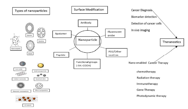
Nanotechnology to Improve Chemotherapy, Radiation Therapy, Immune Therapy and Gene Therapy (Figure 1)
Although most commonly used in clinics, chemotherapy is dissatisfactory in many instances due to nonspecific drug distribution, multidrug resistance, lack of solubility etc. The advent of nanotechnology in chemotherapy has provided better opportunities to address these issues Zhao et al. [66]. Active targeting of chemotherapeutic drugs using nanocarriers with specificity for receptors overexpressed by cancer cells such as folate receptor, transferrin receptor, Luteinizing Hormone-Releasing Hormone (LHRH) receptor expressed by breast cancer, ovarian cancer and prostate cancers have improved specific targeting [52].
Figure 1: Nanoparticles in Cancer diagnosis and therapy
Nanoparticles such as dendrimers and liposomes which can encapsulate hydrophobic drugs in micelles have effectively solved the lack of solubility of certain anticancer drugs Liu et al. [31]. Drug resistance caused by p -glycoprotein which pumps out drugs from cancer cells reducing intracellular concentration has been successfully mitigated by the codelivery of anti p glycoprotein agents such as verapamil or cyclosporine using Nano constructs. Chemo-chemotherapy combination therapy with two or more drugs using nano platforms have shown successful results in preclinical /clinical investigations with effective tumor recession.
Radiation therapy received by more than 60% of all cancer patients too is challenged by a tradeoff between efficacy and safety Mi et al. [34]. Nanoparticles due to their inherent properties including high atomic number that enhances Compton and photoelectric effects of conventional radiation therapy allows to use them as enhancement agents for radiation therapy and photodynamic therapy, they have been used to sensitize cancer cells and produce localized enhancements in radiation dose. Nanocarrier radioisotope delivery decreases renal clearance and increase the half-life in blood. The half-life of 111 In PEGylated liposomes in blood was 10.2, while the half-life of 111 In-DTPA in blood was no longer than 2 h showing that nanocarriers can prevent opsonization through PEGylation Wang et al. [55]. Nanomaterial base delivery systems have proven to prolong the effects of DNA and RNA-based genetic therapeutics such as small interfering RNAs (siRNAs) and microRNAs (miRNAs) which are sensitive to degradation by increasing stability and allowing controlled release Yao et al. [60]. Gene silencing therapeutics, siRNAs, have been reported to have significantly extended half-lives when delivered either encapsulated or conjugated to the surface of nanoparticles. Nano enabled immunotherapy has proven successful for the delivery of immunostimulatory or immunomodulatory molecules in combination with chemo- or radiotherapy or as adjuvants to other immunotherapies [16]. Nanoparticle vaccines allowing co delivery of antigens and adjuvants and allowing prolonged immune stimulation are known to raise sufficient T cell response to eradicate tumors. Immune depots placed in or near tumors for in situ vaccination and artificial antigen presenting cells has also being investigated in nano enabled immunotherapy [58]. Photothermal Therapy (PTT) and photodynamic therapy (PDT), are two minimally invasive methods having promising potential in the diagnosis and treatment of cancer [59]. In PTT, photothermal agent is stimulated by specific band light and vibrational energy/heat releases to selectively target cancer cells [26] while in PDT, photosensitizer drugs generate reactive oxygen species through phytochemical reactions triggering oxidative stress leading to cell death via lipid peroxidation, DNA damage etc. Dolmans et al. [15]. Recently, precious metal nanomaterials such as Au nanocages and Au nanohexapods Yucai et al. [64], carbon nanomaterials Liu et al. [31], upconversion nanoparticles which can convert light from long wave lengths to short wavelengths through the excitation of near infrared light which can penetrate deep tissues eliminating cancer, transition metal sulfide nanomaterials such as CuS and CuSe based nanoparticles have shown promise as candidate materials in phototherapy for diagnosis and treatment of cancer [11,59].
Nanoparticles Frequently used in Cancer Diagnosis and Therapy (Table 1) Quantum dots: Quantum dots (QDs) are luminescent semiconductor nanocrystals with unique optical and chemical properties [19]. Conjugated with biomolecules such as peptides and antibodies, they can be used to target tumors, thus making them invaluable tools in cancer detection and therapy Tripathi et al. [54]. High stability, resistance to photobleaching, tunable wavelength makes them a unique fluorophore in place of conventional organic dyes for imaging Babu and Paira [1]. A striking property of QDs is that their optical properties change as a function of size and shape, therefore they can be designed with multiplex analysis capabilities for simultaneous detection of multiple targets Bilan et al. [4]. QDs emitting in the near infrared red have become an attractive tool for deep-tissue mono- and multiphoton in vivo imaging Bilan et al. [4]. QDs can be used to quantitatively measure the level of specific molecular targets and guide targeted cancer therapy. QDs as a traceable drug carrier have shown successful results in anticancer drug and gene delivery and also in photo dynamic therapy [7,30]. However, potential toxicity is still considered as a major limitation. Enhancing stability in biological environment, improving drug loading, release and tissue targeting, actual clinical utility could be further improved Tripathi et al. [54].
Gold Nanoparticles Although recently revived extensive attention in the medical field, gold nanoparticles (AuNP) have been incorporated by artisans in the stained glasses found in Old Church’s infrastructure during the medieval ages for their optical properties [10]. Along with its different metal properties AuNP is explored in tumor detection, drug delivery, anti-angiogenesis, photothermal and photodynamic therapy [33,45]. AuNP are used as signal enhancers for CT-guided radiotherapy. Gold nanoparticles due to their non-toxic and non-immunogenic nature and the high permeability and retention effect provide additional benefits by enabling easy penetration and accumulation of drugs at the tumor sites [64,65].
Carbon nanotubes Carbon Nanotubes (CNT) are composed of thin sheets of benzene ring carbons rolled up into the shape of a seamless tubular structure. Single Walled (SWNTs) consist of one layer of cylinder graphene and multi-walled (MWNTs) contain several concentric graphene sheets [48]. Owing to the unique physical, chemical and mechanical properties including electrical and thermal conductivity CNT are well suited for cancer diagnosis and therapy. SWNTs have flourished as contrast agents in multiple imaging modalities while CNTs have been promising in biomarker detection, drug delivery, lymphatic targeted chemotherapy as well as Photodynamic therapy [23,48].
Table 1: Examples of Nanoparticle formulations in cancer diagnosis and therapy
Fluorescence imaging and MRI prostate cancer, immuno assays, Nano sensor
Zhao and Zhu, [67], Bose et al. [5]
Gold nanoparticles
CYT-6091
TNF alpha PEGylated colloidal gold particle
Drug delivery
(Bose et al., 2014)
Carbon nanotubes
Paclitaxel Doxorubicin
Advanced breast cancer
Elhissi et al. [17]
Protein nanosphere
Abraxane (Paclitaxel albumin)
Metastatic breast cancer
Jain et al. [24]
Liposomes Liposomes, which are nanoscale spheres composed of natural or synthesized phospholipid bilayer membrane and water phase nuclei are one of the most successful nanocarriers that have made their way to the market Slingerland et al. [46]. Liposomes are biocompatible and allow hydrophilic and hydrophobic drug delivery protecting the drug from degradation while reducing drug related nonspecific toxicity. Under appropriate pH, ultrasonic and electromagnetic field, liposomes can release the drug through passive or active ligand-mediated mechanisms Kopeckova et al. [29]. Doxorubicin for ovarian cancer, Paclitaxel for non-small cell lung cancer, liposomal daunorubicin for acute myeloid leukemia are approved liposome formulations for clinical therapy [7,48].
Theranostics Platforms Theranostics, as the name suggests is the combination of the two terms diagnostics and therapy [43]. By the means of nanoplatforms, efforts are being made to combine both in to clinical formulations. Theranostic nanoparticles are multifunctional nanosystems, well-designed for reporting biochemical and morphological characteristics of cancer and efficiently deliver a sufficient amount of drug on-demand Chen et al. [8]. Therapeutic and diagnostic agents are physically bound or else bound with polymers which are exclusively designed together to form nanocarriers Chen et al. [8]. Gold, silver, silica, zinc oxide and iron oxide nanoplatforms have been engineered into theranostic platforms showing successful results [32]. Latest developments in the field of cancer theranostics are bioinspired nanomaterials including protein nanoparticles, liposomes, biosynthesized metal nanoparticles, viral nanoparticles [22]. Although complexities are associated with the scale up and clinical translation, nanotheranostics provide positive outlook to be realized in the future Madamsetty et al. [32].
Drawbacks, Challenges and Future Prospects During the past years, great progress has been made in cancer diagnostics and therapy with the advent of nanotechnology. Future envision is towards the development of noninvasive methods that could be used in cancer screening, early detection of precancerous and neoplastic lesions to ensure diagnosis at the most treatable stage and targeted therapy with minimum side effects. Although nanotechnology offers the potential to reach these ideal scenarios, however several challenges need to be addressed. For instance, as for conventional agents, Nano vectored imaging and therapeutic agents too face biological and biophysical barriers as well as immune response during their journey to the target Zhang et al. [65]. Developing nanoparticles to overcome these barriers with improved target efficacy is important. Unwanted toxicity from nanomaterials, formation of aggregates with other biomolecules such as a protein corona and stability issues are few drawbacks of nanomaterials influencing their pharmacological performance [2,39]. Rather than using cell culture multiwell plates which lacks complexity of biological tissues and control over fluid flow, biomimetic ‘organ/tumor-on-a-chip’ tools could be advantageous for evaluation of nanoparticles for cell interactions and safety Shi et al. [44]. Development of models of human tumor metastasis will be invaluable for the evaluation of EPR and NP penetration and targeting in metastatic tissues compared with primary tumors Shi et al. [44]. Research should focus on development of new drug delivery systems reformulating existing drugs to enhance effectiveness and reduce cost. Complexity in the chemistry, Reproducibility, manufacturing and controls (CMC) encountered during scale up should be addressed for smooth transition from preclinical to clinical development. Medical conditions vary from patient to patient, therefore advancement towards personalized Nano oncology by integrating strategies such as Feedback System Control (FSO) where outcomes are used to suggest new possible combinations need to be explored to help personalize dosing [39,49]. Therefore, by addressing limitations and improving existing technologies, Nano enabled diagnosis and next generation nanomedicines are expected to shift the paradigm of cancer management to improve patient outcomes in the foreseeable future.
REFERENCES
Babu, L.T. and P. Paira. "Current Application of Quantum Dots (QD) in cancer therapy: A review." Mini-Reviews in Medicinal Chemistry, vol. 17, no. 14, 2017. https://doi.org/10.2174/1389557517666170315125504.
Bertrand, N., et al. "Mechanistic understanding of in vivo protein corona formation on polymeric nanoparticles and impact on pharmacokinetics." Nature Communications, vol. 8, no. 1, 2017, pp. 1–8. https://doi.org/10.1038/s41467-017-00600-w.
Bhana, S. et al. "Nanotechnology for enrichment and detection of circulating tumor cells." Nanomedicine, vol. 10, no. 12, 2015, pp. 1973–1990. Future Medicine Ltd., https://doi.org/10.2217/nnm.15.32.
Bilan, R. et al. "Quantum dot-based nanotools for bioimaging, diagnostics and drug delivery." ChemBioChem, vol. 17, no. 22, 2016, pp. 2103–2114. https://doi.org/10.1002/cbic.201600357.
Bose, T. et al. "Overview of nano-drugs characteristics for clinical application: The journey from the entry to the exit point." Journal of Nanoparticle Research, vol. 16, no. 8, 2014, pp. 1–25. https://doi.org/10.1007/S11051-014-2527-7.
Chaturvedi, V.K. et al. "Cancer nanotechnology: A new revolution for cancer diagnosis and therapy." Current Drug Metabolism, vol. 20, no. 6, 2019, pp. 416–429.
Chen, A.A. et al. "Quantum Dots to monitor RNAi delivery and improve gene silencing." Nucleic Acids Research, vol. 33, no. 22, 2005. https://doi.org/10.1093/nar/gni188.
Choi, Y.E. et al. "Nanotechnology for early cancer detection." Sensors (Basel, Switzerland), vol. 10, no. 1, 2010, pp. 428. https://doi.org/10.3390/S100100428.
Chugh, H., et al. "Role of gold and silver nanoparticles in cancer nano-medicine." Artificial Cells, Nanomedicine and Biotechnology, vol. 46, sup1, 2018, pp. 1210–1220. https://doi.org/10.1080/21691401.2018.1449118.
CM, H., et al. "Copper selenide nanocrystals for photothermal therapy." Nano Letters, vol. 11, no. 6, 2011, pp. 2560–2566. https://doi.org/10.1021/NL201400Z.
Dagogo-Jack, I. and A.T. Shaw. "Tumour heterogeneity and resistance to cancer therapies." Nature Reviews Clinical Oncology, vol. 15, 2018, pp. 81–94. https://doi.org/10.1038/nrclinonc.2017.166.
Dharmawickreme, R. and Witharana. "Bacterial protein azurin and tumour suppressor p53 in cancer regression." Advances in Human Biology, vol. 11, no. 0, 2021, pp. 147–151. https://doi.org/10.4103/aihb.aihb_69_20.
Dolmans, D., D. Fukumura and R.J. Cancer. "Photodynamic therapy for cancer." Nature Reviews Cancer, 2003, pp. 380–387.
E, B., et al. "Nano-enhanced cancer immunotherapy: Immunology Encounters Nanotechnology." Cells, vol. 9, no. 9, 2020, Article ID 2102. https://doi.org/10.3390/CELLS9092102.
Elhissi, A.M.A., et al. "Carbon nanotubes in cancer therapy and drug delivery." Journal of Drug Delivery, vol. 2012, 2012, pp. 1–10. https://doi.org/10.1155/2012/837327.
Fang, C., M. Z. "Multifunctional magnetic nanoparticles for medical imaging applications." Journal of Materials Chemistry, vol. 19, no. 35, 2009, pp. 6258–6266.
Fang, M., et al. "Applications of Quantum Dots in Cancer Detection and Diagnosis: A Review." Journal of Biomedical Nanotechnology, vol. 13, no. 1, 2017, pp. 1–16. https://doi.org/10.1166/jbn.2017.2334.
Foster, C., et al. "Improved targeting of cancers with nanotherapeutics." Methods in Molecular Biology, vol. 1530, 2017, pp. 13–37. https://doi.org/10.1007/978-1-4939-6646-2_2.
Gmeiner, W.H. and S. Ghosh. "nanotechnology for cancer treatment." Nanotechnology Reviews, vol. 3, no. 2, 2015, pp. 111–122.
Guo, Q., et al. "Carbon nanotubes-based drug delivery to cancer and brain." Current Medical Science, vol. 37, no. 5, 2017, pp. 635–641. https://doi.org/10.1007/S11596-017-1783-Z.
Jain, A., et al. "Recent advances in galactose-engineered nanocarriers for the site-specific delivery of siRNA and anticancer drugs." Drug Discovery Today, vol. 23, no. 5, 2018, pp. 960–973. https://doi.org/10.1016/J.DRUDIS.2017.11.003.
Jiang, T., et al. "Nanoparticles for tumor targeting." Biopolymer-Based Composites: Drug Delivery and Biomedical Applications, 2017, pp. 221–267.
Jianhua, L., et al. "In vivo near-infrared photothermal therapy and computed tomography imaging of cancer cells using novel tungsten-based theranostic probe." Nanoscale, vol. 6, no. 11, 2014, pp. 5770–5776. https://doi.org/10.1039/C3NR06292A.
Jin, C., et al. "Application of nanotechnology in cancer diagnosis and therapy - A mini-review." International Journal of Medical Sciences, vol. 17, no. 18, 2020, pp. 2964. https://doi.org/10.7150/IJMS.49801.
K, X., et al. "Biomimetic immuno-magnetosomes for high-performance enrichment of circulating tumor cells." Advanced Materials, vol. 28, no. 36, 2016, pp. 7929–7935. https://doi.org/10.1002/ADMA.201601643.
Kopeckova, K., et al. "Nanodrugs used in cancer therapy." Biomedicine Papers of the Medical Faculty of the University Palacky, Olomouc, Czech Republic, vol. 163, no. 2, 2019, pp. 122–131.
Li, J., et al. "In vitro study of drug accumulation in cancer cells via specific association with CdS nanoparticles." Bioorganic and Medicinal Chemistry Letters, vol. 16, no. 18, 2006, pp. 4808–4812. https://doi.org/10.1016/j.bmcl.2006.06.069.
Liu, Z., et al. "PEGylated nanographene oxide for delivery of water-insoluble cancer drugs." Journal of the American Chemical Society, vol. 130, no. 33, 2008, pp. 10876–10877. https://doi.org/10.1021/JA803688X.
Madamsetty, V.S., A. Mukherjee and S. Mukherjee. "Recent trends of the bio-inspired nanoparticles in cancer theranostics." Frontiers in Pharmacology, vol. 1264, 2019. https://doi.org/10.3389/FPHAR.2019.01264.
Ming, Y., et al. "Circulating tumor cells: From theory to nanotechnology-based detection." Frontiers in Pharmacology, vol. 0, February 2017, Article ID 35. https://doi.org/10.3389/FPHAR.2017.00035.
Misra, R., et al. "Cancer nanotechnology: Application of nanotechnology in cancer therapy." Drug Discovery Today, vol. 15, issues 19–20, 2010, pp. 842–850. https://doi.org/10.1016/j.drudis.2010.08.006.
Moritz, K., et al. "A multimodal nanoparticle for preoperative magnetic resonance imaging and intraoperative optical brain tumor delineation." Cancer Research, vol. 63, no. 23, 2003, pp. 8122–8125.
Mukherjee, B., et al. "Current status and future scope for nanomaterials in drug delivery." Application of Nanotechnology in Drug Delivery, 2014. https://doi.org/10.5772/58450.
Navya, P.N., et al. "Current trends and challenges in cancer management and therapy using designer nanomaterials." Nano Convergence, vol. 6, no. 1, 2019, pp. 1–30. https://doi.org/10.1186/S40580-019-0193-2.
Q, M., et al. "Nanoparticles for imaging and treatment of metastatic breast cancer." Expert Opinion on Drug Delivery, vol. 14, no. 1, 2017, pp. 123–136. https://doi.org/10.1080/17425247.2016.1208650.
Riggio, C., et al. "Nano-oncology: Clinical application for cancer therapy and future perspectives." Journal of Nanomaterials, vol. 2011, 2011. https://doi.org/10.1155/2011/164506.
Roy and B. Saikia. "Cancer and cure: A critical analysis." Indian Journal of Cancer, vol. 53, no. 3, 2016, pp. 441–442. https://doi.org/10.4103/0019-509X.200658.
Sarkar, B. and P. Paira. "Theranostic Aspects: Treatment of cancer by nanotechnology." Mini-Reviews in Medicinal Chemistry, vol. 18, no. 11, 2018, pp. 969–975. https://doi.org/10.2174/1389557518666171129214336.
Shi, J., et al. "Cancer nanomedicine: Progress, challenges and opportunities." Nature Reviews Cancer, vol. 17, no. 1, 2017, pp. 20–37. https://doi.org/10.1038/NRC.2016.108.
Singh, P., et al. "Gold nanoparticles in diagnostics and therapeutics for human cancer." International Journal of Molecular Sciences, vol. 19, no. 7, 2018, Article ID 1979. https://doi.org/10.3390/ijms19071979.
Slingerland, M., et al. "Liposomal drug formulations in cancer therapy: 15 Years Along the Road." Drug Discovery Today, vol. 17, no. 3, 2011. https://doi.org/10.1016/j.drudis.2011.09.015.
Smith, L., et al. "Nanoparticles in cancer imaging and therapy." Journal of Nanomaterials, vol. 2012, 2012. https://doi.org/10.1155/2012/891318.
SR, J., et al. "Carbon nanotubes in cancer diagnosis and therapy." Biochimica et Biophysica Acta, vol. 1806, no. 1, 2010, pp. 29–35. https://doi.org/10.1016/J.BBCAN.2010.02.004.
Stephen, B.J., et al. "Cancer nanotechnology in medicine: A promising approach for cancer detection and diagnosis." Critical Reviews in Therapeutic Drug Carrier Systems, vol. 37, no. 4, 2020, pp. 375–405. https://doi.org/10.1615/CritRevTherDrugCarrierSyst.2020032634.
Sun, T., et al. "Engineered nanoparticles for drug delivery in cancer therapy." Angewandte Chemie International Edition, vol. 53, no. 46, 2014, pp. 12320–12364. https://doi.org/10.1002/ANIE.201403036.
Sung, H., et al. "Global cancer statistics 2020: GLOBOCAN Estimates of incidence and mortality worldwide for 36 cancers in 185 countries." CA: A Cancer Journal for Clinicians, vol. 71, no. 3, 2021, pp. 209–249. https://doi.org/10.3322/caac.21660.
Sutradhar, K.B. and M.L. Amin. "Nanotechnology in cancer drug delivery and selective targeting." ISRN Nanotechnology, vol. 2014, 2014, pp. 1–12. https://doi.org/10.1155/2014/939378.
Toy, R., et al. "Targeted nanotechnology for cancer imaging." Advanced Drug Delivery Reviews, vol. 76, no. 1, 2014, pp. 79–97. https://doi.org/10.1016/j.addr.2014.08.002.
Wang, H.E., et al. "Internal radiotherapy and dosimetric study for 111In/177Lu-pegylated liposomes conjugates in tumor-bearing mice." Nuclear Instruments and Methods in Physics Research, Section A: Accelerators, Spectrometers, Detectors and Associated Equipment, vol. 569, no. 2 SPEC. ISS., 2006, pp. 533–537. https://doi.org/10.1016/J.NIMA.2006.08.124.
Whitaker, K. "Earlier diagnosis: The importance of cancer symptoms." The Lancet Oncology, vol. 21, no. 1, 2020, pp. 6–8. https://doi.org/10.1016/S1470-2045(19)30658-8.
Y, B. "Doxil®--The first fda-approved nano-drug: Lessons Learned." Journal of Controlled Release, vol. 160, no. 2, 2012, pp. 117–134. https://doi.org/10.1016/J.JCONREL.2012.03.020.
Y, Z., et al. "Nanovaccines for Cancer Immunotherapy." Wiley Interdisciplinary Reviews: Nanomedicine and Nanobiotechnology, vol. 11, no. 5, 2019. https://doi.org/10.1002/WNAN.1559.
Yang, Z., et al. "Advances in nanomaterials for use in photothermal and photodynamic therapeutics (Review)." Molecular Medicine Reports, vol. 20, no. 1, 2019, pp. 5–15. https://doi.org/10.3892/MMR.2019.10218.
Yao, Y., et al. "Nanoparticle-based drug delivery in cancer therapy and its role in overcoming drug resistance." Frontiers in Molecular Biosciences, vol. 193, 2020. https://doi.org/10.3389/FMOLB.2020.00193.
Yu, M.K. et al. "Targeting Strategies for Multifunctional Nanoparticles in Cancer Imaging and Therapy." Theranostics, vol. 2, no. 1, 2012, pp. 3. https://doi.org/10.7150/THNO.3463.
Yu, Z., et al. "Nanoparticles: A New Approach to Upgrade Cancer Diagnosis and Treatment." Nanoscale Research Letters, vol. 16, no. 1, 2021, Article ID 88. https://doi.org/10.1186/s11671-021-03489-z.
Yucai, W., et al. "Comparison Study of Gold Nanohexapods, Nanorods and Nanocages for Photothermal Cancer Treatment." ACS Nano, vol. 7, no. 3, 2013, pp. 2068–2077. https://doi.org/10.1021/NN304332S.
Zhang, Y., et al. "Nanotechnology in Cancer Diagnosis: Progress, Challenges and Opportunities." Journal of Hematology & Oncology, vol. 12, no. 1, 2019, Article ID 137. https://doi.org/10.1186/S13045-019-0833-3.
Zhao, C.Y., et al. "Nanotechnology for Cancer Therapy Based on Chemotherapy." Molecules: A Journal of Synthetic Chemistry and Natural Product Chemistry, vol. 23, no. 4, 2018. https://doi.org/10.3390/MOLECULES23040826.
Zhao, M.X. and B.J. Zhu. "The research and applications of quantum dots as nano-carriers for targeted drug delivery and cancer therapy." Nanoscale Research Letters, vol. 11, no. 1, 2016, Article ID 1. https://doi.org/10.1186/S11671-016-1394-9e.
License
Creative Commons Attribution-NonCommercial-NoDerivatives 4.0 International License
All papers should be submitted electronically. All submitted manuscripts must be original work that is not under submission at another journal or under consideration for publication in another form, such as a monograph or chapter of a book. Authors of submitted papers are obligated not to submit their paper for publication elsewhere until an editorial decision is rendered on their submission. Further, authors of accepted papers are prohibited from publishing the results in other publications that appear before the paper is published in the Journal unless they receive approval for doing so from the Editor-In-Chief.
Himalayan Journal of Applied Medical Sciences and Research open access articles are licensed under a Creative Commons Attribution-Share A like 4.0 International License. This license lets the audience to give appropriate credit, provide a link to the license, and indicate if changes were made and if they remix, transform, or build upon the material, they must distribute contributions under the same license as the original.
Advertisement
Recommended Articles
Research Article
Systemic Inflammatory Markers in Patients with Combined Otitis Media and Rhinosinusitis
Mohammed Qasim Mohammed,
...
Abdulkareem Hussien Dabi
Published: 28/01/2026
Download PDF
Cite
x
APA
Qasim Mohammed, M., A. Mohammed Ali, W. & Hussien Dabi, A. (2026). Systemic Inflammatory Markers in Patients with Combined Otitis Media and Rhinosinusitis. Himalayan Journal of Applied Medical Sciences and Research, 7(1), 1-5.
MLA
Qasim Mohammed, Mohammed, Watheq A. Mohammed Ali and Abdulkareem Hussien Dabi. "Systemic Inflammatory Markers in Patients with Combined Otitis Media and Rhinosinusitis." Himalayan Journal of Applied Medical Sciences and Research 7.1 (2026): 1-5.
Chicago
Qasim Mohammed, Mohammed, Watheq A. Mohammed Ali and Abdulkareem Hussien Dabi. "Systemic Inflammatory Markers in Patients with Combined Otitis Media and Rhinosinusitis." Himalayan Journal of Applied Medical Sciences and Research 7, no. 1 (2026): 1-5.
Harvard
Qasim Mohammed, M., A. Mohammed Ali, W. and Hussien Dabi, A. (2026) 'Systemic Inflammatory Markers in Patients with Combined Otitis Media and Rhinosinusitis' Himalayan Journal of Applied Medical Sciences and Research 7(1), pp. 1-5.
Vancouver
Qasim Mohammed M, A. Mohammed Ali W, Hussien Dabi A. Systemic Inflammatory Markers in Patients with Combined Otitis Media and Rhinosinusitis. Himalayan Journal of Applied Medical Sciences and Research. 2026 Jan;7(1):1-5.
Download PDF
Research Article
Association Between Oxidative Stress Biomarkers and Age-Related Hearing Loss: A Hospital-Based Case–Control Study
Watheq A. Mohammed Ali,
...
Mohammed Qasim Mohammed
Published: 24/01/2026
Download PDF
Cite
x
APA
Mohammed Ali, W. A., Hussien Dabi, A. & Qasim Mohammed, M. (2026). Association Between Oxidative Stress Biomarkers and Age-Related Hearing Loss: A Hospital-Based Case–Control Study. Himalayan Journal of Applied Medical Sciences and Research, 7(1), 1-5.
MLA
Mohammed Ali, Watheq A., Abdulkareem Hussien Dabi and Mohammed Qasim Mohammed. "Association Between Oxidative Stress Biomarkers and Age-Related Hearing Loss: A Hospital-Based Case–Control Study." Himalayan Journal of Applied Medical Sciences and Research 7.1 (2026): 1-5.
Chicago
Mohammed Ali, Watheq A., Abdulkareem Hussien Dabi and Mohammed Qasim Mohammed. "Association Between Oxidative Stress Biomarkers and Age-Related Hearing Loss: A Hospital-Based Case–Control Study." Himalayan Journal of Applied Medical Sciences and Research 7, no. 1 (2026): 1-5.
Harvard
Mohammed Ali, W. A., Hussien Dabi, A. and Qasim Mohammed, M. (2026) 'Association Between Oxidative Stress Biomarkers and Age-Related Hearing Loss: A Hospital-Based Case–Control Study' Himalayan Journal of Applied Medical Sciences and Research 7(1), pp. 1-5.
Vancouver
Mohammed Ali WA, Hussien Dabi A, Qasim Mohammed M. Association Between Oxidative Stress Biomarkers and Age-Related Hearing Loss: A Hospital-Based Case–Control Study. Himalayan Journal of Applied Medical Sciences and Research. 2026 Jan;7(1):1-5.
Download PDF
Research Article
Bones in Balance: Awareness of Rickets and Nutritional Deficiencies Among the Hamirpur Community
Vishal Dhatwalia,
Swati Chandel
Published: 10/12/2024
Download PDF
Cite
x
APA
Dhatwalia, V. & Chandel, S. (2024). Bones in Balance: Awareness of Rickets and Nutritional Deficiencies Among the Hamirpur Community. Himalayan Journal of Applied Medical Sciences and Research, 5(2), 1-5.
MLA
Dhatwalia, Vishal and Swati Chandel. "Bones in Balance: Awareness of Rickets and Nutritional Deficiencies Among the Hamirpur Community." Himalayan Journal of Applied Medical Sciences and Research 5.2 (2024): 1-5.
Chicago
Dhatwalia, Vishal and Swati Chandel. "Bones in Balance: Awareness of Rickets and Nutritional Deficiencies Among the Hamirpur Community." Himalayan Journal of Applied Medical Sciences and Research 5, no. 2 (2024): 1-5.
Harvard
Dhatwalia, V. and Chandel, S. (2024) 'Bones in Balance: Awareness of Rickets and Nutritional Deficiencies Among the Hamirpur Community' Himalayan Journal of Applied Medical Sciences and Research 5(2), pp. 1-5.
Vancouver
Dhatwalia V, Chandel S. Bones in Balance: Awareness of Rickets and Nutritional Deficiencies Among the Hamirpur Community. Himalayan Journal of Applied Medical Sciences and Research. 2024 Jul;5(2):1-5.
Download PDF
Research Article
Clinical and Functional Outcomes of Interlocking and Küntscher Nailing in the Management of Femoral Shaft Fractures: A Prospective Study
Ahmed Abdalzahra Mohaisen ,
...
Mohammed Saad Abdulzahra
Published: 10/01/2026
Download PDF
Cite
x
APA
None, A. A. M., None, H. A. A. & None, M. S. A. (2026). Clinical and Functional Outcomes of Interlocking and Küntscher Nailing in the Management of Femoral Shaft Fractures: A Prospective Study. Himalayan Journal of Applied Medical Sciences and Research, 7(1), 1-7.
MLA
None, Ahmed Abdalzahra Mohaisen, Hassan Ali Abid and Mohammed Saad Abdulzahra . "Clinical and Functional Outcomes of Interlocking and Küntscher Nailing in the Management of Femoral Shaft Fractures: A Prospective Study." Himalayan Journal of Applied Medical Sciences and Research 7.1 (2026): 1-7.
Chicago
None, Ahmed Abdalzahra Mohaisen, Hassan Ali Abid and Mohammed Saad Abdulzahra . "Clinical and Functional Outcomes of Interlocking and Küntscher Nailing in the Management of Femoral Shaft Fractures: A Prospective Study." Himalayan Journal of Applied Medical Sciences and Research 7, no. 1 (2026): 1-7.
Harvard
None, A. A. M., None, H. A. A. and None, M. S. A. (2026) 'Clinical and Functional Outcomes of Interlocking and Küntscher Nailing in the Management of Femoral Shaft Fractures: A Prospective Study' Himalayan Journal of Applied Medical Sciences and Research 7(1), pp. 1-7.
Vancouver
Ahmed Abdalzahra Mohaisen AAM, Hassan Ali Abid HAA, Mohammed Saad Abdulzahra MSA. Clinical and Functional Outcomes of Interlocking and Küntscher Nailing in the Management of Femoral Shaft Fractures: A Prospective Study. Himalayan Journal of Applied Medical Sciences and Research. 2026 Jan;7(1):1-7.
B.L. Dharmawickreme, R. & Witharana, C. (2021). Advances in Nanotechnology for Cancer Diagnosis and Therapy. Himalayan Journal of Applied Medical Sciences and Research, 2(2), 1-7.
MLA
B.L. Dharmawickreme, R. and C. Witharana. "Advances in Nanotechnology for Cancer Diagnosis and Therapy." Himalayan Journal of Applied Medical Sciences and Research 2.2 (2021): 1-7.
Chicago
B.L. Dharmawickreme, R. and C. Witharana. "Advances in Nanotechnology for Cancer Diagnosis and Therapy." Himalayan Journal of Applied Medical Sciences and Research 2, no. 2 (2021): 1-7.
Harvard
B.L. Dharmawickreme, R. and Witharana, C. (2021) 'Advances in Nanotechnology for Cancer Diagnosis and Therapy' Himalayan Journal of Applied Medical Sciences and Research 2(2), pp. 1-7.
Vancouver
B.L. Dharmawickreme R, Witharana C. Advances in Nanotechnology for Cancer Diagnosis and Therapy. Himalayan Journal of Applied Medical Sciences and Research. 2021 Jul;2(2):1-7.