Mycosis fungoides is the commonest cutaneous T cell lymphoma. Clinically it is characterized by patch, plaque, tumour nodules; erythrodermic and poikilodermas stages which may overlap. Based on clinical symptoms, histology and immunostaining, these patients are then diagnosed with Mycosis Fungoides. As it has similar clinical features with other diseases so early diagnosis in these patients is often difficult. In patients with a history of chronic and progressive dermatosis, regular observation and repeat biopsy is necessary.
Key findings:
The abstract highlights Mycosis fungoides as the most common cutaneous T-cell lymphoma, presenting clinically with various stages including patch, plaque, tumor nodules, erythrodermic, and poikilodermic stages. Diagnosing Mycosis fungoides can be challenging due to overlapping clinical features with other diseases, necessitating regular observation and repeat biopsy in patients with chronic and progressive dermatosis for early detection and accurate diagnosis.
What is known and what is new?
The key findings of the abstract emphasize Mycosis fungoides as the most prevalent cutaneous T-cell lymphoma, manifesting with various clinical stages including patch, plaque, tumor nodules, erythrodermic, and poikilothermic stages. Diagnosis relies on clinical symptoms, histology, and immunostaining. Early diagnosis is challenging due to overlapping clinical features with other diseases, necessitating regular observation and repeat biopsy in patients with chronic and progressive dermatosis.
What is the implication, and what should change now?
The abstract underscores the challenge of early diagnosis of Mycosis fungoides due to overlapping clinical features with other diseases. Clinicians should prioritize regular observation and repeat biopsy in patients with chronic and progressive dermatosis to facilitate timely diagnosis and appropriate management. Enhanced awareness and diagnostic strategies are crucial for improving outcomes in these cases.
Cutaneous T-cell lymphoma (CTCL) includes a wide range of lymphoproliferative disorders that appear in the skin [1]. CTCL itself is one of the manifestations of Extra nodal Non-Hodgkin's Lymphoma (NHL) [2,3]. Non-Hodgkin's Lymphoma manifestations in other organs are known as extra nodal NHL and on the skin known as Primary Cutaneous Lymphoma. The major subtypes of CTCL include Mycosis Fungoides (MF) and Sezary Syndrome (SS). Mycosis Fungoides is commonest among two and is described by a malignant T-cell population that is limited to the skin [1]. However the pathogenesis of this variant is still unclear [4], it is assumed that T-cell proliferation, caused by long-term antigenic stimulation, may lead to the appearance of a malignant clone [5].
A 63 years old male with skin lesions on hands, back and legs was admitted to the department of dermatology in September 2018. On examination, vitals, laboratory and radiological investigations were normal. The histopathological examination of excisional biopsy revealed an infiltration of lymphocytes into the epidermal tissue. Based on peripheral blood smear (no Sezary cell was identified) and immunohistochemistry (IHC) findings (LCA, CD20, CD3, CD45, CD8 and Ki67 were positive but CD4 and CD30 were negative), the final diagnosis was CTCL and according to the modified tumor-node-metastasis-blood (TNMB) classification its type was the mycosis fungoides syndrome with IB stage (T2 N0 M0 B0). Thus, the patient started on interferon-α and showed a relatively good response. Patient was well for around 1 year and then defaulted for the next 1 year. Patient then presented in 2020 with stage IIA disease and was referred to the department of radiotherapy for further management. Here the patient was subjected to a combination chemotherapy of cyclophosphamide, doxorubicin, vincristine and prednisone (CHOP) and till now the patient has received 4 cycles. At the moment, the patient has a promising condition and is undergoing our therapeutic courses.
Fig: 1 and Fig: 2
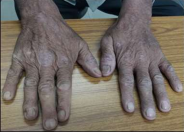
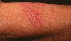
Cutaneous T cell lymphoma (CTCL) is a category of varied non-Hodgkin lymphomas which demonstrates the malignancy of T cells in the skin [7]. This lymphoma includes two subtypes: mycosis fungoides (>65%) and Sezary syndrome6. The risk of MF in male: female is 2:1, the average age of the diagnosis is 55 years and incidence of this disease is about 0.4 per 100,000 person-years [7,8]. However, the main pathogenesis of this disease is still unknown, but according to recent studies, many factors can be involved in the pathogenesis of this disease, including dysfunction of some genes such as TOX or disturbance in signaling pathways such as Notch or the SMARC gene family affecting changes in histones and DNA methylation [2].
In early-stage disease, skin directed therapies are used which includes PUVA, topical bexarotene, UVB (ultraviolet B), radiotherapy, topical corticosteroids, phototherapy, nitrogen mustard (such as ifosfamide), carmustine etc. However in advanced-stage disease HDACi (histone deacetylase Inhibitors), localized radiotherapy, systemic chemotherapy, monoclonal antibodies and TSEB (total skin electron beam) are the only valid treatment options available [9]. Interferon-α can be used in both early and advanced stages of the disease [9].
Funding: No funding sources.
Conflict of interest: None declared.
Ethical approval: The study was approved by the Institutional Ethics Committee of Tanda, (HP).
1. Alibert, Jean-Louis. Description des maladies de la peau observées à l'Hôpital Saint-Louis... 1806.
2. Murphy, G. F., and R. Schwarting. "Cutaneous lymphomas and leukemias." Lever's histopathology of the skin. 9th ed. Philadelphia: Lippincott Williams & Wilkins (2005): 927-978.
3. van Doorn, Remco, et al. "Mycosis fungoides: disease evolution and prognosis of 309 Dutch patients." Archives of dermatology 136.4 (2000): 504-510. https://jamanetwork.com/journals/jamadermatology/article-abstract/190143
4. Vergier, Beatrice, et al. "Transformation of mycosis fungoides: clinicopathological and prognostic features of 45 cases." Blood, The Journal of the American Society of Hematology 95.7 (2000): 2212-2218. https://doi.org/10.1182/blood.V95.7.2212
5. Kim, Youn H., and Richard T. Hoppe. "Mycosis fungoides and the Sezary syndrome." Seminars in oncology. Vol. 26. No. 3. [New York] Grune & Stratton., 1999. https://api.taylorfrancis.com/content/chapters/edit/download?identifierName=doi&identifierValue=10.1201/b13424-92&type=chapterpdf
6. Nagatani, Tetsuo, et al. "Comparative study of cutaneous T‐cell lymphoma and adult T‐cell leukemia/lymphoma: Clinical, histopathologic, and immunohistochemical analyses." Cancer 66.11 (1990): 2380-2386. https://acsjournals.onlinelibrary.wiley.com/doi/abs/10.1002/1097-0142(19901201)66:11%3C2380::AID-CNCR2820661122%3E3.0.CO;2-H
7. Barcos, Maurice. "Mycosis fungoides: diagnosis and pathogenesis." American journal of clinical pathology 99.4 (1993): 452-458. https://academic.oup.com/ajcp/article-abstract/99/4/452/1797692
8. Glusac, Earl J. "Criterion by criterion, mycosis fungoides." The American journal of dermatopathology 25.3 (2003): 264-269. https://journals.lww.com/amjdermatopathology/fulltext/2003/06000/Criterion_by_Criterion,_Mycosis_Fungoides.00014.aspx
9. Zackheim, Herschel S., et al. "Prognosis in cutaneous T-cell lymphoma by skin stage: long-term survival in 489 patients." Journal of the American Academy of Dermatology 40.3 (1999): 418-425. https://www.sciencedirect.com/science/article/pii/S0190962299704913