Contents
Download PDF
pdf Download XML
388 Views
390 Downloads
Share this article
Research Article | Volume 2 Issue 2 (July-Dec, 2021)
Seroprevalence of Human T-Lymphotropic Virus 1/2 among HIV Patients Attending Infectious Diseases Clinic in Sokoto State, Nigeria
 ,
1
Medical Microbiology Department School of Medical Laboratory Science, Usmanu Danfodiyo University, Sokoto, Nigeria, 840212 - 840283
2
Microbiology and Parasitology Department, Usmanu Danfodiyo University Teaching Hospital, Sokoto, Nigeria, 840212 - 840283
Under a Creative Commons license
Open Access
Received
April 10, 2021
Revised
April 22, 2021
Accepted
April 20, 2021
Published
Oct. 31, 2021
Abstract

Human T-lymphotropic viruses (HTLV) and human immunodeficiency viruses (HIV) are two distinct family members of Retroviridae causing significant infections worldwide. These viruses have common modes of transmission and share an in vivo tropism for cells of the immune system particularly T lymphocytes, resulting into co–infections of a number of individuals worldwide. The study was conducted to determine the seroprevalence of HTLV 1/2 infection among HIV patients. ELISA kit (Nanjing Pars Biochem, China) was used to detect the presence of HTLV 1/2 antibodies. A total number of 381 HIV patients consisting of males (123) and females (258) aged 18-59 years were tested. Of the 381 HIV patients tested, 288 (75.6%) were positive for HTLV 1/2 using ELISA kit. The prevalence of HTLV 1/2 was significantly higher among patients in the 30-39 years’ age group (24.4%). The prevalence of HTLV 1/2 was significantly higher among female’s HIV patients 195 (51.2%) than males 93 (24.4%). This study indicates a high prevalence of Human T-Lymphotropic virus 1/2 infection among HIV patients although there was a limitation in this study because a confirmatory test using a molecular test was not performed due to lack of financial capability and time factor. Finding from this study may justify the need for a further research and study of HTLV 1/2 among HIV patients to know if there is co-infection between the two. And also a more sensitive and specific assay should be used to confirm it like PCR or Western Blot or LIA test.

Keywords
None

Key findings:

The study identified a high seroprevalence of HTLV 1/2 among HIV patients, notably higher in females and the 30-39 age group. Using ELISA kits, 75.6% of HIV patients tested positive for HTLV 1/2. Lack of confirmatory molecular testing due to resource constraints underscores the need for further investigation and more sensitive assays.


What is known and what is new?

Human T-lymphotropic viruses (HTLV) and human immunodeficiency viruses (HIV) co-infections are recognized, sharing immune cell tropism. This study unveils a high seroprevalence of HTLV 1/2 among HIV patients, particularly in females aged 30-39. It underscores the necessity for further research utilizing more sensitive assays to confirm co-infection and explore clinical implications.


What is the implication, and what should change now?

The study reveals a substantial co-infection rate of HTLV 1/2 among HIV patients, particularly among females aged 30-39. However, the absence of confirmatory molecular testing due to financial constraints highlights the need for more comprehensive investigations. Future studies should employ sensitive assays like PCR or Western Blot to confirm co-infection and explore clinical implications thoroughly.

INTRODUCTION:

Human T-lymphotropic viruses (HTLV) and human immunodeficiency viruses (HIV) are two distinct family members of Retroviridae causing significant infections worldwide. These viruses have common modes of transmission and share an in vivo tropism for cells of the immune system particularly T lymphocytes, resulting in co–infections of a number of individuals worldwide [1, 2]. HTLV is made up of two major types associated with disease in humans which are HTLV-1 and HTLV-2. Two other types have been discovered which are HTLV-3 and HTLV-4 [3]. HTLV-1 is endemic in many well–defined geographic areas like Sub–Saharan Africa in which Nigeria is included. However, there is no recent representative data regarding prevalence of HTLV-1 among HIV-1 infected individuals in Nigeria but majority of serological studies carried out were on blood donors [4-8]. Research has shown that HIV-1/HTLV-1 and HIV-1/HTLV-2 co-infection probably occur more frequently than physicians are aware of since routine testing for HTLV-1/2 is not usually performed. HIV infection is sexually transmitted therefore people with HIV disease are at risk of having HTLV infection because both have same routes of transmission and similar cell tropism [9]. Therefore, co–infection of both viruses will likely influence the pattern of progression to AIDS, since both viruses preferentially infect CD4+ T-cells. The common link between HTLV–1/2 in HIV–1 patients is that both have been linked to normal or high CD4+ T cell count hinder proper treatment as a result of delay in introduction of highly active anti–retroviral therapy (HAART) in co–infected patients. It has also been reported that co–infection of HTLV–1/2 and HIV, may accelerate progression to AIDS and significantly shorten survival time in such individuals [10].


A major issue in developing countries including Nigeria is lack of routine screening practice for HTLV, thus there exists a gap in knowledge and awareness of transmission of HTLV infection from both healthy individuals and those who are co–infected with HIV. Little is known about the pattern of sero-prevalence of HTLV–1/2 in Sokoto; this study is therefore designed to determine the sero-prevalence of HTLV–1/2 among HIV infected individuals attending the infectious diseases clinic of UDUTH, Sokoto.


There is paucity of information on this clinical entity in Nigeria [11]. The transmission of HIV and HTLV-1 are similar, infection can occur by sexual contact with an infected individual, through sharing of contaminated needles and syringes by intravenous drug users, or as a result of a transfusion of contaminated blood. Mother to child transmission may happen through perinatal exposure or most often through breast-feeding. HTLV–1 or 2 co–infection in individuals infected with HIV can lead to increased morbidity. The shared routes of transmission of HTLV with HIV may increase the prevalence of HTLV among HIV infected populations and subsequently affect patient’s management [9] hence the need for this study. 

MATERIALS AND METHODS:

Study Area

The study was conducted in the Sokoto metropolis. Sokoto metropolis lies between longitudes 0.5 ° .11’ to 13 ° .03’ east and latitude 13 ° .00 to 13 ° .06 north [12]. Sokoto State is at the extreme part of North-western region of Nigeria, sharing a border with Niger Republic to the North with a total land area of 32,000 square kilometers and a population of 4,602,298 million. There are two seasons in Sokoto; the dry season and the raining seasons in which the harmattan season ranges from October to February [13]. Farming and animal husbandry are the major occupation in this region, comprising Hausa and Fulani [14].


Study Population and Design

This descriptive study was conducted at the infectious diseases clinic among HIV seropositive patients 18 years and above that visited Usmanu Danfodiyo University Teaching Hospital (UDUTH), Sokoto, Sokoto State, Nigeria by analyzing the serum samples collected. At the clinic an average of 10 to 20 HIV patients are seen daily and between 25 to 70 patients are seen weekly including male and female patients from different works of life. And their CD4+ cells are monitored too.


Ethical Considerations

The participants gave their written informed consent before they were enrolled in the study. All data were analyzed anonymously throughout the study. Ethical approval was obtained from the UDUTH Health Research Ethics Committee Sokoto (UDUTH/HREC/2021/1052/V3) and Sokoto State Ministry of Health (SHREK/068/2021).


Inclusion and Exclusion Criteria

All HIV seropositive patients (male and female) attending IDC of UDUTH Sokoto, Sokoto State, who gave written informed consent to participate in the study were included in the study. Non HIV, non-adult and patients who did not give written informed consent to participate in the study were excluded.


Sample Size Determination

Sample size was estimated using fisher’s formula [15] adopting a prevalence rate of  45.4 % as follows; N = Z2pq/d2 where ‘N’ is the required sample size, ‘Z’ is the confidence interval at 95% (1.96), ‘p’ is estimated prevalence from previous studies = 45.4 % , ‘q’ is 1 – p, and ‘d’ is the degree of accuracy set at 0.05.Substituting;   N= 1.962 x 0.454 x 0.546/0.052   and  this gives a sample size of 381.


Sample Collection and Analysis

Patients were consecutively recruited after giving informed consents. Five (5) ml amount of whole blood samples was collected aseptically into two (2) ml amount of ethylenediaminetetraacetic (EDTA) acid-preserved blood sample bottle and was spun at 12000/g for 10 minutes to harvest their serum samples for HTLV-1/2 serological tests. HIV surface antigen was determined using HIV Test Strip (determine kit). The foil of the strip was pulled off and placed on the bench top then a drop of serum sample was placed on the sample pad on the strip using a pasteur pipette. The strip was allowed to stand for 15 minutes. Then the result was read. Anti-HTLV IgM and IgG antibodies in serum of HIV-1 patients were determined by HTLV-1+2 ELISA Test Kits (Nanjing Pars Biochem, China).


Determination of HIV Surface Antigen Using HIV Test Strip (Determine HIV Early Detect)

The foil of the strip was pulled off and placed on the bench top then a drop of serum sample was placed on the sample pad on the strip using a pasteur pipette. The strip was allowed to stand for 15 minutes. Then the result was read (Abbott Diagnostic Medical Co. Ltd, Matsudo, Japan).


Determination of Anti–HTLV Igm and Igg Antibodies in Serum of HIV Patients

Analysis of IgG and IgM Antibodies to HTLV were detected using human T–lymphotropic virus sandwich ELISA. The micro–ELISA strip plates were pre–coated with an antigen specific to HTLV IgG and IgM antibodies respectively. Standards test samples were added to appropriate micro–ELISA strip plate wells and combined to specific antigen. Then a horseradish peroxidase (HRP)-conjugated antigen specific to HTLV was added to each micro-ELISA strip plate well and incubated. Free components were washed away. The TetraMethylBenzidine (TMB) contained HTLV substrate solution was added to each well. Only those wells that contained HTLV-IgG or IgM in different micro-ELISA strip plates respectively and HRP conjugate HTLV antigen appeared blue in colour and then yellow after addition of the stop solution. The optical density (O.D) was measured using a spectrophotometer at a wavelength of 450nm. The presence of anti-HTLV IgG or IgM antibodies in different micro- ELISA strip plates, respectively, was determined by comparing the O.D of the sample to the CUT OFF value of the plate according to manufacturers’ instructions. The reagents and samples were allowed to reach ambient temperature (25°C) for 30 minutes. Wells was numbered including one Blank, three for the Negative control and two for the positive control. Fifty (50) µl of Positive control, negative control, and test sera was added into their respective wells except the blank. A separate disposal pipette tip was used for each specimen, negative control and positive control to avoid cross-contamination. Fifty (50) µl HRP-Conjugate (Horseradish peroxidase-conjugated anti-HBS) was added to each well except the Blank, and mixed by tapping the plate gently. The plate was covered with plate cover and incubated for 60 minutes at 37°C. Plate cover was removed and discarded. The plate was emptied by aspiration and washed each well 5 times with a diluted Wash buffer. After the washing cycle, the plate was dried by turning them upside down onto blotting paper. Fifty (50) µl of chromogen A (Urea peroxide solution) and fifty (50) µl chromogen B (tetramethylbenzidine) was dispensed into each well including the Blank, and mixed by tapping the plate gently. The plate was incubated at 37°C for 15 minutes avoiding light. The enzymatic reaction between the chromogen solutions and HRP-Conjugate produces blue colour in positive control and HTLV 1/2 positive sample well. Multichannel pipette was used to stop the reaction by adding fifty µl stop solution (diluted sulphuric acid solution 0.5 M H2SO4) into each well and was mixed gently. Intensive yellow colour develops in positive control and HTLV 1/2 positive sample wells. The plate reader was calibrated with the blank well and the absorbance was read at 450 nm within 15 minutes. Specimens giving absorbance less than the cut-off value are negative for this assay, which indicates that no HTLV 1/2 virus surface antigen has been detected with this HTLV 1/2 ELISA. Specimens giving an absorbance equal to or greater than the cut-off value are considered initially reactive, which indicates that HTLV 1/2 virus surface antigen could probably be detected using this HTLV 1/2 ELISA (Nanjing Pars Biochem, China).   


Statistical Analysis

All generated data was analyzed using SPSS software version 26.0 (2016, IBM California, USA). The prevalence of HTLV-1/2 will be expressed in simple proportions and percentages for the study groups. Chi-square contingency table will be used to determine the relationship between seroprevalence of HTLV-1/-2 infections and HIV in relation to age group, gender and ethnicity. A confidence interval of 95%, p-values <0.05 were considered statistically significant.
 

RESULT AND DISCUSSION:

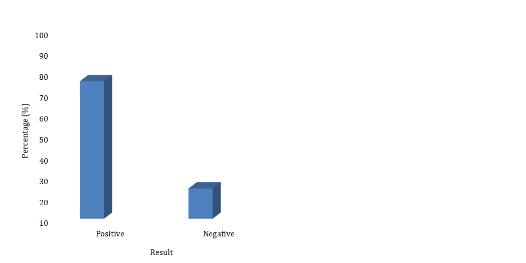
Out of the 381 HIV patients that participated in the study, the prevalence of HTLV 1/2 antibodies was 75.6% (228/381) (Figure 1). There was a marked difference in the distribution of HTLV 1/2 by age group. Individuals who were 30-39 years had the highest prevalence (24.4%: 93/381) of HTLV 1/2 antibodies, followed by 21-29 years with prevalence of (21.3%: 81/381), 40-49 years old (14.2%: 54/381), 50-59 years (8.7%: 33/381) and 18-20 years old with prevalence of (7.1%: 27/381). However, there was a significant difference observed between age group (chi square= 16.005 and p value= 0.001) (Table 1). Out of tested 381 HIV patients which consist of 123 males and 258 females, indicating 32.3% and 67.7% males and females representative respectively. The prevalence of HTLV 1/2 according to gender (Table 2) was significant (chi square= 7.658 and p value= 0.001). However, higher prevalence was recorded among the female HIV patients (51.2%: 195/381) than male HIV patients (24.4%: 93/381). Out of 381 subjects tested, 333 were Hausa representing (87.4%), 9 were Igbo (2.4%), 15 were Yoruba (3.9%) and those from Other ethnic groups were 24 representing (6.3%) respectively. The prevalence of HTLV 1/2 according to ethnicity (Table 3) was not significant.


 In this study, it was discovered that a higher percentage of the participants infected were females, with 51.2% of them having antibodies to HTLV 1/2 compared to males having 24.4%. This agrees with the work done in University of Illorin Teaching Hospital [9] having a prevalence of 60.6% and 52.5% for male and female respectively. Although the number of females recruited in this study was more than the males, this could be because there is more efficient transmission from men to women during sexual intercourse. The HTLV 1/2 seroprevalence among HIV infected individuals in this study is higher than the prevalence reported in previous studies, and this may be due to the reluctance in adapting to behaviours that are less risky especially amongst those with HIV in Sokoto. Also, the high seroprevalence rate may be as a result of the method employed in this study.  There was a report of an overall seroprevalence of 4.9% for HIV/HTLV 1/2 co-infection among ART naïve patients in Abuja using PCR to confirm.  HTLV 1/2 prevalence of 1.9% was reported in HIV patients in Brazil using the Polymerase Chain Reaction assay [16]. Rego et al. [17] reported the prevalence of HTLV/HIV co-infection to be 1.8% in KwaZulu-Natal, South Africa. In another study conducted in rural Guinea-Bissau among HIV infected women, overall HTLV prevalence of 5.2% was reported [18]. The difference is most likely due to the difference in the design, population and method of detection of HTLV 1/2. Molecular methods were used to confirm HTLV proviral DNA in the studies mentioned, whereas antibody detection was solely used in this study which was a limitation as a result of time factor and also lack of financial resources.


However, in this study, the highest prevalence of HTLV 1/2 antibodies was among the age group 30-39 years. This was in contrast with Nasir et al. (Nasir et al., 2015) study who reported the highest prevalence of HTLV 1/2 among the age group 21-30 years. This could be due to the fact that the population of those mostly recruited falls among this age group in our study. Also, there is a presumption that the accumulation of sexual exposures with age in women of this population might contribute to the results obtained in this study. Low prevalence rates have been obtained from different populations in other studies like in healthy blood donors at a tertiary center in Lagos and among pregnant women at the university of Nigeria teaching hospital, Enugu [19, 20], as well as moderate among pregnant women in Ibadan and among blood donors in Osogbo [21, 22] and high prevalence among pregnant women in Ibadan [21].


There are reports that CD4+ lymphocyte count cannot always be considered a reliable marker of immunological competence in HIV infected people, especially in patients co–infected with HTLV [3, 23, 24]. There was no significant association between CD4+ cell count in HTLV/HIV co-infected patients compared to HIV infected individuals (p=0.163). Hence, CD4+ count was not carried out in this study [9]. CD4+ cell count is one of the important criteria used to determine eligibility for HAART in HIV infected individuals especially in resource–limited settings nevertheless in the event of HTLV/HIV co-infections, CD4 cell counts may not be reliable [9].


Figure 1: Seroprevalence of HTLV 1/2 Antibodies among HIV Patients in Sokoto, Nigeria.


Table 1: Prevalence of HTLV 1/2 Antibodies According to Age Group

Age RangeHTLV 1/2 StatusTotal N=381 (%)X2p-value
Positive (%)Negative (%)
18-2027(7.1)6(1.6)33(8.7)16.0050.001
21-2981(21.3)24(6.3)105(27.6)  
30-3993(24.4)42(11.0)135(35.4)  
40-4954(14.2)15(3.9)69(18.1)  
50-5933(8.7)6(1.6)39(10.3)  

        HTLV 1/2 =Human T-Lymphotropic Virus 1 and 2

        N=Number of Participants


Table 2: Prevalence of HTLV 1/2 among Gender

GenderHTLV 1/2 StatusTotal N=381 (%)X2p-value
Positive (%)Negative (%)
Male93(24.4)30(7.9)123(32.3)7.6580.001
Female195(51.2)63(16.5)258(67.7)  

        HTLV 1/2 =Human T-Lymphotropic Virus 1 and 2

        N=Number of Participants


Table 3: Prevalence of HTLV 1/2 Antibodies According to Ethnicity

EthnicityHTLV 1/2 StatusTotal N=381 (%)X2p-value
Positive (%)Negative (%)
Hausa249(65.4)84(22.0)333(87.4)0.5650.573
Igbo6(1.6)3(0.8)9(2.4)  
Yoruba15(3.9)0(0.0)15(3.9)  
Others18(4.7)6(1.6)24(6.3)  

      HTLV 1/2 =Human T-Lymphotropic Virus 1and 2

      N=Number of Participants


CONCLUSION:

Our findings in this study revealed that there is high seroprevalence of HTLV 1/2 infection among HIV patients in sokoto. The high rate of co-infection supports routine screening for HTLV-1/2 coinfection among HIV infected individuals in Sokoto, Nigeria so that the purpose of HAART treatment and monitoring of patients to prevent progression to AIDS will not be aborted. There is also a need to educate patients visiting the hospital on the modes of transmission and prevention. The low specificity of ELISA test suggests that a more effective confirmatory test such as Western blot or INNO-LIA™ or PCR confirmatory assay which include DNA extraction from the blood be performed alongside to verify the HTLV 1/2 antibody detected from plasma where present. Treatment modality in individuals co-infected with HIV and HTLV-1/2 is recommended because these patients present normal or unexpectedly high CD4+ T cell counts which do not account for the suppression of the immune response experienced.


Funding: No funding sources 


Conflict of interest: None declared


Ethical approval: The study was approved by the Institutional Ethics Committee of Usmanu Danfodiyo University


REFERENCES:
  1. Araujo, Abelardo, et al. "Concomitant Infections with Human Immunodeficiency Virus Type 1 and Human T‐Lymphotropic Virus Types 1 and 2." Polymicrobial diseases (2002): 75-97. https://doi.org/10.1128/9781555817947.ch5 
  2. Roy, Upal, et al. "Upregulation of HTLV‐1 and HTLV‐2 expression by HIV‐1 in vitro." Journal of medical virology 80.3 (2008): 494-500. https://doi.org/10.1002/jmv.21089 
  3. Mazanderani, AF Haeri, and Osman Ebrahim. "Progressive HIV infection in the presence of a raised CD4+ count: HIV/HTLV-1 co-infection." Southern African Journal of HIV Medicine 14.2 (2013): 92-94. https://www.ajol.info/index.php/sajhivm/article/view/89445 
  4. Mazanderani, AF Haeri, and Osman Ebrahim. "Progressive HIV infection in the presence of a raised CD4+ count: HIV/HTLV-1 co-infection." Southern African Journal of HIV Medicine 14.2 (2013): 92-94. https://www.ajol.info/index.php/sajhivm/article/view/89445 
  5. Iyalla, Caroline, et al. "Seroprevalence study of HTLV-1 and 2 in prospective blood donors and pregnant women in Port Harcourt, Nigeria." African Journal of Infectious Diseases 9.2 (2015): 57-60. https://www.ajol.info/index.php/ajid/article/view/116554 
  6. Durojaiye, Idris, et al. "Seroprevalence of human T lymphotropic virus antibodies among healthy blood donors at a tertiary centre in Lagos, Nigeria." Pan African Medical Journal 17.1 (2014). https://www.ajol.info/index.php/pamj/article/view/131133 
  7. Yuguda, S., et al. "Seroprevalence of human T-cell lymphoma/Leukemia virus type-1 (HTLV-1) antibodies among blood donors at Ibadan, Nigeria." J Hum Virol Retrovirol 5.5 (2017): 00169. https://www.researchgate.net/profile/Saleh-Yuguda/publication/321867914_Seroprevalence_of_Human_T-Cell_LymphomaLeukemia_Virus_Type-1_HTLV-1_Antibodies_among_Blood_Donors_at_Ibadan_Nigeria/links/5b8ef78fa6fdcc1ddd0fd633/Seroprevalence-of-Human-T-Cell-Lymphoma-Leukemia-Virus-Type-1-HTLV-1-Antibodies-among-Blood-Donors-at-Ibadan-Nigeria.pdf 
  8. Okoye, Augustine Ejike, et al. "Seroprevalence of human T-cell lymphoma/leukemia virus type-1 (HTLV-1) antibodies among blood donors at Enugu, Nigeria." Journal of blood medicine (2015): 31-36. https://www.tandfonline.com/doi/abs/10.2147/JBM.S65556 
  9. Adeoye, O. A., et al. "Sero-prevalence of human T-lymphotrophic virus 1/2 among HIV-1 infected individuals in Ilorin, Nigeria." African Journal of Clinical and Experimental Microbiology 20.3 (2019): 202-208. https://www.ajol.info/index.php/ajcem/article/view/186729 
  10. Brites, Carlos, et al. "Co-infection with HTLV-1 is associated with a shorter survival time for HIV-1-infected patients in Bahia, Brazil." Aids 15.15 (2001): 2053-2055. https://journals.lww.com/aidsonline/fulltext/2001/10190/Co_infection_with_HTLV_1_is_associated_with_a.23.aspx 
  11. Babayo, Adamu, et al. "Serosurvey and Cellular Immune Status of HTLV-1/2 and HIV Co-infections in Bauchi State, Nigeria." (2019). https://repozytorium.ur.edu.pl/handle/item/5175 
  12. Collins Maps. "Geographical Information for Sokoto." Collins Maps, 2014, www.collinsmaps.com. Accessed 28 June 2019.
  13. Sokoto State Business Directory. A Publication of the Commerce Department, Ministry of Commerce, Industry and Tourism. SSBD, 2007.
  14. Okwesili, A. N., et al. "Prevalence of hepatitis B surface antigen among biomedical students of African descent in Usmanu Danfodiyo university, Sokoto, Nigeria." Human antibodies 23.3-4 (2015): 57-62. https://content.iospress.com/articles/human-antibodies/hab282 
  15. Araoye, Margaret Olabisi. "Research methodology with statistics for health and social sciences." Ilorin: Nathadex Publisher 115.9 (2003): 25-120.
  16. Galetto, Leonardo Ramos, et al. "Prevalence and risk factors for human T cell lymphotropic virus infection in Southern Brazilian HIV-positive patients." AIDS research and human retroviruses 30.9 (2014): 907-911. https://www.liebertpub.com/doi/abs/10.1089/aid.2013.0210 
  17. Rego, Filipe FA, Luiz CJ Alcantara, and Tulio de Oliveira. "Human T cell lymphotropic virus type 1 (HTLV-1) infection increases the CD4 count in human immunodeficiency virus type 1 (HIV-1) infected patients submitted to antiretroviral therapy (ARV)." Retrovirology 11.Suppl 1 (2014): P62. https://link.springer.com/article/10.1186/1742-4690-11-S1-P62 
  18. Holmgren, Birgitta, et al. "Dual infections with HIV-1, HIV-2 and HTLV-I are more common in older women than in men in Guinea-Bissau." Aids 17.2 (2003): 241-253. https://journals.lww.com/aidsonline/fulltext/2003/01240/Trends_and_interaction_of_HIV_1_and_HIV_2_in.00015.aspx 
  19. Durojaiye, Idris, et al. "Seroprevalence of human T lymphotropic virus antibodies among healthy blood donors at a tertiary centre in Lagos, Nigeria." Pan African Medical Journal 17.1 (2014). https://www.ajol.info/index.php/pamj/article/view/131133 
  20. Okoye, Augustine Ejike, et al. "Seroprevalence and correlates of human T-cell lymphoma/leukemia virus type 1 antibodies among pregnant women at the University of Nigeria Teaching Hospital, Enugu, Nigeria." International Journal of Women's Health (2014): 849-855. https://www.tandfonline.com/doi/abs/10.2147/IJWH.S64199 
  21. Olaleye, David O., et al. "Evidence of serological cross-reactivities with human immunodeficiency virus types 1 and 2 and human T-lymphotropic virus types I and II in sera of pregnant women in Ibadan, Nigeria." International journal of epidemiology 24.1 (1995): 198-203. https://doi.org/10.1093/ije/24.1.198 
  22. Terry, A. A., et al. "Seroprevalence of HTLV-I/II amongst Blood Donors in Osogbo, Nigeria." Sudan Journal of Medical Sciences 6.3 (2011): 177-182.  https://www.ajol.info/index.php/sjms/article/view/73630 
  23. Scapellato, Pablo Gustavo, et al. "CD4 cell count among HIV-infected patients with an AIDS-defining disease: higher count in patients coinfected than in those not coinfected with human T-cell lymphotropic virus type I." JAIDS Journal of Acquired Immune Deficiency Syndromes 33.2 (2003): 279-280. https://journals.lww.com/jaids/citation/2003/06010/CD4_Cell_Count_Among_HIV_Infected_Patients_With_an.28.aspx 
  24. Gudo Jr, E. S., et al. "Performance of absolute CD4+ count in predicting co-infection with human T-lymphotropic virus type 1 in antiretroviral-naive HIV-infected patients." International journal of STD & AIDS 23.10 (2012): 717-723. https://journals.sagepub.com/doi/abs/10.1258/ijsa.2012.011446 


Advertisement
Recommended Articles
Research Article
Role of Hydroxychloroquine Therapy in COVID 19 Patients. A Systematic Review and Meta Analysis
...
Published: 30/05/2022
Download PDF
Original Article
Role of Audits in Helathcare associated Infections
Published: 20/06/2024
Download PDF
Research Article
Thyroid Health in Shimla: Recognizing and Addressing Common Disorders
...
Published: 29/05/2024
Download PDF
Research Article
Exploring Public Awareness of Eating Disorders Among Adolescents in District Solan: A Study on Anorexia and Bulimia
Published: 30/07/2024
Download PDF
Chat on WhatsApp
Flowbite Logo
Najmal Complex,
Opposite Farwaniya,
Kuwait.
Email: kuwait@iarcon.org

Editorial Office:
J.L Bhavan, Near Radison Blu Hotel,
Jalukbari, Guwahati-India
Useful Links
Order Hard Copy
Privacy policy
Terms and Conditions
Refund Policy
Others
About Us
Contact Us
Online Payments
Join as Editor
Join as Reviewer
Subscribe to our Newsletter
Follow us
MOST SEARCHED KEYWORDS
scientific journal
 | 
business journal
 | 
medical journals
 | 
Scientific Journals
 | 
Academic Publisher
 | 
Peer-reviewed Journals
 | 
Open Access Journals
 | 
Impact Factor
 | 
Indexing Services
 | 
Journal Citation Reports
 | 
Publication Process
 | 
Impact factor of journals
 | 
Finding reputable journals for publication
 | 
Submitting a manuscript for publication
 | 
Copyright and licensing of published papers
 | 
Writing an abstract for a research paper
 | 
Manuscript formatting guidelines
 | 
Promoting published research
 | 
Publication in high-impact journals
Copyright © iARCON Internaltional LLP . All Rights Reserved.