Contents
Download PDF
pdf Download XML
1691 Views
312 Downloads
Share this article
Research Article | Volume 2 Issue 1 (Jan-June, 2021) | Pages 1 - 14
The Impact of Rotavirus Vaccination on Disease Burden among the Lebanese Infants and Children under 5 Years Old who have been Diagnosed with Acute Gastroenteritis from Year 2016 to 2017
 ,
 ,
 ,
 ,
 ,
 ,
1
Lebanese University-Faculty of Medical Sciences, Lebanon
Under a Creative Commons license
Open Access
Received
Oct. 7, 2020
Revised
Nov. 22, 2020
Accepted
Dec. 14, 2020
Published
Jan. 20, 2021
Abstract

Background: Worldwide, diarrhea is the record leading cause of deaths in children, with rotavirus being the most common diarrheal pathogen irrespective of the country’s level of development. It’s recognized that hospitalization for Rotavirus Gastroenteritis (RV GE) represents the severe end of the spectrum of the disease, for that, the vaccine trial was introduced in our country. Our main objective was to study the impact of the vaccination program for Rotavirus (RV) on disease burden among the Lebanese infants and children under 5 years of age who have been diagnosed with acute gastroenteritis from 2016 to 2017. In addition to estimating the effectiveness of both included vaccines in our country. Methods: This was a retrospective database study between years 2016-2017, wherein data on infants and children younger than 5 years who have been diagnosed with acute Gastroenteritis (GE), has been collected from the electronic admission database or other records from the participating hospitals. Results: In Lebanon, the rate of hospitalized patients with RV GE was found around 31.9% during 2016-2017. Overall, 30.4% were vaccinated against RV among hospitalized children for all GE. Therefore, vaccination against RV was associated with reduction of the incidence of disease, as well as the clinical severity and number of hospitalizations among immunized patients compared to those non-immunized. Same effectiveness was found between the 2 types of vaccines. Conclusion: The findings of our study confirm the positive impact of RV vaccines on the control of RV diarrheal disease in Lebanon and the reduction of the disease burden upon introduction of the RV vaccine into the routine immunization schedule in children. This result suggests that maximizing vaccine coverage may lead to further reductions in GE RV mortality and hospitalizations.

Keywords
INTRODUCTION

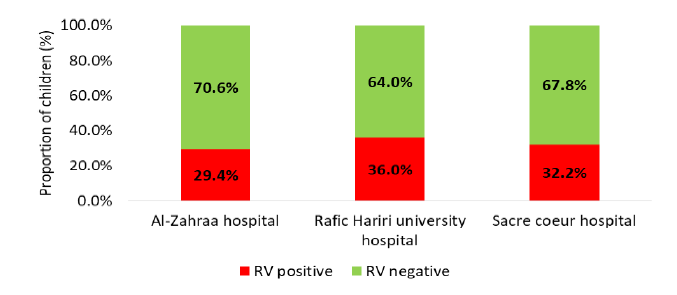
General Overview: Acute Gastroenteritis (GE) is one of the most common infectious diseases of all age groups worldwide, mainly in the pediatric population and it is a leading global cause of morbidity and mortality among children under 5 years of age in developing countries where complications include electrolyte disturbances and malnutrition that necessitates hospitalization [1]. The condition can be caused by some different microorganisms, but viruses are increasingly recognized as predominant causative factors. To note that acute GE is usually acquired by fecal-oral route, also through contaminated surfaces or from water sources. Identified symptoms vary between watery diarrhea, nausea, vomiting, fever and abdominal pain [1].However, viruses have become more frequently identified mainly with the advances in the ability to diagnose viral infections. Among them, Rotavirus (RV) is the leading cause of acute GE in infants and young children worldwide [1]. Although the first human RV was discovered in 1973; each year, about 25 million episodes of GE due to RV are reported in children worldwide, of which 2 million require hospitalizations and 180000-450,000 deaths occur less than 5 years of age, mostly in developing countries. In Lebanon, prevalence of RV infection has been estimated between 27.7 and 30.6% [1] (Figure 1).


The spectrum of RV illness ranges from mild, watery diarrhea of limited duration to severe diarrhea with vomiting and fever. The dehydration that can result can be more serious than the infection itself and is the most common cause of death in rotavirus cases [1]. Because the clinical features of RV related GE are similar to other viral and bacterial GE, confirmation of RV infection by laboratory testing of fecal specimens is necessary. The RV is shed in high concentration in the stool of children with gastroenteritis (i.e., 1012 viruses/G), so the best inexpensive and available method is antigen detection in the stool by an Enzyme Immunoassay (EIA) directed at an antigen common to all rotaviruses. Also other techniques can be cited but mainly used in research settings ex: electron microscopy, reverse transcription-polymerase chain reaction RT-PCR, nucleic acid hybridization, sequence analysis and culture [2]. Since 2006, two rotavirus vaccines are currently licensed for infants worldwide for routine immunization and have been introduced in both developed and developing countries leading to herd immunity in several states. So since RV vaccines were implemented, studies have been undertaken to assess the effects of vaccination on RV disease [3]. This study will discuss the impact of the RV vaccination program on relative reductions of RV disease burden and RV related GE hospitalizations among Lebanese infants and children aged less than 5 years in 3 different hospitals, using file or electronic records for data collection.


Review of literature

Epidemiology: RV remains the world’s commonest cause of GE among children predominantly in developing countries; globally RV alone is responsible for almost 40% of GE related hospitalization. In developing countries, Rotavirus is responsible for around half a million deaths annually. In addition, diarrhea caused by RV infection plays a major role in malnutrition among children and predisposes them to other infectious diseases which lead to increase in childhood morbidity and mortality. It is prevalent in all geographical regions of the world and almost all socioeconomic groups. Three of the seven RV groups (A to G) are known to infect humans. Among them, the most dominant is group A, which causes diarrheal diseases worldwide. 


Rotavirus structure: The name RV derives from the characteristic wheel-like appearance of the virus when observed by electron microscopy, which is derived from the Latin word “Rota” meaning “Wheel”. This pathogen was initially discovered in diarrheic cattle, mice and monkeys and finally in infants and young children in 1973 by Bishop and Flewett [4]. RV is a triple layered, non-enveloped virus within the family of Reoviridae. The genome of this virus consists of 11 double-stranded RNA segments. The nucleic acid is surrounded by 2 layers of capsid–inner capsid (VP6) and outer capsid (VP7). In addition to VP4, the spike protein, this is a cell surface receptor [5]. This 11 segmented double-stranded RNA (dsRNA) encoding six structural Proteins (VP) and 6 Non-Structural Proteins (NSP). The viral proteins VP1, VP2, VP3, VP4, VP6 and VP7 are, as we mentioned, the structural proteins that form the virion. The non-structural proteins (produced by the cell infected by rotavirus) are NSP1, NSP2, NSP3, NSP4, NSP5 and NSP6 [5] (Figure 2 and 3).


Rotavirus Genotype

RV is classified into P and G serotypes, respectively. The genetic diversity of the virus is due to the segmented nature of its genome. Of the 11 genomic segments of RV, segment 6 codes for the most abundant viral protein, the VP6 protein, this is the major antigenic determinant of group A reactivity. Also, based on the VP-6 capsid gene, the virus has been classified into the seven major genogroups A to G. The Segments 7, 8 or 9 codes for the major glycoprotein, VP7, which is the basis for RV serotyping. Additionally, Segment 4 codes for a protease-sensitive protein, VP4, which is the basis for P-serotyping. So based on the sequence of VP7 genes (segment 9 mainly) and VP4 genes (segment 4), RVAs are classified into G and P-genotypes, respectively. Currently, there are 14 G serotypes and 11 of them have been detected in humans. The majority of human RVs belong to serotypes G1–G4 and to the newly emerged G9. Among the P types, P[8] was the most common type found across all G types [6]. To note that the genotype combinations which causing 90% infections in humans are G1P[8], G2P[4], G3P[8], G4P[8], G9P[8] and G12P[8] [7]. So based on that, the two live-attenuated oral rotavirus vaccines licensed for infants <6 months old contains human G1P[8] serotype for Rotarix while RotaTeq contains a mixture of five human serotypes G1–G4 and P[8] [7]. 


Distribution of the Fraction of Diarrhea-Associated Mortality... | Download  Scientific Diagram


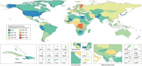
Figure 1: Rotavirus Related Mortality Worldwide. Adapted and Reprinted from Troeger C, Khalil IA, Rao PC, Cao S, Blacker BF, Ahmed T, Armah G, Bines JE, Brewer TG, Colombara DV, Kang G, Kirkpatrick BD, Kirkwood CD, Mwenda JM, Parashar UD, Petri WA Jr, Riddle MS, Steele AD, Thompson RL, Walson JL, Sanders JW, Mokdad AH, Murray CJL, Hay SI, Reiner RC Jr. Rotavirus Vaccination and the Global Burden of Rotavirus Diarrhea Among Children Younger Than 5 Years. JAMA Pediatr. 2018 Oct 1; 172 (10):958-965. Copyright © 2018 Troeger et al. JAMA Pediatrics

https://www.onlinebiologynotes.com/wp-content/uploads/2017/05/rota-virus2.jpg


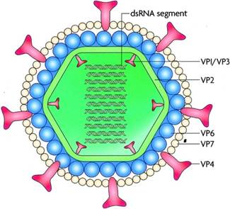
Figure 2: Rotavirus Structure. The Genetic Material is found inside a Complex 70-Nanometre Viral Nucleocapsid with three Concentric Shells: An Inner Core, an Internal Capsid vp6 and an Outer Capsid vp7. Sixty Spikes vp4 between 10 and 12 nm in Length Protrude from the outer Capsid. Adapted and Reprinted from Shaheen MNF. Rotavirus Gastroenteritis among Hospitalized Children under 5 years of age in the Eastern Mediterranean Region: A Review. East Mediterr Health J. 2019 Aug 19; 25(6):422-430. Copyright © World Health Organization (WHO) 2019


https://www.onlinebiologynotes.com/wp-content/uploads/2017/05/rotavirus-genome.jpg


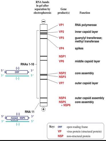
Figure 3: 11 Segments of the Genome of Rotavirus. Adapted and Reprinted from Shaheen MNF. Rotavirus Gastroenteritis among Hospitalized Children under 5 Years of Age in the Eastern Mediterranean Region: A Review. East Mediterr Health J. 2019 Aug 19; 25(6):422-430. Copyright © World Health Organization (WHO) 2019

https://www.onlinebiologynotes.com/wp-content/uploads/2017/05/replication-of-rotavirus.jpg


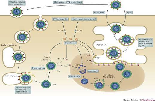
Figure 4: Replication Cycle of Rotavirus. Adapted and Reprinted from Online Biology Notes. Guarab Karki. Rotavirus-Classification, Structure composition and Properties, Replication, Mode of Transmission, Pathogenesis, Clinical Symptoms, Laboratory Diagnosis, Treatment, Prevention and Control. Available from: Rotavirus-Classification, Structure Composition and Properties, Replication, Mode of Transmission, Pathogenesis, Clinical Symptoms, Laboratory Diagnosis, Treatment, Prevention and Control - Online Biology Notes


Replication Cycle of Rotavirus in a Host Cell

The proteins VP4 and VP7, which compose the most outer layer, are important for the first interactions of the virus with the host cell surface by the attachment of the VP4 to the cell surface receptor. Rotavirus enters cells by endocytosis mediated by this receptor. During this entry, uncoating occurs in lysosomes leading to a Double Layer Particle (DLP) from the initial Triple Layer Particle (TLP) by losing the external protein layer. The transcriptionally active DLP starts transcription of the viral genome and the process is mediated by endogenous virus dependent RNA polymerase (transcriptase). The viral mRNAs direct the production of the viral structural and non-structural proteins by translation of this viral transcript and also it serves as RNA templates for the synthesis of negative sense RNA strand with capsid to form the double stranded RNA (dsRNA). The new viral proteins and RNAs accumulate in protected sites of the cytoplasm called viroplasms, where the viral genome replicates an double-layer intermediate replication particles assemble and form an inner capsid. The interaction of newly formed polymerase complexes with the core capsid protein triggers genome replication, which is followed by addition of the intermediate protein layer of the virion. These newly formed DLPs start budding into Rough Endoplasmic Reticulum (RER) and acquiring a pseudo envelope followed by subsequent removal of this transient lipid envelope which will be replaced by an outer capsid in RER to finally yield mature infectious TLPs. After release of TLPs through cell lysis or trafficking, the attachment spike must be cleaved by trypsin-like proteases in the intestinal lumen to activate the virus for subsequent infection [8] (Figure 4).


Transmission of Rotavirus

The primarily documented mode of RV transmission is fecal-oral route, directly from person to person, or indirectly via contaminated fomites or surfaces. A respiratory mode of transmission has also been proposed but it is not the usual mode of transmission [9]. In addition, Community wide water borne epidemics of group A RV (G-A RV) have been documented in Turkey in 2011 [10]. The young infant can get infected either from older siblings or patients with subclinical infection. And as known, Rotaviruses are highly contagious and a low viral load may trigger a human infection (<100 virus particles is enough to cause infection) [11].


Incubation Period

The primarily documented mode of Rotavirus transmission is faeco-oral route, directly from person to person, or indirectly via contaminated fomites or surfaces. A respiratory mode of transmission has also been proposed but it is not the usual mode of transmission [9]. In addition, Community wide water borne epidemics of group A Rotavirus (G-A RV) have been documented in Turkey in 2011 [10]. The young infant can get infected either from older siblings or patients with subclinical infection. And as known, Rotaviruses are highly contagious and need low doses for human infection (<100 virus particles is enough to cause infection) [11]).


Distribution

RV is distributed throughout the world and they are the major causative agent of severe diarrhea with dehydration in infants and young children. According to WHO reports, there are more than 25 million outpatient visits and more than 2 million hospitalizations that were attributed to RV infections. It also estimated that 82% of global Rota viral deaths occur in poorest countries such as Africa, Asia and Latin America [9].


Seasonal Variation

Before the introduction of vaccination, RV showed marked seasonal variation in developed countries with the epidemic peaks occurring in the cooler months of the year, but the seasonal peaks of the infections can vary broadly and occur from autumn to spring. Following the introduction of Rotavirus vaccination, the seasonal pattern has decreased markedly. On the other hand, in the tropics, less or minimal seasonal variations have been noted even before vaccination. This may be due to high birth rates as well as high transmission rates found in developing countries when many of these countries lie in the tropical belt [12] (Figure 5).


Risk Factors

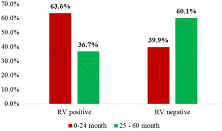
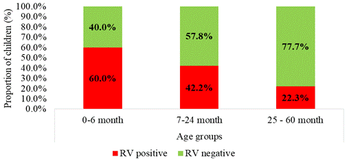
The Age has been identified as an important risk factor for RV infection: In developed countries, RV GE mainly affects infants and young children between 6 months to 2 years of age followed by the infants younger than 6 month of age and it is generally asymptomatic in neonates due to maternal antibodies which are transferred transplacentally during pregnancy. Concerning the Sex, male children were found to be two times more susceptible and more hospitalized than females [13,14] (Figure 6).

For the other risk factors, such as Socioeconomic Status (SES), the association is less clear, with certain results observed among studies that reported higher rates of RV infections among lower socioeconomic groups because of poverty, suboptimal living conditions and low uptake of the optional vaccines [15]. In addition, illiteracy, low birth weight, inadequate breastfeeding, malnutrition is also linked with higher incidence.


Pathogenesis

The pathogenesis of Rotavirus is multifactorial and incompletely understood. The virion infects villous enterocytes and causes cell destruction. The possible role of Rotavirus in pathogenesis of gastroenteritis is related to 3 main factors: Viral enterotoxin (mainly the NSP4), malabsorption due to intestinal mucosal (absorptive enterocytes) damage and virus-induced down-regulation of the expression of absorptive enzymes, leading mainly to depletion of the enzyme ‘disaccharidases’. Although chemokine secreted by infected enterocytes plays an important active role in the pathogenesis [16] (Figure 7).


Immunity

The immune response to rotavirus infection is complex and not completely understood. Innate, cellular and humoral immunity contributes to eliminating the infection. Probably, antibodies against VP7 and VP4 are important for protection from the disease. This Humoral immunity has a dominant role to prevent severe disease on successive infection or on primary infection after immunization. Concerning Cell-mediated immunity, it probably plays a role in recovery from infection and in protection in addition to local immune factors such as secretory IgA or Interferon. Also, the duration of immunity against RV infection is limited and reinfection occurs in both children and adults but generally with less severity [17]. 


WHO EMRO | Rotavirus gastroenteritis among children aged under 5 years in  Al Karak, Jordan | Volume 16, issue 10 | EMHJ volume 16, 2010


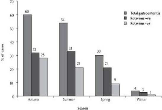
Figure 5: Seasonal Distribution of Rotavirus Infection. Adapted and Reprinted from Nafi O. Rotavirus Gastroenteritis among Children Aged Less Than 5 Years in Al Karak, Jordan. East Mediterr Health J. 2010 Oct; 16(10):1064-9. Copyright © World Health Organization (WHO), 2010

Age distribution of children positive for rotavirus in countries with... |  Download Scientific Diagram


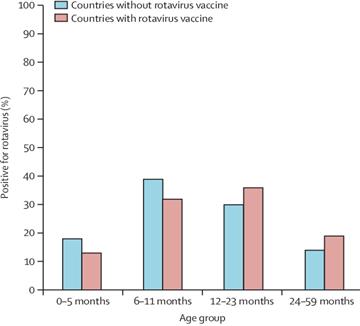
Figure 6: Age Distribution of Children Positive for Rotavirus in Countries with and without Rotavirus Vaccine Introduction. Adapted and Reprinted from Aliabadi N, Antoni S, Mwenda JM, Weldegebriel G, Biey JNM, Cheikh D, Fahmy K, Teleb N, Ashmony HA, Ahmed H, Daniels DS, Videbaek D, Wasley A, Singh S, de Oliveira LH, Rey-Benito G, Sanwogou NJ, Wijesinghe PR, Liyanage JBL, Nyambat B, Grabovac V, Heffelfinger JD, Fox K, Paladin FJ, Nakamura T, Agócs M, Murray J, Cherian T, Yen C, Parashar UD, Serhan F, Tate JE, Cohen AL. Global Impact of Rotavirus Vaccine Introduction on rotavirus Hospitalizations among Children under 5 years of age, 2008-16: Findings from the Global Rotavirus Surveillance Network. Lancet Glob Health. 2019 Jul; 7(7):e893-e903. Copyright © 2019 Published by Elsevier Ltd


Dr Sherazi: Gastroenteritis


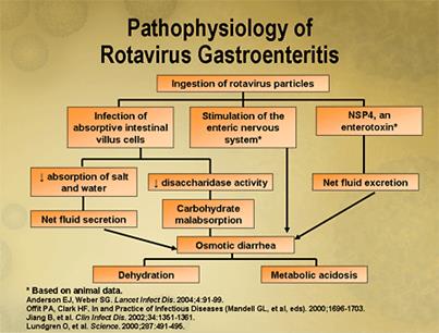
Figure 7: Rotavirus Pathogenesis. Adapted and Reprinted from Dr. Sherazi. Gastroenteritis. Available from Dr Sherazi (drsherazi110.blogspot.com)


Clinical Findings and Complications

RV is responsible for the major cause of diarrheal diseases in children below five years. The classical clinical features include watery diarrhea, fever, vomiting and abdominal pain. Infected children may also have a cough or runny nose. Also, RV infection may be asymptomatic, may cause self-limited watery diarrhea, or may result in dehydration (due to diarrhea, vomiting and fever) which may be severe and life threatening due to hypovolemia and circulatory collapse. In immunocompromised children, rotavirus infection can cause prolonged and chronic diarrhea. Generally, symptoms resolving occur between 3 to 7 days in mild and moderate infections. It has been reported that frequent RV infection has been associated with coeliac disease in genetically predisposed children [17] (Figure 8).

sings of dehydration


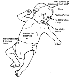
Figure 8: Signs of Dehydration. Adapted and Reprinted from Nationwide Children’s Hospital. Dehydration. Available from: Dehydration: Overview and Hydration Recommendations (nationwidechildrens.org)


Diagnosis

It is difficult to differentiate RV GE from other causes of GE by clinical spectrum alone. Also early diagnosis in hospitalized patients will decrease the morbidity and mortality considerably and avoid inappropriate use of antibiotics in pediatric patients. The most used method for diagnosis of RV infection is detection of rotavirus antigen in stool specimens by Enzyme ImmunoAssay (EIA) which is simple to use, inexpensive and with good sensitivity and specificity. RV antigen can be identified in the serum of patients 3–7 days after disease onset, but still the routine diagnostic testing is based primarily on testing of fecal specimens. In addition, the virus can be detected from stool by other techniques, used mainly in research settings such as Electron-microscopy, Immunoelectrophoresis, Flow cytometry, Latex Agglutination Test (LAT) and newly the Reverse Transcription Polymerase Chain Reaction (RT PCR), nucleic acid hybridization, Immunochromatography and more others [18]. 


Treatment

Since RV is stable in room temperature and highly contagious isolation of the patient is necessary. Treatment of RV GE is mainly by supportive measures, either by intravenous route or orally, in order to correct the loss of water and electrolytes that may lead to dehydration, acidosis, shock and death. In addition, symptomatic treatment of fever and vomiting is needed. Also, it is important to know that nor antibiotics or antivirals will help during a rotavirus infection [19]. The other key element for treatment of RV GE is nutritional support and early introduction to a normal diet. A Randomized controlled trial has demonstrated that other treatments such as probiotics, zinc, are effective in the management of acute gastroenteritis [18]. 


Preventive Measures

General Measures: As the main mode of transmission of Rotavirus is through faeco-oral route, proper hand hygiene, washing with soap and water after defecation and use of safe drinking water are mandatory. In addition, vitamin A supplementation is recommended in children since it reduces the incidence of diarrhea and associated deaths in children aged six months to five years [20]. 


Vaccine

Despite improvements in sanitation, limited effect on Rotavirus incidence is shown. So it is important to introduce safe and effective immunization to decrease the impact of RV infection in the community and reduce the worldwide burden of rotavirus disease. The first Rotavirus vaccine– ‘Rota-shield’ was introduced in United States (1998) by Wyeth and approved by Food and Drug Administration (USFDA) and Advisory Committee on Immunity Practices (ACIP) with an affectivity of 80-100% in preventing severe gastroenteritis caused by group A. However, one year later, the vaccine was removed from the market by the manufacturer since it was associated with increased risk of intussusception (1 in 12000 vaccinated infants) among vaccinated children [21]. Then, two new vaccines (Rotarix and RotaTeq) against Rotavirus Group A infection were introduced in 2006: 1. Rotarix® is an oral live attenuated vaccine containing the attenuated monovalent G1, P[8] human rotavirus strain and is recommended to be orally administered in two doses to infants at ages 2 months and 4 months.(or separated for at least 4 weeks), 2. RotaTeq® is a live attenuated, multivalent bovine-human reassortant rotavirus vaccine containing the most common rotavirus antigens seen in humans (G1, G2, G3, G4 and P[8]) and is recommended to be orally given in three doses, often at ages 2 months, 4 months and 6 months [22,23]. Therefore, WHO has recommended inclusion of RV vaccination in all national immunization programs? Then, the incidences and severity of Rotavirus infection has declined dramatically in countries that have adopted this recommendation and early estimates of the reduction in all-cause diarrhea hospitalizations in high-income countries have ranged from 20-50% and from 17 to 55% in middle-income countries. Also, the reduction in rotavirus hospitalizations has ranged from 49-92% in high-income countries, 54–59% in middle-income countries and 69–81% in low-income countries. These reductions have been sustained for several years after vaccine introduction [24]. In Lebanon, the Rotarix was introduced in late 2006 and RotaTeq was first available in 2009 but they have yet to be included in the National Immunization Program (NIP) [25] (Figure 9).

Returning to the risk of intussusception related to RV vaccines, a post licensure evaluation review (on 2012) concluded that Rotarix and RotaTeq were the effective vaccines and they were not associated with risk of intussusception in immunized children. The benefits of the vaccine far outweigh the small risk of intussusception [24]. 

Rotavirus Vaccine | UPSC Medico


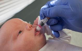
Figure 9: Rotavirus Vaccine Administration. Adapted and Reprinted from Available from: UPSC Medical Sciences. Rotavirus Vaccine. Available from: Rotavirus Vaccine| UPSC Medico (medcampus.io)


In this context, it is important to mention that FDA recommends that the rotavirus vaccine should not be given to children who have a history of intussusception because they are more likely to have intussusception recurrence after receiving the RV vaccine. Additional RV vaccine contraindications include: Severe allergic reaction to a vaccine component (including latex) or following a prior dose of vaccine (latex rubber is contained in the RV1 oral applicator). Severe Combined Immunodeficiency (SCID). Finally, since the rotavirus vaccines became available, studies have shown that they prevent thousands of children from becoming sick and being hospitalized each year which highlight the importance of implantation of RV vaccine in the national immunization program in each country [26].


Objectives

Primary Objective: to evaluate the impact of vaccination program for rotavirus on disease severity among Lebanese infant’s population under 5 years of age who have been diagnosed with acute GE or RV GE from year 2016 to 2017 in 3 different hospitals localized in Beirut: RHUH, Al-Zahraa hospital and Sacré Coeur hospital.


Secondary Objectives

To estimate the rate of hospitalization related to RV in Lebanon, to determine the average age among RV associated hospitalizations and to compare the vaccine effectiveness between RV1 and RV5.


MATERIALS AND METHODS

Study Design and Population: The methodological approach was a retrospective hospital-based review of database during 2016-2017 in 3 hospitals at Beirut, wherein data on children younger than 5 years of age who have been diagnosed with acute GE or RV related GE, was collected from the archives of participating hospitals. An episode of acute GE was defined as diarrheal disease of rapid onset over the last 2 days, with or without accompanying symptoms and signs, such as vomiting, fever or abdominal pain. 


Inclusion Criteria

Children are eligible for enrolment in our study if they me et al. l of the following criteria: Both male and female infants or children, age ≤ 5 years of old and who have been diagnosed with acute GE or RV GE from year 2016 to 2017 if they provided a stool sample for rotavirus test within 48h of admission.


Exclusion Criteria were children who were previously participants in any kind of RV clinical trial and patient with a known admission diagnosis explaining the ongoing GE; in addition to healthy volunteers. 


Data Collection

After obtaining the approval to access the medical electronic records from the administration of the 3 participating hospitals: Sacré Coeur Hospital (SGH), Al Zahraa University Medical Center (ZHMUC) and Rafic Hariri University Hospital (RHUH), data was collected from computerized medical records of this hospitals between 2016 and 2017 using a questionnaire designed for the study. These included questions about demographic characteristics (age ...), clinical spectrum (before and during hospitalization), result of stool testing for RV, total days of stay in hospital, immunization status for RV and type of vaccine if taken. Medical records of patients were analyzed thoroughly, upon ensuring they all met the inclusion criteria. A total of 401 subjects were enrolled from these sites and included in the final analysis. A descriptive analysis of the collected data was carried out and results were converted to tables and column charts using the Microsoft office excel program. Finally, the data was analyzed using the statistical program Statistical Package for the Social Science (SPSS).


Data Analysis

Exposure Variables: In our study, children admitted for GE younger than 5 years of age were first divided according to the result of RV stool test detection into RV-positive patients and RV–negative patients. Immunization history for RV was collected for all those patients when available in medical records to determine if any type of RV vaccine was given. Medical history was also collected; it included information about demographics (age mainly), GE symptoms and severity and total days of hospitalization. Regarding to the vaccinated subjects, the type of vaccine was also reported when noticed.


Statistical Analysis 

Descriptive analyses of demographics, clinical and immunization status against RV information was performed to describe the children enrolled in the study as described earlier. Trends in RV-related GE were compared for vaccinated and non-vaccinated cases concerning the severity of clinical spectrum and the day of hospitalization. The distribution of RV-associated GE was assessed and compared in percentages among vaccinated and non-vaccinated patients. To evaluate the prevalence of RV-related GE, the proportion of positive-RV stool test was calculated. Then for evaluating the variation of distribution of RV infection between immunized and non-immunized cases, the proportion of rotavirus positivity between the 2 populations was compared. All statistical analyses were performed using IBM SPSS statistics, version 23. Categorical variables were summarized as frequency and percentage. Continuous variable were presented as mean and standard deviation. Categorical variables were compared using chi-square test or fisher exact test. Mann-Whitney U test was used to compare median age, duration of hospitalization and duration of vomiting by vaccination status. All tests were two-sided and p-value <0.05 was considered to be statistically significant.

RESULTS

The distribution of the demographic characteristics of children hospitalized for GE according to their vaccination status is showed in Table 1. Four hundred and one children hospitalized for Gastroenteritis were enrolled in the study. About 180 children (44.9%) were enrolled in Al-Zahraa hospital, 100 children (24.9%) were in Rafic Hariri university hospital and 121 children (30.2%) were in Sacré Coeur hospital. The median age of children was 28.54 ± 14.17 month (range: 5-60 months). Of the total, 190 (47.4%) of patients diagnosed with GE were under 2 years of age.


Among the 401 children hospitalized for GE, 30.4% (n = 122) were vaccinated against RV. Of the 122 vaccinated children, the highest rate of vaccination (43.8%, 53 children) was found in Sacré Coeur hospital whereas the highest rate of unvaccinated children was found in Rafic Hariri university hospital (98.0%; 98 children) when vaccination rate was only 2% (n = 2) (p-value <0.001). The highest rate of vaccination (27%) was in the category of 7-24 months whereas, understandably, the highest rate of unvaccinated children diagnosed for GE was in the category of 0-6 months. There was no statistically significant difference in the rate of vaccination between age groups (p-value = 0.136).


The prevalence of RV infection in children hospitalized for GE was 31.9% (n = 128). The rate of RV GE in the vaccinated group (13.1%) was significantly lower than the unvaccinated group (40.1%) (p-value <0.001); So vaccinated patients may have lower RV disease severity due to immunity provided by the vaccine which reflects the lower rate of hospitalization.


The highest rate of RV GE (36.0%) was recorded in Rafic Hariri university hospital reasoned by the fact that lower rate of vaccination (2%) was found at RHUH (Table 1 and 3). Among the different age groups, rate of RV infection is significantly higher in the 7-24 months age group (n = 78) compared to other groups (p-value <0.001) (Figure 10).


The RV infection was more prevalent in children under than 24 months of age (total of 63.6%) than those 25–60 months of age (36.7%), when the majority of RV GE hospitalized children were under 2 years of age (Figure 11 and 12).


Clinical Characteristics of RV GE Hospitalization

Among 128 children infected with rotavirus, using the Vesikari severity score, 26 children (20.3%) had mild GE, 87 children (68.0%) had Moderate GE and 15 Children (11.7%) had severe GE with dehydration. The majority of mild cases of RV GE (75%) were found in vaccinated children. Also higher rate of severe cases was found in non-vaccinated (12.5%) children compared to those vaccinated (6.3%). Associated Symptoms among children infected with RV are summarized in table 4. Almost, all of children with positive rotavirus experienced diarrhea, 72.7% were associated with vomiting, 76.6% were associated with fever and overall 11.7% experienced dehydration. Between dehydrated cases (11.7%), the majority was found in non-vaccinated children (73.3%) compared to those vaccinated. The associated GE symptoms occurred in children with positive rotavirus (fever, vomiting) were similar in incidence along vaccinated and not vaccinated groups without statistical significance (Table 4). Clinically, the incidence of fever was lower in vaccinated group (56,3%) compared to unvaccinated group (79.5%) (p-value = 0.057)The overall median duration of hospitalization for children infected with rotavirus was 4 days (range: 1 – 10 days) and the median duration of vomiting in those children was 3 days (range: 1 – 10 days). There was a statistically significant difference between vaccinated and non-vaccinated children with RVGE by duration of hospitalization and then duration of vomiting and diarrhea which will indicate the length of stay. The median duration of hospitalization in vaccinated group was 2 days whereas 4 days in non-vaccinated group (p-value <0.001). We reasoned that vaccinated patients might have lower disease burden resulting in short stay at hospital.


Figure 10: Proportion of Children with Positive Rotavirus GE by Hospital


Figure 11: Distribution of Age Groups by Rotavirus Test Result


Figure 12: Proportion of Children with Positive Rotavirus GE by Age Group


Table 1: Demographic Characteristics by Vaccination Status

Parameters


Overall (N = 401)

Vaccinated (n = 122)

Not Vaccinated (n = 279)

p-value

Hospital

Al-Zahraa 

180 (44.9%)

67 (37.2%)

113 (62.8%)

<0.001*


Rafic Hariri University 

100 (24.9%)

2 (2.0%)

98 (98.0%)


Sacré Coeur

121 (30.2%)

53 (43.8%)

68 (56.2%)


Age (Month)†

Valid answers

401

122

279

0.051


Mean ± SD

28.54 ± 14.17

30.91 ± 15.40

27.50 ± 13.49


Median

27.0

30.0

25.0


Min - Max

5 - 60

6 – 60 

5 - 60


Age Groups

0-6 month

5 (1.2%)

1 (20.0%)

4 (80.0%)

0.316


7-24 month

185 (46.1%)

50 (27.0%)

135 (73.0%)


25-60 month

211 (52.6%)

71 (33.6%)

140 (66.4%)


*:p<0.05 is statistically significant; p-value was calculated using Fisher’s exact test, †: p-value was calculated using Mann-Whitney test


Table 2: Vaccination Status According to Rotavirus Status

Parameters


Overall (N = 401)

Vaccinated (n = 122)

Not Vaccinated (n = 279)

p-value

Rotavirus test result 

Negative

273 (68.1%)

106 (86.9%)

167 (59.9%)

< 0.001*


Positive

128 (31.9%)

16 (13.1%)

112 (40.1%)


*:p-value <0.05 is statistically significant; p-value was calculated using Fisher’s exact test


Table 3: Distribution of Age Groups by Rotavirus Test Result


Overall (n = 401)

RV Positive (n = 128)

RV Negative (n = 273)

p-value

Hospital

Al-Zahraa hospital

180 (44.9%)

53 (29.4%)

127 (70.6%)

0.528


Rafic Hariri university hospital

100 (24.9%)

36 (36.0%)

64 (64.0%)


Sacré Coeur hospital

121 (30.2%)

39 (32.2%)

82 (67.8%)


Age Groups

0 - 6 month

5 (1.2%)

3 (60.0%)

2 (40.0%)

< 0.001*


7 – 24 month

185 (46.1%)

78 (42.2%)

107 (57.8%)


25-60 month

211 (52.6%)

47 (22.3%)

164 (77.7%)


*:p<0.05 is statistically significant; p-value was calculated using Fisher’s exact test

Table 4: GE Severity and Symptoms by Vaccination Status among Children with Positive Rotavirus

Parameters


Overall (N = 401)

Vaccinated (n = 122)

Not Vaccinated (n = 279)

p-value

GE Severity

Mild

26 (20.3%)

12 (75.0%)

14 (12.5%)

<0.001*


Moderate

87 (68.0%)

3 (18.8%)

84 (75.0%)


Severe

15 (11.7%)

1 (6.3%)

14 (12.5%)


Duration of Hospitalization (days) †

Valid answers

128

16

112

<0.001*

Mean ± SD

3.77±1.51

2.38±2.16

3.96±1.29


Median

4.00

2.0

4.0


Min – Max

1 – 10

1 – 10

2 – 8


Duration of Vomiting (days) †


Valid answers

86

11

75

0.018*

Mean ± SD

3.58±1.70

2.45±2.58

3.75±1.49


Median

3.0

2

3


Min - Max

1 - 10

1 - 10

2 - 8


Associated GE Symptoms


Vomiting

93 (72.7%)

12 (75.0%)

81 (72.3%)

1.000


 Fever

98 (76.6%)

9 (56.3%)

89 (79.5%)

0.057

Severe Dehydration 


15 (11.7%)

4 (26.6%)

11 (73.3%)

0.095

*:p-value <0.05 is Statistically Significant; p-value was Calculated using Fisher’s Exact Test, †:p-value was Calculated using MANN-Whitney Test


Table 5: Vaccination Status and Type of Vaccine by Rotavirus Status

Parameters


Overall (n = 401)

RV Positive (n = 128)

RV Negative (n = 273)

p-value

Vaccination Status

Not Vaccinated

279 (69.6%)

112 (87.5%)

167 (61.2%)

<0.001*

Vaccinated

122 (30.4%)

16 (12.5%)

106 (38.8%)


Rotavirus vaccine

(17unknown vaccine)

Rotarix  

64 (61.0%)

12 (18.8%)

52 (81.3%)

0.153

RotaTeq  

41 (39.0%)

3 (7.3%)

38 (92.7%)


*:p<0.05 is statistically significant; p-value was calculated using Fisher’s exact test


Table 6: Vaccine Effectiveness

Parameters


Rotavirus test (Ref = positive)

Vaccine Effectiveness [95% CI]


OR (95% CI)

p-value

Vaccination status (Ref = Not vaccinated)

Vaccinated

4.443 [2.494-7.917]

<0.001

81.63% [71.38%-88.79%]


Vaccination Status of Patients Diagnosed with GE and Vaccine Effectiveness

Similar to previous result (Table 2), the incidence of RV GE in the vaccinated group was significantly lower than in the unvaccinated group (12.5% vs. 87.5%, p-value<0.001) (Table 5). Also children with negative rotavirus (38.8%) were more likely to be vaccinated as compared to the children infected with rotavirus (12.5%) (p-value <0.001).Concerning vaccinated children (n = 122), 64 (61.0%) were vaccinated with Rotarix and 41 (39.0%) were vaccinated with RotaTeq. The type of vaccine given was remained unknown in 17 patients vaccinated for RV. Occurrence of Rotavirus infection in vaccinated group was clinically more prevalent in children vaccinated with Rotarix (18.8%) than those who vaccinated with RotaTeq (7.3%) (Table 5).


Statistically, there was no significant difference in the rate of rotavirus disease by type of Rotavirus vaccine (p-value = 0.153). Finally, the overall Vaccine Effectiveness (VE) against RV hospitalizations was 81.63% [71.38%-88.79%], which is statistically significant (p-value <0.001) (Table 6).

DISCUSSION

The main objective for this retrospective database study was to evaluate the impact of RV vaccination program on disease severity among infant’s population. Our selected population was children under 5 years of age hospitalized for acute gastroenteritis or rotavirus gastroenteritis in RHUH, Sacré Coeur hospital and Al Zahraa hospital during 2016-2017. The secondary objective was to estimate the rate of hospitalization related to RV, to determine the median age of RV GE hospitalized children and to compare the vaccine effectiveness between Rotarix (RV1) and RotaTeq RV5). In our study, the prevalence of RV GE was significant in Lebanon with the rate of hospitalized GE caused by RV being 31.9% during 2016-2017. This is lower than the findings reported in study done in other neighboring countries in the Middle East. The percentage of RV infection in hospitalized infants was 37% in Erbil (Iraqi Kurdistan) [27], 38.9% in turkey [28], 40% in Kuwait [7], 45.2% in Yemen [29]. The highest rate was detected in Syria 61% [30]. However, as these studies were done with different population, different sampling methods and different season of sampling, comparability is limited as there is no sound and justifiable basis for comparison. Conversely, lower percentages were documented in developed countries such as Belgium where the rate of RV GE hospitalizations was 7.7% mainly in the post-vaccination era [31]. These variations reflect differences in RV GE rates and may be related to vaccination mass against RV. Indeed, high RV vaccination coverage rates were reported in the developed countries where vaccination programs have been implemented (74% in USA, > 85% in Austria and Belgium) [32] leading to a significant drop in rates of RV associated hospitalizations. To be mentioned that in Lebanon, RV vaccination is not included in the national immunization program but it is available in private sectors. In our study, the three hospitals involved serve patients with different range of socioeconomic status, which is reflected by the vaccination rates observed in each hospital that ranged from 2% at RHUH (public/governmental hospital), 37.2% at Al Zahraa (Private) and 43.8% at Sacré Coeur (Private). The lowest rate recorded at RHUH is explained by the poor category visiting this public hospital resulting in low vaccination rate for RV seen between their children. Overall, 30.4% were vaccinated against RV among hospitalized children which is higher than rate of vaccination seen in other previous Lebanese studies [20]. The vaccination and the reduction of RV infection rates were significantly correlated (p-value <0.001), as between the positive cases, the rate of RV infection in the vaccinated group (12.5%) was significantly lower than in the unvaccinated group (75.5%) which confirms a significant reduction of RV cases among the vaccinated population. In addition, our analysis demonstrated that less severe RV infection was observed among vaccinated children (6.3%), as compared with non-vaccinated children (12.5%), (p-value <0.001). Furthermore, the median duration of hospitalization was significantly lower in the vaccinated patients (= 2 days) compared to the unvaccinated one (= 4 days) (p-value <0.001) as well as duration of vomiting (p-value = 0.018) indicating that vaccinated patients recovered faster. So our study highlights that RV vaccination reduces the burden of RV disease which confirm the positive impact of vaccination program for RV on RV disease severity on the Lebanese population with vaccine effectiveness of 81.63%. These findings are similar to previous studies findings done in Brazil as well as U.S [33,34]. As for age, nearly 63.3% of the RV GE cases occurred in children under 2 years of age, this result is concordant with previous study conducted in Lebanon by Dbaibo et al. [35]. Also according to our study, the majority was aged 6-24 months (60.9%), which may be explained by the protection given by maternal antibodies in the first six months of the child’s life. Indeed, the vaccine effectiveness of 81.63% observed in our study was in same range with that of other studies assessing the effectiveness of RV1 and RV5 vaccines in different countries [36,37,38]. Finally, concerning the type of used vaccine, the majority of patients in our study were vaccinated with Rotarix (61%) and statistically, there was no significant difference in the rate of rotavirus GE by type of Rotavirus vaccine (p-value = 0.153).

CONCLUSION

This study provides updated information on the extent of RV related hospitalizations and the impact of vaccination against RV in Lebanon. The results indicate that GE caused by RV in Lebanon is a significant health problem mainly in children aged less than 2 years. Also, our findings reveal a significant impact of RV vaccination in decrease burden of disease concerning incidence, clinical severity and hospitalizations stay and maximizing vaccine coverage remain an important health challenge. However, our data showed a rate of vaccination of 30.4% the included population. Furthermore, both type of vaccine demonstrated same effectiveness against RV related GE. Currently, alternate RV vaccine schedules have been proposed worldwide to improve vaccine effectiveness and other vaccines are currently under development which may overcome certain barriers in order to maximize the impact of current vaccines. Finally, this study would help inform the health policymakers about impact of RV GE in children and assess the need for National Immunization Programs to be inserted all over the country. Additionally, implantation of these RV vaccine related programs helps to limit the healthcare cost by reducing hospitalization, morbidity and mortality.


Limitations

Some limitations of this study needed to be mentioned. First, this review is designed to assess only hospitalized cases, so no clinic data was used to describe the home managed GE patients and role of vaccination in this population. The difference between male and female prevalence concerning RV infection was not conducted in this study as well as the clinical spectrum of RV-negative gastroenteritis. Additionally, as data was collected for only 2016-2017, it was difficult to obtain conclusions about seasonality of RV GE. Also the detection of RV cases based on testing of stool specimens by enzyme immunoassay, which is widely used in hospital’s lab, may have missed low levels of rotavirus excretion.


Strengths

Importantly and despite some limitations, our study was designed to evaluate both the impact of RV vaccines on the burden of RV infection and vaccine effectiveness of both type of vaccine: RV1 and RV5. Also, many other strengths support our results: the adequate sample size, the absence of selection bias in addition to the similarity in results in our study when compared to other international studies.


Study Perspectives

RV vaccines have been shown to reduce severe RV disease in a range of socioeconomics settings. The needed impact has not been fully realized as RV vaccine have not been universally introduced into National Immunization Programs (NIP) so concerted effort is needed in this domain. In other hand, vaccination effectiveness is shown to be lower in low-and middle –income countries compared to high income countries, there are several hypothesized reason for this difference. Then the suspicions that poor nutritional status reduces RV vaccination effectiveness have been supported and additional review is needed to fully understand the impact of malnutrition and to develop specific interventions.

REFERENCES
  1. Zaraket, R. et al. “Prevalence, risk factors and clinical characteristics of rotavirus and adenovirus among Lebanese hospitalized children with acute gastroenteritis.” Heliyon, vol. 6, no. 6, 2020, e04248. PubMed PMID: 32613122. Available from: https://pubmed.ncbi.nlm.nih.gov/32613122/.

  2. Parashar, U.D. et al. “Prevention of rotavirus gastroenteritis among infants and children: Recommendations of the Advisory Committee on Immunization Practices (ACIP).” MMWR Recommendations and Reports, vol. 55, 2006, pp. 1–13. PubMed PMID: 16902398. Available from: https://pubmed.ncbi.nlm.nih.gov/16902398/.

  3. Burnett, E. et al. “Global impact of rotavirus vaccination on childhood hospitalizations and mortality from diarrhea.” The Journal of Infectious Diseases, vol. 215, no. 11, 2017, pp. 1666–1672. PubMed PMID: 28430997. Available from: https://pubmed.ncbi.nlm.nih.gov/28430997/.

  4. Tomislav, M. “Rotavirus structure and classification.” News-Medical, 13 May 2019. Retrieved on December 22, 2020, from https://www.news-medical.net/health/Rotavirus-Structure-and-Classification.aspx.

  5. Karki, G. “Rotavirus-classification, structure composition and properties, replication, mode of transmission, pathogenesis, clinical symptoms, laboratory diagnosis, treatment, prevention and control.” Online Biology Notes, 21 May 2017. Retrieved on December 23, 2020.

  6. Marmash, R.W. et al. “Genotypic characterization of rotaviruses and prevalence of serotype-specific serum antibodies in children in Kuwait.” Epidemiology & Infection, vol. 135, no. 8, 2007, pp. 1331–1337. PubMed PMID: 17291379. Available from: https://pubmed.ncbi.nlm.nih.gov/17291379/.

  7. Sadiq, A. et al. “Molecular characterization of human group A rotavirus genotypes circulating in Rawalpindi, Islamabad, Pakistan during 2015–2016.” PLoS One, vol. 14, no. 7, 2019, e0220387. PubMed PMID: 31361761.

  8. López, T. et al. “Replication of the rotavirus genome requires an active ubiquitin-proteasome system.” Journal of Virology, vol. 85, no. 22, 2011, pp. 11964–11971. PubMed PMID: 21900156. Available from: https://pubmed.ncbi.nlm.nih.gov/21900156/.

  9. World Health Organization (WHO). “Rotavirus.” 2020. Retrieved on December 23, 2020.

  10. Köroğlu, M. et al. “A waterborne outbreak of epidemic diarrhea due to group A rotavirus in Malatya, Turkey.” New Microbiologica, vol. 34, no. 1, 2011, pp. 17–24. PubMed PMID: 21344142. Available from: https://pubmed.ncbi.nlm.nih.gov/21344142/.

  11. Centers for Disease Control and Prevention (CDC). “Rotavirus home.” 5 Nov. 2019. Retrieved on December 23, 2020.

  12. Levy, K. et al. “Seasonality of rotavirus disease in the tropics: A systematic review and meta-analysis.” International Journal of Epidemiology, vol. 38, no. 6, 2009, pp. 1487–1496. PubMed PMID: 19056806. Available from: https://pubmed.ncbi.nlm.nih.gov/19056806/.

  13. Nafi, O. “Rotavirus gastroenteritis among children aged under 5 years in Al Karak, Jordan.” Eastern Mediterranean Health Journal, vol. 16, no. 10, 2010, pp. 1064–1069. PubMed PMID: 21222424. Available from: https://pubmed.ncbi.nlm.nih.gov/21222424/.

  14. Adams, N.L. et al. “Relationship between socioeconomic status and gastrointestinal infections in developed countries: A systematic review and meta-analysis.” PLoS One, vol. 13, no. 1, 2018, e0191633. PubMed PMID: 29360884. Available from: https://pubmed.ncbi.nlm.nih.gov/29360884/.

  15. Badur, S. et al. “Systematic review of the rotavirus infection burden in the WHO-EMRO region.” Human Vaccines & Immunotherapeutics, vol. 15, no. 11, 2019, pp. 2754–2768. PubMed PMID: 30964372. Available from: https://www.ncbi.nlm.nih.gov/pmc/articles/PMC6930073/.

  16. Greenberg, H.B. and M.K. Estes. “Rotaviruses: From pathogenesis to vaccination.” Gastroenterology, vol. 136, no. 6, 2009, pp. 1939–1951. PubMed PMID: 19457420. Available from: https://pubmed.ncbi.nlm.nih.gov/19457420/.

  17. Turin, C.G. and T.J. Ochoa. “The role of maternal breast milk in preventing infantile diarrhea in the developing world.” Current Tropical Medicine Reports, vol. 1, no. 2, 2014, pp. 97–105. PubMed PMID: 24883263. Available from: https://pubmed.ncbi.nlm.nih.gov/24883263/.

  18. Parashar, U.D et al. “Diagnosis, management and prevention of rotavirus gastroenteritis in children.” BMJ, vol. 347, 2013, f7204. PubMed PMID: 24379214. Available from: https://pubmed.ncbi.nlm.nih.gov/24379214/.

  19. Mayo Clinic. “Rotavirus.” 12 Mar. 2019. Retrieved on December 23, 2020.

  20. Keusch, G.T. et al. “Diarrheal diseases.” Disease Control Priorities in Developing Countries. 2nd Edn., edited by D.T. Jamison et al., World Bank, 2006, Chapter 19. Available from: https://www.ncbi.nlm.nih.gov/books/NBK11764/.

  21. Schwartz, J.L. “The first rotavirus vaccine and the politics of acceptable risk.” Milbank Quarterly, vol. 90, no. 2, 2012, pp. 278–310. PubMed PMID: 22709389. Available from: https://www.ncbi.nlm.nih.gov/pmc/articles/PMC3460207/.

  22. World Health Organization (WHO). Detailed review paper on rotavirus vaccines. 17 Mar. 2009. Available from: https://www.who.int/vaccine_safety/committee/reports/rotavirus_vaccines_review.pdf.

  23. Dennehy, P.H. “Rotavirus vaccines: An overview.” Clinical Microbiology Reviews, vol. 21, no. 1, 2008, pp. 198–208. PubMed PMID: 18202442. Available from: https://pubmed.ncbi.nlm.nih.gov/18202442/.

  24. Burnett, E. et al. “Rotavirus vaccines: Current global impact and future perspectives.” Future Virology, vol. 11, no. 10, 2016, pp. 699–708. PubMed PMID: 27840654. Available from: https://pubmed.ncbi.nlm.nih.gov/27840654/.

  25. Harastani, H.H. et al. “Genetic diversity of human rotavirus A among hospitalized children under 5 years in Lebanon.” Frontiers in Immunology, vol. 11, 2020, 317. PubMed PMID: 32174920. Available from: https://www.ncbi.nlm.nih.gov/pmc/articles/PMC7054381/.

  26. Infection Control Today (ICT). “CDC offers tips to prevent rotavirus.” 29 Dec. 2016. Retrieved on December 23, 2020.

  27. Ahmed, H.M. et al. “Molecular characterization of rotavirus gastroenteritis strains, Iraqi Kurdistan.” Emerging Infectious Diseases, vol. 12, no. 5, 2006, p. 824. PubMed PMID: 16704845. Available from: https://www.ncbi.nlm.nih.gov/pmc/articles/PMC3374452/.

  28. Ozkaya-Parlakay, A. and H. Tezer. “Burden of rotavirus in hospitalized children in Turkey.” The Pediatric Infectious Disease Journal, vol. 33, no. 9, 2014, pp. 992–993. PubMed PMID: 00006454-201409000-00029. Available from: https://journals.lww.com/pidj/Fulltext/2014/09000/Burden_of_Rotavirus_in_Hospitalized_Children_in.29.aspx.

  29. Al-Badani, A. et al. “Rotavirus diarrhea among children in Taiz, Yemen: Prevalence, risk factors and detection of genotypes.” International Journal of Pediatrics, 2014, 928529. PubMed PMID: 25197286. Available from: https://www.ncbi.nlm.nih.gov/pmc/articles/PMC4145802/.

  30. Khoury, H. et al. “Burden of rotavirus gastroenteritis in the Middle Eastern and North African pediatric population.” BMC Infectious Diseases, vol. 11, no. 1, 2011, 9. PubMed PMID: 21214934. Available from: https://pubmed.ncbi.nlm.nih.gov/21214934/.

  31. Sabbe, M. et al. “Sustained low rotavirus activity and hospitalisation rates in the post-vaccination era in Belgium, 2007 to 2014.” Eurosurveillance, vol. 21, no. 27, 2016, 30273. PubMed PMID: 27418466. Available from: https://pubmed.ncbi.nlm.nih.gov/27418466/.

  32. Ai, C.E et al. “Disease burden and seasonal impact of improving rotavirus vaccine coverage in the United States: A modeling study.” PLoS One, vol. 15, no. 2, 2020, e0228942. PubMed PMID: 32059029. Available from: https://www.ncbi.nlm.nih.gov/pmc/articles/PMC7021296/.

  33. Justino, M.C.A. et al. “Clinical severity and rotavirus vaccination among children hospitalized for acute gastroenteritis in Belém, northern Brazil.” Journal of Tropical Pediatrics, vol. 62, no. 4, 2016, pp. 276–281. PubMed PMID: 26884442. Available from: https://pubmed.ncbi.nlm.nih.gov/26884442/.

  34. Baker, J.M. et al. “Effects of the rotavirus vaccine program across age groups in the United States: Analysis of national claims data, 2001–2016.” BMC Infectious Diseases, vol. 19, no. 1, 2019, 186. PubMed PMID: 30795739. Available from: https://pubmed.ncbi.nlm.nih.gov/30795739/.

  35. Dbaibo, G. et al. “Hospital-based surveillance study of rotavirus gastroenteritis in children under 5 years of age in Lebanon.” Trials in Vaccinology, vol. 2, 2013, pp. 25–30. doi:10.1016/j.trivac.2013.08.002. Available from: https://www.researchgate.net/publication/259555679_Hospital_based_surveillance_of_rotavirus_gastroenteritis_in_children_5_years_of_age_in_Lebanon.

  36. Adlhoch, C. et al. “Rotavirus vaccine effectiveness and case-control study on risk factors for breakthrough infections in Germany, 2010–2011.” The Pediatric Infectious Disease Journal, vol. 32, no. 2, 2013, pp. e82–e89. PubMed PMID: 23334342. Available from: https://pubmed.ncbi.nlm.nih.gov/23334342/.

  37. Cortese, M.M. et al. “Effectiveness of monovalent and pentavalent rotavirus vaccine.” Pediatrics, vol. 132, no. 1, 2013, pp. e25–e33. PubMed PMID: 23776114. Available from: https://www.ncbi.nlm.nih.gov/pmc/articles/PMC4074617/.

  38. Chang, W.C. et al. “Effectiveness of 2 rotavirus vaccines against rotavirus disease in Taiwanese infants.” The Pediatric Infectious Disease Journal, vol. 33, no. 3, 2014, pp. e81–e86. PubMed PMID: 24569388. Available from: https://pubmed.ncbi.nlm.nih.gov/24569388/.

License
CC BY-NC-ND
Creative Commons Attribution-NonCommercial-NoDerivatives 4.0 International License
The Impact of Rotavirus Vaccination on Disease Burden among the Lebanese Infants and Children under 5 Years Old who have been Diagnosed with Acute Gastroenteritis from Year 2016 to 2017 © 2026 by Lana Khater, Laudy Abou Reda, Kamal Kanso, Gladys Gemayel, Rouwayda Dana, Bassem Abou Merhi, Zeinab Hussein Kalach licensed under CC BY-NC-ND 4.0
All papers should be submitted electronically. All submitted manuscripts must be original work that is not under submission at another journal or under consideration for publication in another form, such as a monograph or chapter of a book. Authors of submitted papers are obligated not to submit their paper for publication elsewhere until an editorial decision is rendered on their submission. Further, authors of accepted papers are prohibited from publishing the results in other publications that appear before the paper is published in the Journal unless they receive approval for doing so from the Editor-In-Chief.
Himalayan Journal of Applied Medical Sciences and Research open access articles are licensed under a Creative Commons Attribution-Share A like 4.0 International License. This license lets the audience to give appropriate credit, provide a link to the license, and indicate if changes were made and if they remix, transform, or build upon the material, they must distribute contributions under the same license as the original.
Advertisement
Recommended Articles
Research Article
Study of the Role of Serum Interleukin-6 in Inflammation among Pregnant Women with COVID-19
Published: 27/07/2021
Download PDF
Research Article
Evaluation of Serum Heat Shock Protein 70 as a Diagnostic Biomarker for Ectopic Pregnancy: A Comparative Case-Control Study
Published: 28/02/2026
Download PDF
Research Article
Assessing First Trimester Maternal Serum Pentraxin-3 Levels in Primary Unexplained Recurrent Pregnancy Loss: A Case-Control Study
Published: 28/02/2026
Download PDF
Research Article
Bones in Balance: Awareness of Rickets and Nutritional Deficiencies Among the Hamirpur Community
Published: 10/12/2024
Download PDF
Chat on WhatsApp
Flowbite Logo
Najmal Complex,
Opposite Farwaniya,
Kuwait.
Email: kuwait@iarcon.org

Editorial Office:
J.L Bhavan, Near Radison Blu Hotel,
Jalukbari, Guwahati-India
Useful Links
Order Hard Copy
Privacy policy
Terms and Conditions
Refund Policy
Others
About Us
Team Members
Contact Us
Online Payments
Join as Editor
Join as Reviewer
Subscribe to our Newsletter
Follow us
MOST SEARCHED KEYWORDS
scientific journal
 | 
business journal
 | 
medical journals
 | 
Scientific Journals
 | 
Academic Publisher
 | 
Peer-reviewed Journals
 | 
Open Access Journals
 | 
Impact Factor
 | 
Indexing Services
 | 
Journal Citation Reports
 | 
Publication Process
 | 
Impact factor of journals
 | 
Finding reputable journals for publication
 | 
Submitting a manuscript for publication
 | 
Copyright and licensing of published papers
 | 
Writing an abstract for a research paper
 | 
Manuscript formatting guidelines
 | 
Promoting published research
 | 
Publication in high-impact journals
Copyright © iARCON Internaltional LLP . All Rights Reserved.