Contents
Download PDF
pdf Download XML
1751 Views
352 Downloads
Share this article
Case Report | Volume 4 Issue 1 (Jan-June, 2023) | Pages 1 - 3
A Case Report of Epidermoid Cyst in Lower Lip Mucosa in Male Adult
1
MS ENT, Medical Officer , ENT Department, Dr.Yashwant Singh Parmar Government Medical College, Nahan, Sirmaur, Himachal Pradesh
Under a Creative Commons license
Open Access
Received
Nov. 19, 2022
Revised
Dec. 2, 2022
Accepted
Jan. 10, 2023
Published
Feb. 2, 2023
Abstract

Epidermoid cysts represent the most common cutaneous cysts. While these may occur anywhere on the body, they occur most frequently on the face, scalp, neck and trunk. When present in the oral cavity, these cysts are seen in decreasing frequency in the following order: floor of mouth, tongue, sublingual, and submental. We reported a case of a 30-year-old patient having Epidermoid cyst in lower lip mucosa on right side.

Keywords
INTRODUCTION

Epidermoid cysts are rare, slow-growing, benign, developmental cysts that are derived from aberrant ectodermal tissue. An epidermoid cyst is defi ned as “a simple cyst lined with stratifi ed squamous epithelium, and the lumen is fi lled with cystic fluid or keratin and no other specialized structure [1-2].


An epidermoid cyst is an intradermal sac-like formation that is bordered by keratohyaline granule-containing squamous epithelium. An epidermal cyst develops from the epidermis and is made up of a cystic enclosure of epithelium in the dermis that fills with debris that is rich in keratin and lipids. Epidermoid cysts have also been known as follicular infundibular cysts, epidermal cysts, and epidermal inclusion cysts in the past. The epidermoid cyst caused by the implantation and growth of epidermal components in the dermis is specifically referred to as an epidermal inclusion cyst. The more broad name epidermoid is more accurate because the majority of these lesions develop from the follicular infundibulum [1-3].


Epidermoid cysts are mostly asymptomatic lesions that manifest as firm, spherical, movable subcutaneous nodules that range in colour from flesh to yellow. Size ranges typically between 0.5 and 5 cm. There might be a central pore or punctum. These lesions may enlarge and tenderise as a result of secondary infection or inflammation. Epidermoid cysts can develop at any age and are roughly twice as common in males as in women, however they most frequently appear in the fourth decade of life [2-3].


Epidermoid cysts are usually present in the midline of the head and neck region. The floor of the mouth and extraorally in the periorbital area are where they are most frequently seen when they are present in the oral cavity. Epidermoid cysts on the face can be easily detected by a clinical examination; no laboratory testing or imaging studies are required because these cysts only appear very superficially on the skin [1-3].


Since epidermoid cysts are lesions that grow very slowly, patients usually do not visit a doctor if the lesion is asymptomatic. Surgery is used to remove the cyst and cyst wall through the surgical defect, either through excision or incision. Histologically, granular squamous epithelium with a central lumen filled with laminated keratin lines the inside of epidermoid cysts. Older cysts might exhibit calcification [2-4].


We report a case of adult male with Epidermoid Cyst in lower lip mucosa of right side.           


Figure-1-3: A Case Report of Epidermis Cyst in Lower Lip Mucosa in Male Adult


Case Description

A 30-year-old patient presented to ENT OPD with swelling below lower lip on right side. While the patient had swelling from past 2 months, he suffered blunt trauma 6 years ago. Swelling was not associated with pain or discharge. Patient was a smoker.


On inspection, facial asymmetry on right side below lower lip was observed. Skin over the swelling was normal. Intra oral examination revealed a smooth surface swelling of size around 4x4x3cm on the mucosal surface of the lower lip extending to the buccal mucosa on right side. Overlying mucosa was normal. On palpation, swelling was found non tender, mobile, and firm in consistency. There was no palpable lymph-adenopathy in the neck. Thus, initial differential diagnosis was kept as a benign minor salivary gland tumour.


Fine Needle Aspiration Cytology was suggestive of epidermal cyst. Biopsy under local anaesthesia was planned. A horizontal incision was made on the mucosa over the swelling andblunt dissection was done. Acyst of size 3.5x2x2 cm was excised. Specimen was sent for Histopathological examination which revealed a cyst wall lined by stratified squamous epithelium with intact granular layer with keratin flakes in the lumen. Extensive giant cell reaction was seen, suggesting epidermal inclusion cyst. The patient returned after 7 days for review, the wound was healthy, and patient is on regular follow up.

DISCUSSION

An epidermal inclusion cyst is the most common cutaneous cyst. Epidermal inclusion cysts are also known as epidermal cysts, epidermoid cysts, infundibular cysts and keratin cysts.Epidermal inclusion cysts are sometimes referred to as “sebaceous cysts,” however this is a misnomer as this lesion does not involve the sebaceous gland. The term “sebaceous cyst” should only be used in association with steatocystoma multiplex [2,3].


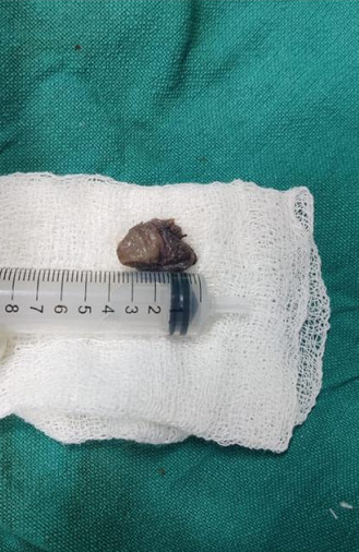
An epidermal inclusion cyst typically appears as a slowly enlarging, mobile, dome-shaped lump, filled with keratin material and located just below the surface of the skin. They can range in size from 0.5 cm to several centimeters. Often times, there is a “punctum,” or small dark-colored opening on the surface of the epidermal inclusion cyst, which connects to the cyst located below skin’s surface. Through the opening, the keratin material may be drained out.Epidermal inclusion cysts are generally considered benign (non-cancerous), although there have been rare cases of malignancy arising within the cyst[2-4].


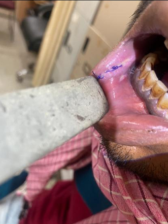
Epidermal inclusion cysts are the most common type of skin cyst. They can be located anywhere, but are most common on the face, followed by the chest or back, scalp, neck, legs, arms and/or genitalia.Epidermal inclusion cysts are often asymptomatic. However as the keratin accumulates within the cyst, the cyst may increase in size, become sore or painful, and eventually rupture, leading to the drainage a foul-smelling, white to yellow “cheese-like” material [3-5]. 


Epidermal inclusion cysts are common and typically harmless, but rarely can become malignant (cancerous). Approximately 1% have been shown to transform into malignancy.An epidermal inclusion cyst may concerning if it has Signs of infection, including pain, redness, swelling and/or drainage, A fast rate of growth and a diameter larger than five centimeters [4,5].


Although they can appear at any age, epidermal inclusion cysts most frequently occur during early to mid-adulthood (ages 20-60s). Epidermal inclusion cysts rarely appear before puberty. They are one and a half times more common in males than in females.An epidermal inclusion cyst does not typically go away by itself completely unless removed surgically, by an excision. However, an epidermal inclusion cyst may shrink in size and stay asymptomatic for quite some time, and no treatment may be necessary.Most epidermal inclusion cysts occur at random [3,4].


Some genetic conditions may be associated with the development of multiple epidermal inclusion cysts like Gardner Syndrome (Familial Colorectal Polyposis), Gorlin Syndrome (Basal Cell Nevus Syndrome) and Favre-Racouchot syndrome [1,2].


Epidermal inclusion cysts are formed from the follicular infundibulum, which is part of the hair follicle. Epidermal inclusion cysts form when the follicular infundibulum is disrupted, or when the surface of the skin becomes implanted below the skin through an injury or trauma in the area, such as a scratch, surgical wound or a skin condition like acne [2,4].


The main symptom of an epidermal inclusion cyst is a small moveable lump under the skin. The lump is usually not painful. In some cases, however, cysts can get inflamed and become tender to the touch. The skin on the area of the cyst may be red and/or warm. Usually, healthcare provider can diagnose an epidermal inclusion cyst with a simple examination of the skin. In some cases, the healthcare provider will perform a biopsy to rule out other skin growths [2,3].


If healthcare provider suspects that your epidermal inclusion cyst is, he or she may order, the tests including an ultrasound may help determine the contents of the cyst, A punch biopsy. This is where a small amount of the tissue from the cyst is removed and examined for signs of cancer. A CT scan may be helpful in confirming the diagnosis of a large epidermal inclusion cyst and help the surgeon determine the best plan for removal [4,5].


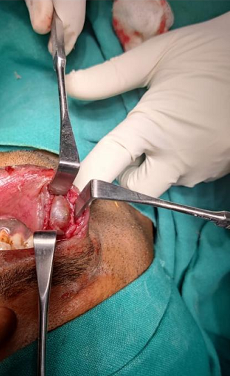
In many cases, epidermal inclusion cysts can be left untreated and monitored, as they are not dangerous, and especially if they are not causing any symptoms.If a cyst becomes inflamed or painful, your healthcare provider may consider treating the epidermal inclusion cyst with Injection of steroid medication - to reduce swelling, which may help temporarily. Surgical excision - The cyst is usually removed using an injection of local anesthetic to numb your skin. This procedure will leave a small line scar after the cyst is removed.Risks of surgical excision of a cyst include a small chance of bleeding, infection and likely a small scar over the area where the cyst is removed [3,4].

CONCLUSION

Epidermoid cysts of the lower lip area are quite rare and need to be differentially diagnosed from several other diseases and conditions of the area. For diagnosis, a detailed clinical examination of their size and anatomical location is important. Furthermore, valuable assistance is provided by the imaging techniques. Surgical enucleation provides a good treatment with excellent prognosis. The final confirmatory diagnosis of the disease is always through histopathologic examination.

REFERENCE
  1. Ashok, S. et al. "Periorbital epidermoid cyst in an elderly male." Medical Journal of Dr. D.Y. Patil University, vol. 7, 2014, pp. 388–391.

  2. Kim, C.S. et al. "Epidermoid cyst: A single-center review of 432 cases." Archives of Craniofacial Surgery, vol. 21, no. 3, June 2020, pp. 171–175.

  3. Cleveland Clinic. "Epidermal inclusion cyst." Cleveland Clinic, https://my.clevelandclinic.org/health/diseases/ 21465-epidermal-inclusion-cyst. Accessed 15 January 2023.

  4. Zito, P.M. and R. Scharf. "Epidermoid cyst." StatPearls [Internet], StatPearls Publishing, Treasure Island (FL), January 2022–, https://www.ncbi.nlm.nih.gov/books/NB K499974/. Accessed 14 January 2023.

  5. Narain, S. et al. "Epidermoid cysts of face: Clinicopathological presentation and a report of four cases." International Journal of Clinical Dental Science, vol. 3, no. 1, March 2012, pp. 30–34.

License
CC BY-NC-ND
Creative Commons Attribution-NonCommercial-NoDerivatives 4.0 International License
A Case Report of Epidermoid Cyst in Lower Lip Mucosa in Male Adult © 2026 by Sarita Negi licensed under CC BY-NC-ND 4.0
All papers should be submitted electronically. All submitted manuscripts must be original work that is not under submission at another journal or under consideration for publication in another form, such as a monograph or chapter of a book. Authors of submitted papers are obligated not to submit their paper for publication elsewhere until an editorial decision is rendered on their submission. Further, authors of accepted papers are prohibited from publishing the results in other publications that appear before the paper is published in the Journal unless they receive approval for doing so from the Editor-In-Chief.
Himalayan Journal of Community Medicine and Public Health open access articles are licensed under a Creative Commons Attribution-Share A like 4.0 International License. This license lets the audience to give appropriate credit, provide a link to the license, and indicate if changes were made and if they remix, transform, or build upon the material, they must distribute contributions under the same license as the original.
Recommended Articles
Research Article
Study of fish quality in Hor Abu Zark 2021-2022
Published: 24/01/2024
Download PDF
Research Article
Etiological Analysis of the Incident of Computer Vision Syndrome (Cvs) in Students of the Faculty of Medical Muhammadiyah University Semarang
...
Published: 20/01/2026
Download PDF
Research Article
Otolaryngologic Perspective on Endoscopic Sellar Reconstruction after Transsphenoidal Pituitary Surgery
...
Published: 31/12/2025
Download PDF
Research Article
Pharmacological Prophylaxis of Deep Venous Thrombosis in Lower Limbs Long Bones Compound Fractures caused by Missile Injuries, in Comparison with no Prophylaxis
...
Published: 10/01/2026
Download PDF
Chat on WhatsApp
Flowbite Logo
Najmal Complex,
Opposite Farwaniya,
Kuwait.
Email: kuwait@iarcon.org

Editorial Office:
J.L Bhavan, Near Radison Blu Hotel,
Jalukbari, Guwahati-India
Useful Links
Order Hard Copy
Privacy policy
Terms and Conditions
Refund Policy
Others
About Us
Team Members
Contact Us
Online Payments
Join as Editor
Join as Reviewer
Subscribe to our Newsletter
Follow us
MOST SEARCHED KEYWORDS
scientific journal
 | 
business journal
 | 
medical journals
 | 
Scientific Journals
 | 
Academic Publisher
 | 
Peer-reviewed Journals
 | 
Open Access Journals
 | 
Impact Factor
 | 
Indexing Services
 | 
Journal Citation Reports
 | 
Publication Process
 | 
Impact factor of journals
 | 
Finding reputable journals for publication
 | 
Submitting a manuscript for publication
 | 
Copyright and licensing of published papers
 | 
Writing an abstract for a research paper
 | 
Manuscript formatting guidelines
 | 
Promoting published research
 | 
Publication in high-impact journals
Copyright © iARCON Internaltional LLP . All Rights Reserved.