Contents
Download PDF
pdf Download XML
2667 Views
211 Downloads
Share this article
Research Article | Volume 2 Issue 2 (July-Dec, 2021) | Pages 1 - 4
Evaluation of clinical characteristics of bullous pemphigoid and its effect on Neurological
 ,
 ,
1
Iraqi Ministry of Health and Environment-Karbala Health Directorate, AL-Hussein Medical City, Karbala, Iraq
Under a Creative Commons license
Open Access
Received
July 12, 2021
Revised
Aug. 14, 2021
Accepted
Aug. 27, 2021
Published
Sept. 10, 2021
Abstract

A descriptive and retrospective study was conducted in the hospital, which included all age groups with a clinical diagnosis between January 1, 2019 and June 30, 2020. Ninety patients were collected from AL-Hussein Medical City, Karbala, Iraq, where the study aims to describe the clinical and epidemiological profile of patients with diseases. Bullous pemphigoid is an autoimmune skin disease that occurs mainly in the elderly. It is estimated that it affects between seven and thirteen people per million. Neurological symptoms often start very subtly and the onset of autoimmune diseases may contribute to a delay in neurological diagnosis", who consider it reasonable to speculate that some neurological disorders may predispose to bullous pemphigoid. “The association between bullous pemphigoid and neurodegenerative disease has been the subject of several prominent clinical cases, but a subset of these patients has not been characterized. We have shown a significant association between the two and that neurodegenerative disease can be a predisposing factor. Pemphigus can coexist with central nervous system disorders, particularly dementia, epilepsy and Parkinson's disease. It is present in CNS neurons (and in all epithelial cells) and an immune cross-reactivity between CNS and epithelial isoforms has been suggested.

Keywords
INTRODUCTION

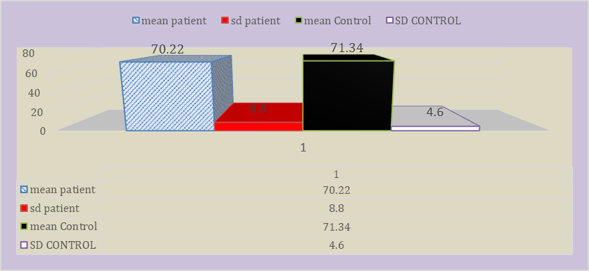
Bullous pemphigoid is a skin disease that attacks the autoimmune and generates bullous eruptions that cause itching and affect more in older patients. It is rare for any thousand to occur in the mucous membranes [1].


Some neurological problems or disorders may result before bullous pemphigoid or some psychological problems such as dementia or intracranial bleeding may occur [2,3].


The first symptom of pemphigus is itching. Skin lesions may not develop for several years. Bullae may appear on normal-looking skin or may be preceded by erythematous or urticarial-like plaques [6,7,8]. Often, characteristic tensed bubbles form on the skin of the trunk, in the areas of bends and diaper rashes. Bullae may appear on normal-looking skin or may be preceded by erythematous or urticarial-like plaques [9]. 


The disease can be localized at the sites of infection, in the oral cavity, in the anogenital and distal areas of the pelvic extremities. Bubbles usually do not rupture, but blebs that do rupture usually heal quickly [10].


Polymorphous annular dark red, edematous lesions with/without peripheral vesicles may occur. Rarely, small blisters appear on the mucous membrane. Leukocytosis and eosinophilia are common, but fever is rare [11]. Nikolsky's symptom is negative in which the upper layers of the epidermis peel off with slight pressure or friction of the skin adjacent to the blisters.


Skin lesions in bullous pemphigoid can be localized or generalized. Rashes are more often localized on the limbs, abdomen, groin-femoral folds and inner thighs. It is known that bullous pemphigoid has two periods of development - prodromal (non-bullous) and bullous [12]. The non-bullous phase of pemphigoid, as a rule, manifests itself often nonspecifically and is manifested by severe itching, accompanied by excoriation, schematization and erythematous, popular and urticarial eruptions. True and false (evolutionary) polymorphism of the rash is possible. 


Itching and nonspecific rashes may remain the only symptom of the disease for a long time (up to 5 years) [4,13]. In the future, in the presence of a specific picture of bullous pemphigoid (bullous stage), its diagnosis does not cause difficulties. The blisters have a tense, dense cover, round or oval, serous or serous-hemorrhagic contents; as a rule, they are located on an erythematous background or intact skin. Formed at the site of erosion bubbles, quickly epithelialize in the absence of secondary infection, not prone to peripheral growth. Nikolsky's phenomenon is negative. When pressing on the bladder, its diameter can increase due to subepithelial perifocal detachment - the Asbo-Hansen phenomenon is positive. The mucous membranes are affected in 10-25% of patients [5,14].


However, in 20% of cases, the picture of bullous pemphigoid takes on a nonspecific character, the diagnosis of which is based on morphological and immunohistochemically research methods. 


The names of the variants of pemphigoid are different and depend on its clinical picture. Thus, the presence of papules and vesicles, which tend to cluster with the formation of arcuate outlines and tense blisters on the skin of the back, limbs, buttocks, is considered as vesicular-bullous pemphigoid. The erythroderma form of bullous pemphigoid occurs against the background of chronic dermatoses (for example, psoriasis), when tense blisters are located throughout the skin, up to the upper arm of the head, against the background of erythroderma.


The pruriginous form of bullous pemphigoid is represented by pruriginous elements located on the lower extremities and back skin. The localized bullous pemphigoid is characterized by a local (limited) location of blisters on the skin with their spontaneous resolution and periodic appearance of tense blisters in the same place. In cases of paraneoplasia, the picture of bullous pemphigoid may resemble the vulgar form of autoimmune pemphigus, Stevens-Johnson syndrome and Lyell's syndrome [11,12,13].


The severity of the course of bullous pemphigoid is determined by the number of vesicular elements appearing. Bullous pemphigoid is defined as severe when more than ten blisters appear per day for three consecutive days, as light - when ten or fewer blisters appear per day [15]. 


The Main Types of Bullous Pemphigoid


  • Bullous Pemphigoid (Primarily a skin disease)

  • Mucous Membrane Pemphigoid (primarily disease Mucosa)

  • Pemphigus viremia (herpes) (a skin disease primarily in pregnant women

MATERIALS AND METHODS

A descriptive and retrospective study was conducted in the hospital, which included all age groups with a clinical diagnosis between January 1, 2019 and June 30, 2020. 


As 90 patients were collected from AL-Hussein Medical City, Karbala, Iraq, Where the study aims to describe the clinical and epidemiological profile of patients with diseases.


Bullous lesions and their effect on the nervous system general specifications were taken to the patients. In addition, the medical history of the neurological disease was recorded. 


BP was manufactured based on positive direct immunofluorescence (DIF) tests, H & E staining, skin biopsies and history. 


Medical and medicines used by patients, as it has been confirmed by specialized doctors that there is no ambiguity in the case of the medical history, it was also established by the control group that there were no previous neurological diseases or symptoms


When typical blisters are present, the diagnosis is suspected clinically. In most cases, the diagnosis is confirmed percutaneously—biopsy of early bullae. The diagnosis can also be made with inflamed skin without blisters.


Bullous pemphigoid pathological examination shows subepidermal cleavage. A skin infiltrating neutrophilia is expected but not always present. Eosinophils may be prominent.


Direct immunofluorescence staining of a skin biopsy next to a light bubble highlights antibodies along the basement membrane between the epidermis and dermis. Blood tests include an indirect immunofluorescence test for BP180 antibody to circulating bullous pemphigoid.


Statistical analysis

All data were analyzed statistically through the use of the statistical analysis program to find out the distribution of data, age and gender, in addition to knowing the type of emerging relationship where the following factors were used:


  • Mean ±SD

  • Correlation

  • P-value 

RESULTS

Through the statistical analysis, it was found that the actual value and the standard regression indicate that the patients' ages are 70.22±8.8 While the ages of the control group were 71.34±4.6, as explained in figure 1.


The samples were divided into 90 patients and 20 control groups and the percentage of patients ranged to 60% male and 40% for women in the patient group. As for the control group, the rate of males was 77% and women 33%, as shown in Figures 2 and 3.

DISCUSSION

The prevalence of neurological diseases is estimated to be between 13-16% of the general population 12. In dementia, the majority among the Spanish population of age 70 years is 10.9%. 13. In cerebrovascular disease, some studies estimate it to be approximately 4.9%14 and 7.5%15 in the population over 65 years of age.


In recent years, some studies that revealed an increase in the prevalence of various neurodegenerative diseases in patients diagnosed with AP studied the majority in patients hospitalized with multiple sclerosis and Parkinson's disease compared with the population hospitalized for trauma causes.


The prevalence of AP was significantly higher in the group of neurodegenerative patients. Another uncontrolled study aimed to determine the prevalence of neurodegenerative disease in a group of 341 patients with Acute Pancreatitis. 


Figure 1: Shown distribution depends on age


Figure 2: Lack of samples


Figure 3: Percentage of patients based on gender


Figure 4 : P-Value


One hundred twenty-three patients (36%) had at least one previously diagnosed neurological morbidity. The most common dementia (20%), the majority was Alzheimer's disease and vascular dementia, followed by stroke (15%) and Parkinson's disease (9%). Multiple sclerosis, epilepsy, amyotrophic lateral sclerosis and syringomyelia develop in less than 5% (table 1).


These results have been confirmed in several controlled studies. In one of them, 46% (42 cases) of neurodegenerative diseases were observed in patients with AP, significantly higher than in the control group. Dementia (13%) and cerebrovascular disease (30%) were closely associated with AP with a probability of 8 and 6 times more, respectively, than the control group9. Another publication observed a proportion of patients with AP and neurodegenerative diseases similar to other studies (42.7%). This was the only variable among those studied that was statistically significant. 


A higher prevalence of bedridden (38.5%), cognitive deficits (42.7%), stroke (25.9%), Parkinson's disease (14.3%) and bipolar disorder (4%) was observed in cases where comorbidities were presented (table 2).


Table 1: Neurological diseases relevance inpatient

Type

Epilepsy

5%

panic syndrome

7%

dementia

5%

CVA

3%

depression

2%

stroke

4%

Multiple sclerosis

3%


Table 2: neurological diseases relevance in group control

Type

Epilepsy

2%

panic syndrome

1%

dementia

3%

CVA

4%

depression

5%

Multiple sclerosis

2%


CONCLUSION

We conclude from this study that there is a close relationship by working on the statistical analysis between neurological disorders and bullous pemphigoid. This may also affect autoimmunity. 

It is possible that the nervous event was a significant cause of the destruction of the blood-brain barrier, leading to an activity in the immune system.

REFERENCE
  1. Taghipour, K. et al. "The association of bullous pemphigoid with cerebrovascular disease and dementia: A case-control study." Archives of Dermatology, vol. 146, no. 11, 2010, pp. 1251–54.

  2. Cordel, N. et al. "Neurological disorders in patients with bullous pemphigoid." Dermatology, vol. 215, no. 3, 2007, pp. 187–91.

  3. Schiavo, A.L. et al. "Bullous pemphigoid: Etiology, pathogenesis and inducing factors: Facts and controversies." Clinics in Dermatology, vol. 31, no. 4, 2013, pp. 391–99.

  4. Rapini, R.P. Practical Dermatopathology. Elsevier Health Sciences, 2012.

  5. Chen, J. et al. "Sera of elderly bullous pemphigoid patients with associated neurological diseases recognize bullous pemphigoid antigens in the human brain." Gerontology, vol. 57, no. 3, 2011, pp. 211–16.

  6. Seppänen, Allan. "Collagen XVII: A shared antigen in neurodermatological interactions?" Clinical and Developmental Immunology, vol. 2013, 2013.

  7. Lloyd-Lavery, A. et al. "The associations between bullous pemphigoid and drug use: A UK case-control study." JAMA Dermatology, vol. 149, no. 1, 2013, pp. 58–62.

  8. Foureur, N. et al. "Bullous pemphigoid in a leg affected with hemiparesis: A possible relation of neurological diseases with bullous pemphigoid?" European Journal of Dermatology, vol. 11, no. 3, 2001, pp. 230–33.

  9. Chen, Y.J. et al. "Comorbidity profiles among patients with bullous pemphigoid: A nationwide population-based study." British Journal of Dermatology, vol. 165, no. 3, 2011, pp. 593–99.

  10. Cordel, N. et al. "Neurological disorders in patients with bullous pemphigoid." Dermatology, vol. 215, no. 3, 2007, pp. 187–91.

  11. Teixeira, V.B. and Cabral R. "Bullous pemphigoid and comorbidities: A case-control study in Portuguese patients." Anais Brasileiros de Dermatologia, vol. 89, 2014, pp. 274–78.

  12. Tarazona, M.J.M. et al. "Bullous pemphigoid and neurological disease: Statistics from a dermatology service." Anais Brasileiros de Dermatologia, vol. 90, 2015, pp. 280–82.

  13. Chevalier, V. et al. "Impact of neurological diseases on the prognosis of bullous pemphigoid: A retrospective study of 178 patients." Annales de Dermatologie et de Venereologie, vol. 143, no. 3, 2016, pp. 179–86.

  14. Chen, J. et al. "Sera of elderly bullous pemphigoid patients with associated neurological diseases recognize bullous pemphigoid antigens in the human brain." Gerontology, vol. 57, no. 3, 2011, pp. 211–16.

  15. Laffitte, E. et al. "Bullous pemphigoid antigen one isoforms: Potential new target autoantigens in multiple sclerosis?" British Journal of Dermatology, vol. 152, no. 3, 2005, pp. 537–40.

License
CC BY-NC-ND
Creative Commons Attribution-NonCommercial-NoDerivatives 4.0 International License
Evaluation of clinical characteristics of bullous pemphigoid and its effect on Neurological © 2026 by Mohammed Ghafil Mohammed Al-Masoodi, Bashar Muslim Salman Al-Obiedy, Hussein Salh Abbas Al-Nakeeb licensed under CC BY-NC-ND 4.0
All papers should be submitted electronically. All submitted manuscripts must be original work that is not under submission at another journal or under consideration for publication in another form, such as a monograph or chapter of a book. Authors of submitted papers are obligated not to submit their paper for publication elsewhere until an editorial decision is rendered on their submission. Further, authors of accepted papers are prohibited from publishing the results in other publications that appear before the paper is published in the Journal unless they receive approval for doing so from the Editor-In-Chief.
Himalayan Journal of Medicine and Surgery open access articles are licensed under a Creative Commons Attribution-Share A like 4.0 International License. This license lets the audience to give appropriate credit, provide a link to the license, and indicate if changes were made and if they remix, transform, or build upon the material, they must distribute contributions under the same license as the original.
Advertisement
Recommended Articles
Research Article
Effect of Land Degradation on Livelihood
Published: 04/01/2024
Download PDF
Research Article
Gastric Cancer: A Comprehensive Review
...
Published: 20/01/2026
Download PDF
Research Article
Mean Platelet Volume, Systemic Immune-Inflammation Index and Procalcitonin as Biomarkers in Streptococcal Acute Tonsillitis and Peritonsillar Abscess
...
Published: 31/12/2025
Download PDF
Research Article
Amniotic Fluid and Umbilical Cord Serum Erythropoietin as Biomarkers of Fetal Hypoxia in Term and Prolonged Pregnancies
Published: 15/01/2026
Download PDF
Chat on WhatsApp
Flowbite Logo
Najmal Complex,
Opposite Farwaniya,
Kuwait.
Email: kuwait@iarcon.org

Editorial Office:
J.L Bhavan, Near Radison Blu Hotel,
Jalukbari, Guwahati-India
Useful Links
Order Hard Copy
Privacy policy
Terms and Conditions
Refund Policy
Others
About Us
Team Members
Contact Us
Online Payments
Join as Editor
Join as Reviewer
Subscribe to our Newsletter
Follow us
MOST SEARCHED KEYWORDS
scientific journal
 | 
business journal
 | 
medical journals
 | 
Scientific Journals
 | 
Academic Publisher
 | 
Peer-reviewed Journals
 | 
Open Access Journals
 | 
Impact Factor
 | 
Indexing Services
 | 
Journal Citation Reports
 | 
Publication Process
 | 
Impact factor of journals
 | 
Finding reputable journals for publication
 | 
Submitting a manuscript for publication
 | 
Copyright and licensing of published papers
 | 
Writing an abstract for a research paper
 | 
Manuscript formatting guidelines
 | 
Promoting published research
 | 
Publication in high-impact journals
Copyright © iARCON Internaltional LLP . All Rights Reserved.