Contents
Download PDF
pdf Download XML
1815 Views
342 Downloads
Share this article
Case Report | Volume 3 Issue 1 (Jan-June, 2022) | Pages 1 - 3
A Case of ANCA-Associated Vasculitis Presenting with Spontaneous Intracerebral Haemorrhage and Hypertrophic Pachymeningitis
 ,
 ,
 ,
 ,
1
DM Neurology, Consultant, Neurology, Health world Hospitals, Durgapur, West Bengal, India
2
DNB Neurosurgery, Consultant, Neurosurgery, Health world Hospitals, Durgapur, West Bengal, India
3
DNB Medicine, Consultant, General Medicine, Health world Hospitals, Durgapur, West Bengal, India
4
MD Geriatric Medicine, Consultant, General Medicine, Health world Hospitals, Durgapur, West Bengal, India
5
MD Biochemistry, Executive Director, Laboratory Services, Health world Hospitals, Durgapur, West Bengal, India
Under a Creative Commons license
Open Access
Received
Oct. 6, 2021
Revised
Nov. 22, 2021
Accepted
Dec. 12, 2021
Published
Jan. 20, 2022
Abstract

Background: Anti-neutrophil cytoplasmic antibody associated vasculitis is a paucity immune systemic small-vessel vasculitis. Objective: If left untreated it carries substantial burden of morbidity and mortality. We report here a case of ANCA-Associated Vasculitis with spontaneous intra cerebral haemorrhage and hypertrophic pachymeningitis. Materials and Methods: CT scan revealed intra cerebral haemorrhages and a dura based intracranial granuloma. Rheumatoid factor and anti-nuclear antibody were negative. Serum PR3 ANCA tested positive with high titer 77.8 IU/ml (negative < 5) and serum MPO ANCA was 1.5 (negative < 5). There was no renal and pulmonary involvement. Result: Keeping high index of suspicion and approach through clinical, radiological and serological parameters, we could diagnose the patient in a nine-day stay at the hospital and initiate specific therapy. Discussion and Conclusion: Anti-neutrophil cytoplasmic antibody associated vasculitis may present with uncommon manifestations like intracerebral haemorrhage and/or hypertrophic pachymeningitis and a high index of suspicion is required for early diagnosis and prompt treatment to prevent adverse outcomes.

Keywords
Background

Anti-Neutrophil Cytoplasm Antibody (ANCA)-Associated Vasculitis (AAV), which includes Microscopic Polyangiitis (MPA), Granulomatosis with Polyangiitis (GPA) (formerly Wegener’s Granulomatosis) and Eosinophilic Granulomatosis with Polyangiitis (EGPA) (formerly Churg-Strauss syndrome), was defined as a group of pauci-immune necrotizing vasculitides [1,2]. Annual incidence rates are 9.8 per million for Wegener’s granulomatosis [3]. An environmental or infectious trigger in a genetically predisposed individual, leading to production of Anti-Neutrophil Cytoplasmic Antibody (ANCA), pathogenic autoantibodies, can lead to this necrotizing disease of small and medium-vessel. The most common symptoms at the time of presentation are ear, nose and throat involvement (70-100%), pulmonary involvement (50-90%) and renal involvement (40-100%) [4]. Central nervous system involvement (7-11%) is rare [5].


Case Presentation              

A 45-year-old male presented to the emergency with sudden onset giddiness one day before admission followed by multiple episodes of vomiting associated with headache. He complained of fever and headache on and off since the last 3 months. At presentation, he had pallor, but no icterus, clubbing, cyanosis, edema, lymphadenopathy or no raised jugular venous pulse. 


Vitals in the Emergency department were as follows: temperature of 98.5 degrees Fahrenheit, pulse rate of 120 per minute, respiratory rate of 22 breaths per minute, blood pressure of 140/60 mmHg and oxygen saturation of 98% in room air. There was a slight slurring of speech,  but the patient was conscious and oriented. There was bilateral air entry in chest, abdomen was soft and non-tender, S1and S2 heart sounds were audible and there was no facial or limb weakness.


Non-contrast Computerized Tomography of the brain on day of admission showed patchy gyral bleed in para sagittal parieto occipital lobe left side with adjoining white matter edema, localised hematoma measuring ~ 32.1 x 18.5 mm in parietal lobe left side with perifocal edema. Thin hyperdense subdural bleed seen along tentorium left side. 15 days later, at follow up, there was marked reduction intra-parenchymal haemorrhage in left parietal region and reduction of Subdural Haemorrhage (SDH) along left fronto-temporo-parietal convexity.


MRI Brain with cerebral angiogram revealed multi focal non-enhancing acute parenchymal hematoma - larger in left parietal and occipital lobe. Thin acute to sub-acute sub dural bleed on the left side with mild patchy diffuse gyral swelling was reported (Figure 1).


Enhancing peripheral dura-based nodule in left parietal region likely to be Granulomatous nodule / Granulomatous angitis was also reported (Figure 2).


Normal course, calibre and branching pattern of vertebro-basillar arteries were obtained in MR angiography. MR venogram was unremarkable. There was no sizeable arterio-venous malformation.


His laboratory results were as follows:  activated partial thromboplastin test 45.1 (26 to 40 seconds), prothrombin time 18.7 (11 to 16 seconds), INR 1.48 (0.8-1.1), haemoglobin 7.6 (13-17 g/dl), RBC count 2.86 (4.5 to 5.5 million cells per microliter), packed cell volume 23.1% (40-50%), MCV 80.8 (83-101femtolitre), MCH 26.6 (27-32 picogram), MCHC 32.9 (31.5-34.5 gram/deciliter) RDW 15.4 (11.5-15 %). Platelet and leucocyte count was within reference range. His erythrocyte sedimentation rate was elevated 120 (0-10 millimeter/hour). Peripheral blood smear showed microcytic hypochromic red cells. His iron level was 34.12 (65-175 ug/dl), total iron binding capacity 168.42 (224-428 ug/dl), iron saturation 20.2 (20-50%), ferritin 320(30-400 ng/ml). Stool for occult blood loss tested negative. Based on clinical and laboratory findings anemia was classified as anemia of chronic disease.


His biochemical tests like electrolytes, liver, lipid profiles were within reference range and serology was non-reactive for HIV, Hepatitis B and C. 


His urinalysis (UA) showed pH 5, specific gravity 1.020, 4-5 Red Blood Cells (RBC) per High Powered Field (HPF), 1-2 WBCs per HPF, few urine bacteria, urine protein 1+ and no ketones, bilirubin, nitrites, glucose, casts.


Based on history of fever and headache on and off, multiple small acute to subacute intra cerebral haemorrhages and a dura based intracranial granuloma in the setting of anemia of chronic disease we thought of inflammatory or infectious small vessel vasculitis. CT chest shows no pulmonary lesion. Mantoux test was weakly positive (14mm); however, Tuberculosis interferon-gamma release assay was negative. Bacterial endocarditis excluded with normal echocardiogram. Syphilis antibody assay tested negative. Sonography of abdomen shows mild hepatosplenomegaly.  Collagen vascular disease excluded with negative rheumatoid factor and Anti-Nuclear Antibody (ANA).


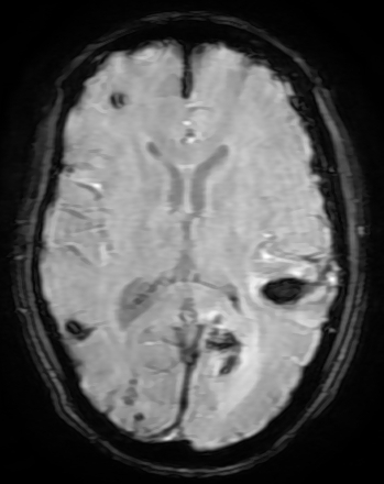
Figure 1: SW MR Image Showing Multiple Acute Haemorrhages


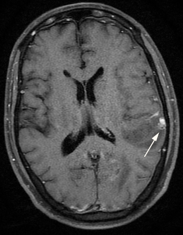
Figure 2: Contrast MR Images Showing Left Parietal Dura Based Enhancing Nodule (Arrow)


Serum PR3 ANCA tested positive with high titer 77.8 IU/ml (negative <5) and serum MPO ANCA was 1.5 (negative <5). 


We diagnosed the case as AAV restricted to CNS based on ANCA being positive, MRI brain findings, in the background of constitutional features, anemia and high ESR. Histopathology of the affected organ is gold standard for diagnosis, which was not feasible in our case. Conditions, which can have similar presentation, have been excluded clinically and with the help of ancillary testing. AAV is systemic vasculitis commonly affected organs are lungs, kidneys, heart, ear, nose, throat, orbit skin and peripheral nerves. At this stage, disease seems to be limited to CNS. According to commonly used classification proposed by Watts’ [6], patient remains unclassifiable. He was started oral prednisolone and cyclophosphamide as induction therapy. He was doing good, discharged after a nine-day stay at the hospital and is currently under follow up.

DISCUSSION

Central nervous system (CNS) vasculitis poses a great diagnostic challenge with its evolving presentation. It occurs either as part of a systemic vasculitis, or a primary disorder restricted to the CNS [7]. Anti-Neutrophil Cytoplasmic Antibody (ANCA) - Associated Vasculitis (AAV), is characterized by pathogenic ANCA production, a systemic small-vessel vasculitis [7]. Neurologic involvement is not uncommon in AAV throughout the disease course. CNS is affected in <15% of patients with AAV [8]. Neurological symptoms can vary including headache, ischemic infarction, intracranial haemorrhage, encephalopathy, seizures, neuropsychiatric disorders and rarely spinal cord symptoms. These symptoms are caused by the involvement of the structures including the dura mater, brain parenchyma, pituitary gland, spinal cord and leptomeninges with decreasing frequency.


78% cases of AAV vasculitis are diagnosed by histopathology and 2% by angiography. In the remaining 20% cases, the diagnosis is made based on typical clinical features of vasculitis in conjunction with a positive ANCA test and/or surrogate markers for granulomatous disease or renal vasculitis [3]. Cerebrovascular disease with AAV is very rare, accounting for 0–3.6% of all AAV cases [9,10] and tends to have a poor prognosis [11]. Sasaki et al. have reported from an autopsy case that cerebral haemorrhage is caused by the rupture of necrotizing angiitis [10]. Multiple cerebral hemorrhages in this case, is assumed to be caused by fragile vessels due to necrotizing angiitis.


Pachymeningeal involvement is more frequent than leptomeninges. The frequency of Hypertrophic Pachymeningitis (HP) in adult AAV ranges from 18 to 35% [12]. HP typically shows linear thickening of dura mater or a bulging mass with enhancement on brain MRI and computed tomography scan [13]. Enhancing peripheral dura-based nodule in our case represents HP.


AAV is responsive to medical treatment with corticosteroids combined with cyclophosphamide or, more recently, rituximab.  Rituximab, a monoclonal antibody directed against B cells, has been advocated for treatment as it reduces the need for maintenance immunosuppression [14].


CONCLUSION

AAV and other vasculitides may present with uncommon manifestations like intracerebral haemorrhage and/or hypertrophic pachymeningitis and a high index of suspicion is required for early diagnosis and prompt treatment to prevent adverse outcomes.

REFERENCES
  1. S. Furuta et al. "Update on eosinophilic granulomatosis with polyangiitis." Allergol Int., vol. 68, no. 4, 2019, pp. 430–436.

  2. D. Geetha et al. "Anca-associated vasculitis: core curriculum 2020." Am J Kidney Dis., vol. 75, no. 1, 2020, pp. 124–137.

  3. A.J. Mohammad et al. "Incidence and survival rates in Wegener’s granulomatosis, microscopic polyangiitis, Churg-Strauss syndrome and polyarteritis nodosa." Rheumatology, vol. 48, no. 12, 2009, pp. 1560–1565.

  4. P.M.K. Lutalo et al. "Diagnosis and classification of granulomatosis with polyangiitis (aka Wegener’s granulomatosis)." J Autoimmun., vol. 48–49, no. 1, 2014, pp. 94–98.

  5. M. Ceri et al. "Massive intracerebral hemorrhage associated with Wegener granulomatosis." Rheumatol Int., vol. 32, no. 6, 2012, pp. 1813–1814.

  6. R. Watts et al. "Development and validation of a consensus methodology for the classification of the anca-associated vasculitides and polyarteritis nodosa for epidemiological studies." Ann Rheum Dis., vol. 66, no. 2, 2006, pp. 222–227.

  7. J.C. Jennette et al. "Revised international Chapel Hill consensus conference nomenclature of vasculitides." Arthritis Rheum., vol. 65, no. 1, 2013, pp. 1–11.

  8. J. Graf "Central nervous system disease in antineutrophil cytoplasmic antibodies–associated vasculitis." Rheum Dis Clin N Am., vol. 43, no. 4, 2017, pp. 573–578.

  9. P.V. Deshpande et al. "Microscopic polyarteritis with renal and cerebral involvement." Pediatr Nephrol., vol. 15, no. 1–2, 2000, pp. 134–135.

  10. A. Sasaki et al. "[An autopsy case of P-ANCA-positive microscopic polyangiitis with multiple cerebral hemorrhagic infarction]." No To Shinkei, vol. 50, no. 1, 1998, pp. 56–60.

  11. F. Mattioli et al. "Frequency and patterns of subclinical cognitive impairment in patients with anca-associated small vessel vasculitides." J Neurol Sci., vol. 195, no. 2, 2002, pp. 161–166.

  12. H.A. Choi et al. "Characteristics of hypertrophic pachymeningitis in patients with granulomatosis with polyangiitis." J Neurol., vol. 264, no. 4, 2017, pp. 724–732.

  13. Y. Zheng et al. "Central nervous system involvement in anca-associated vasculitis: what neurologists need to know." Front Neurol, vol. 9, no. 1, 2019, Available from: https://www.frontiersin.org/article/10.3389/fneur.2018.01166/full

  14. J. Lynch et al. "Granulomatosis with polyangiitis (Wegener’s granulomatosis): evolving concepts in treatment." Semin Respir Crit Care Med., vol. 39, no. 4, 2018, pp. 434–458.

License
CC BY-NC-ND
Creative Commons Attribution-NonCommercial-NoDerivatives 4.0 International License
A Case of ANCA-Associated Vasculitis Presenting with Spontaneous Intracerebral Haemorrhage and Hypertrophic Pachymeningitis © 2026 by Shah UK, Rajaraman K, Nayak PR, Ambastha A, Lodh M licensed under CC BY-NC-ND 4.0
All papers should be submitted electronically. All submitted manuscripts must be original work that is not under submission at another journal or under consideration for publication in another form, such as a monograph or chapter of a book. Authors of submitted papers are obligated not to submit their paper for publication elsewhere until an editorial decision is rendered on their submission. Further, authors of accepted papers are prohibited from publishing the results in other publications that appear before the paper is published in the Journal unless they receive approval for doing so from the Editor-In-Chief.
Himalayan Journal of Medicine and Surgery open access articles are licensed under a Creative Commons Attribution-Share A like 4.0 International License. This license lets the audience to give appropriate credit, provide a link to the license, and indicate if changes were made and if they remix, transform, or build upon the material, they must distribute contributions under the same license as the original.
Advertisement
Recommended Articles
Research Article
Effect of Land Degradation on Livelihood
Published: 04/01/2024
Download PDF
Research Article
Gastric Cancer: A Comprehensive Review
...
Published: 20/01/2026
Download PDF
Research Article
Mean Platelet Volume, Systemic Immune-Inflammation Index and Procalcitonin as Biomarkers in Streptococcal Acute Tonsillitis and Peritonsillar Abscess
...
Published: 31/12/2025
Download PDF
Research Article
Amniotic Fluid and Umbilical Cord Serum Erythropoietin as Biomarkers of Fetal Hypoxia in Term and Prolonged Pregnancies
Published: 15/01/2026
Download PDF
Chat on WhatsApp
Flowbite Logo
Najmal Complex,
Opposite Farwaniya,
Kuwait.
Email: kuwait@iarcon.org

Editorial Office:
J.L Bhavan, Near Radison Blu Hotel,
Jalukbari, Guwahati-India
Useful Links
Order Hard Copy
Privacy policy
Terms and Conditions
Refund Policy
Others
About Us
Team Members
Contact Us
Online Payments
Join as Editor
Join as Reviewer
Subscribe to our Newsletter
Follow us
MOST SEARCHED KEYWORDS
scientific journal
 | 
business journal
 | 
medical journals
 | 
Scientific Journals
 | 
Academic Publisher
 | 
Peer-reviewed Journals
 | 
Open Access Journals
 | 
Impact Factor
 | 
Indexing Services
 | 
Journal Citation Reports
 | 
Publication Process
 | 
Impact factor of journals
 | 
Finding reputable journals for publication
 | 
Submitting a manuscript for publication
 | 
Copyright and licensing of published papers
 | 
Writing an abstract for a research paper
 | 
Manuscript formatting guidelines
 | 
Promoting published research
 | 
Publication in high-impact journals
Copyright © iARCON Internaltional LLP . All Rights Reserved.