Kyphoscoliosis is generally idiopathic and female dominant disease and the prevalence can vary between 0.3-15.3%. Anesthesia for both adult and pediatric patients with scoliosis can present a variety of challenges. Severe spine curvature disorders are commonly associated with multiple pathophysiological challenges to an Anesthetist. These factors are mostly related to the reduced vital capacity and chest wall compliance, along with the misalignment of axes and limitation in neck movement. Careful assessment and planning of alternative strategies by experienced anesthesiologists, appropriate positioning, and proper use of rescue devices can significantly improve the chances of successful intubation.
Keywords
Difficult Intubation
Difficult Airway
Kyphosis
Scoliosis
Spine Curvature Disorders
Anesthesiology
Covid-19
Emergency Medicine
INTRODUCTION
The human spine has a natural degree of the inward curve in the cervical and lumbar region ranging between 35 and 80 degrees and an outward curve in the thoracic region ranging between 30 and 50 degrees [1]. These curves help the spine withstand stress from movement and gravity. When the curvature is misaligned or exaggerated, it results in abnormal convex, concave, or lateral curves, namely, kyphosis, lordosis, and scoliosis [2]. Its prevalence can vary between 0.3-15.3% and has a female:male ratio as 3:1 respectively [3]. Several diseases accompany to scoliosis and anesthesia of adult and pediatric patients with scoliosis can present a variety of challenges. The challenges occur in providing optimal surgical conditions and ensuring adequate oxygenation. It results in pathophysiological changes that present various challenges for the anesthesiologist. The clinical impact of this disorder depends on factors like the degree of axial rotation, the number of vertebrae involved, and the severity and site of the abnormality in curvature. The involvement of the cervical vertebrae may result in difficult airway management and reduced range of neck movements. The difference in anatomical structure may be associated with increased morbidity secondary to prolonged and multiple attempts at laryngoscopy and tracheal intubation [3]. Involvement of the thoracic spine may cause rib cage deformity, which in severe cases leads to poor pulmonary function.In the literature, few cases report the technique of intubation and anesthetic management in patients with kyphoscoliosis [4].
We present a case of a 32year old man COVID-19 positive with severe spine curvature abnormality, low Glasgow Coma Scale (GCS) score (7/15), and low oxygen saturation for emergency intubation.
Case Presentation
A 32 year old man who was 58 kg in weight and 154 cm in height presented to emergency department with altered sensorium. Upon assessment, the patient had a GCS score of 7/15 (E1V2M4), blood pressure of 98/52 mmHg, heart rate of 132 bpm, respiratory rate of 40 breaths/min, and oxygen saturation of 78% while on 15 liters of supplemental oxygen using a non-rebreather mask. On examination, his spine had curvature disorder with moderate cervical kyphosis, severe thoracic kyphoscoliosis, and mild lumbar lordosis resulting in the upper body contorting towards the left side and severe restriction of neck movements. On auscultation, his cardiovascular examination was unremarkable. The chest examination revealed bilateral basal coarse crackles. Patient was found COVID-19 positive by Rapid Antigen Test (RAT) kit. The hematological and biochemical parameters were significant for leukocytosis, metabolic acidosis with a lactate of 2.8 mmol/L, and hypoxemia with a partial pressure of oxygen of 42 mmHg and partial pressure of carbon dioxide of 92 mmHg.
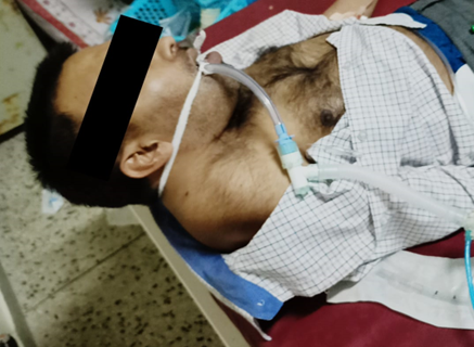
Figure 1: Lateral View of the Patient Showing Severe Thoracic Kyphoscoliosis with Leftward Truncal Deviation and Cervical Spine Rigidity. Patient Is Intubated and Mechanically Ventilated Due to Acute Respiratory Failure
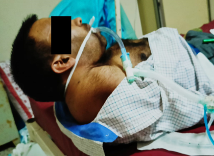
Figure 2: Oblique-Lateral View Highlighting Pronounced Thoracic Deformity and Cervical Kyphosis, Contributing to Restrictive Lung Pathology. The Patient is Seen Intubated Following Decompensation from Covid-19-Associated Respiratory Failure.
Emergency intubation was planned. The airway examination revealed a Mallampati classification of II and a restricted range of neck movement. We prepared the patient, attached the monitors, and checked the equipment for difficult intubation. Besides, the otorhinolaryngology (ENT) department was briefed about the case. Their team prepared for the "can't intubate can't ventilate" scenario and were in the standby position. The patient was propped with cushions below his left shoulder and the left lumbar region to optimize his position. He was given bag-mask ventilation initially but it was not effective as he started to desaturate. We administered 80mcg of fentanyl and 2 mg of midazolam and 100 mg of succinylcholine before intubation. Subsequently, we performed direct laryngoscopy using a size four blade and noted a Cormack-Lehane grade III a view. The endotracheal tube of size 8.0 mm was inserted under vision. The chest was examined for the bilateral rise and auscultated for equal air entry in both lung fields. Due to unavailability, the capnometer was not utilized. Next, the endotracheal tube was fixed at 21cm near the right oral commissure by an OT assistant. Patient was put on ventilator on Assist volume control mode with FiO2 subsequently decrease to 65% to maintain the oxygen saturation of >90% and other supportive treatment was initiated.
DISCUSSION
Severe spine curvature disorders pose various challenges during airway management, secondary to physiological and anatomical factors. Physiologically, the progressive nature of the disorder results in a combination of both obstructive and restrictive patterns of pulmonary disease due to limited vital capacity and chest wall compliance. That results in reduced vital capacity along with ventilation and perfusion ratio mismatch.4 The decline in functional residual capacity reduces the oxygen reserve. This leads to a shorter safe apnea period before the critical hypoxia develops. Second, the morphological features of the patient's anatomy lead to difficult intubation.5 Furthermore, during laryngoscopy, the laryngeal structures are hard to recognize and, the distortion of anatomy makes the successful placement of the endotracheal tube difficult.
Patients with severe spine curvature disorders present various challenges during anesthetic management that may be fatal in some cases. However, the risk of an adverse outcome or alternative invasive strategies can be curbed by employing various factors such as good communication, experienced team, accessibility of rescue devices, careful assessment, proper positioning, and utilization of appropriate airway devices to minimize time and morbidity associated with multiple attempts. Moreover, through new techniques and devices, such as the video laryngoscope, the view of the larynx is improved. This allows successful placement of the endotracheal tube without direct visualization of the vocal cords. Special conditions like kyphoscoliosis may generate extra disadvantages in airway management which makes it difficult. Preparation in the preoperative period becomes more than an issue in such cases.
Declaration of Conflicting Interests
The authors have declared that no competing interests exist.
Kuo, Y.L. et al "Association between spinal curvature disorders and injury: a nationwide population-based retrospective cohort study." BMJ Open, 2019.
Anand, H.K., Ambareesha, M. "Scoliosis and anaesthetic considerations." Indian J. Anaesth., vol. 51, 2007, pp. 486–95.
Kitayama, M. et al "Airway management and rigid spine syndrome." Anesth. Analg., vol. 84, 1997, pp. 690–1.
Janssens, M., Hartstein, G. "Management of difficult intubation." Eur. J. Anaesthesiol., vol. 18, 2001, pp. 3–12.
Advertisement
Recommended Articles
Research Article
Clinicopathological Profile and Disease Presentation Patterns in Colorectal Cancer: A Prospective Observational Study from a Tertiary Care Center in North India
Rahul Rai,
...
Tushar
Published: 05/04/2025
Download PDF
Cite
x
APA
Rai, R., Paul, D. & None, T. (2025). Clinicopathological Profile and Disease Presentation Patterns in Colorectal Cancer: A Prospective Observational Study from a Tertiary Care Center in North India. Himalayan Journal of Medicine and Surgery, 6(1), 1-3.
MLA
Rai, Rahul, Dharam Paul and Tushar . "Clinicopathological Profile and Disease Presentation Patterns in Colorectal Cancer: A Prospective Observational Study from a Tertiary Care Center in North India." Himalayan Journal of Medicine and Surgery 6.1 (2025): 1-3.
Chicago
Rai, Rahul, Dharam Paul and Tushar . "Clinicopathological Profile and Disease Presentation Patterns in Colorectal Cancer: A Prospective Observational Study from a Tertiary Care Center in North India." Himalayan Journal of Medicine and Surgery 6, no. 1 (2025): 1-3.
Harvard
Rai, R., Paul, D. and None, T. (2025) 'Clinicopathological Profile and Disease Presentation Patterns in Colorectal Cancer: A Prospective Observational Study from a Tertiary Care Center in North India' Himalayan Journal of Medicine and Surgery 6(1), pp. 1-3.
Vancouver
Rai R, Paul D, Tushar T. Clinicopathological Profile and Disease Presentation Patterns in Colorectal Cancer: A Prospective Observational Study from a Tertiary Care Center in North India. Himalayan Journal of Medicine and Surgery. 2025 Jan;6(1):1-3.
Download PDF
Review Article
Clinicopathological Profile of Colorectal Cancer Patients: A Contemporary Review
Dharam Pau,
Tushar
Published: 05/04/2025
Download PDF
Cite
x
APA
Pau, D. & None, T. (2025). Clinicopathological Profile of Colorectal Cancer Patients: A Contemporary Review. Himalayan Journal of Medicine and Surgery, 6(1), 1-3.
MLA
Pau, Dharam and Tushar . "Clinicopathological Profile of Colorectal Cancer Patients: A Contemporary Review." Himalayan Journal of Medicine and Surgery 6.1 (2025): 1-3.
Chicago
Pau, Dharam and Tushar . "Clinicopathological Profile of Colorectal Cancer Patients: A Contemporary Review." Himalayan Journal of Medicine and Surgery 6, no. 1 (2025): 1-3.
Harvard
Pau, D. and None, T. (2025) 'Clinicopathological Profile of Colorectal Cancer Patients: A Contemporary Review' Himalayan Journal of Medicine and Surgery 6(1), pp. 1-3.
Vancouver
Pau D, Tushar T. Clinicopathological Profile of Colorectal Cancer Patients: A Contemporary Review. Himalayan Journal of Medicine and Surgery. 2025 Jan;6(1):1-3.
Download PDF
Research Article
Hernia and Its Surgical Management: A Cross-Sectional Study on Awareness Among the General Public of Kangra
Shallabh Sharma,
Ashish Guleria
Published: 05/04/2025
Download PDF
Cite
x
APA
Sharma, S. & Guleria, A. (2024). Hernia and Its Surgical Management: A Cross-Sectional Study on Awareness Among the General Public of Kangra. Himalayan Journal of Medicine and Surgery, 5(1), 1-5.
MLA
Sharma, Shallabh and Ashish Guleria. "Hernia and Its Surgical Management: A Cross-Sectional Study on Awareness Among the General Public of Kangra." Himalayan Journal of Medicine and Surgery 5.1 (2024): 1-5.
Chicago
Sharma, Shallabh and Ashish Guleria. "Hernia and Its Surgical Management: A Cross-Sectional Study on Awareness Among the General Public of Kangra." Himalayan Journal of Medicine and Surgery 5, no. 1 (2024): 1-5.
Harvard
Sharma, S. and Guleria, A. (2024) 'Hernia and Its Surgical Management: A Cross-Sectional Study on Awareness Among the General Public of Kangra' Himalayan Journal of Medicine and Surgery 5(1), pp. 1-5.
Vancouver
Sharma S, Guleria A. Hernia and Its Surgical Management: A Cross-Sectional Study on Awareness Among the General Public of Kangra. Himalayan Journal of Medicine and Surgery. 2024 Jan;5(1):1-5.
Download PDF
Research Article
A Silent Threat: Cervical Cancer Awareness and Prevention Among Women in Shimla
Shivali Ranote,
...
Ashutosh
Published: 05/04/2025
Download PDF
Cite
x
APA
Ranote, S., Sharma, A. & None, A. (2024). A Silent Threat: Cervical Cancer Awareness and Prevention Among Women in Shimla. Himalayan Journal of Medicine and Surgery, 5(1), 1-6.
MLA
Ranote, Shivali, Ajay Sharma and Ashutosh . "A Silent Threat: Cervical Cancer Awareness and Prevention Among Women in Shimla." Himalayan Journal of Medicine and Surgery 5.1 (2024): 1-6.
Chicago
Ranote, Shivali, Ajay Sharma and Ashutosh . "A Silent Threat: Cervical Cancer Awareness and Prevention Among Women in Shimla." Himalayan Journal of Medicine and Surgery 5, no. 1 (2024): 1-6.
Harvard
Ranote, S., Sharma, A. and None, A. (2024) 'A Silent Threat: Cervical Cancer Awareness and Prevention Among Women in Shimla' Himalayan Journal of Medicine and Surgery 5(1), pp. 1-6.
Vancouver
Ranote S, Sharma A, Ashutosh A. A Silent Threat: Cervical Cancer Awareness and Prevention Among Women in Shimla. Himalayan Journal of Medicine and Surgery. 2024 Jan;5(1):1-6.
Singh Raizada, M. & Saphiya, L. (2022). Emergency Intubaton of a Covid Positive Patient with Severe Spine Curvature Abnormality. Himalayan Journal of Medicine and Surgery, 3(1), 1-3.
MLA
Singh Raizada, Maninder and Latesh Saphiya. "Emergency Intubaton of a Covid Positive Patient with Severe Spine Curvature Abnormality." Himalayan Journal of Medicine and Surgery 3.1 (2022): 1-3.
Chicago
Singh Raizada, Maninder and Latesh Saphiya. "Emergency Intubaton of a Covid Positive Patient with Severe Spine Curvature Abnormality." Himalayan Journal of Medicine and Surgery 3, no. 1 (2022): 1-3.
Harvard
Singh Raizada, M. and Saphiya, L. (2022) 'Emergency Intubaton of a Covid Positive Patient with Severe Spine Curvature Abnormality' Himalayan Journal of Medicine and Surgery 3(1), pp. 1-3.
Vancouver
Singh Raizada M, Saphiya L. Emergency Intubaton of a Covid Positive Patient with Severe Spine Curvature Abnormality. Himalayan Journal of Medicine and Surgery. 2022 Jan;3(1):1-3.