Incidence of Scoliosis is approximately 2% and higher in females. Therefore, anaesthesiologist should be familiar with its implications for safe anaesthetic management [1]. Such patients present unique challenges for administration of regional anaesthesia. Reporting one such case with a successful outcome using pre procedural Ultrasound spine for spinal and epidural anaesthesia. A 48 years female patient of height 138 cm, P2L2 diagnosed with abnormal uterine bleeding secondary to leiomyoma with grade III scoliosis and post burn contractures involving neck, was posted for elective total abdominal hysterectomy. Patient underwent surgery successfully under Epidural anaesthesia after accessing spine by ultrasonography for identifying point of insertion, depth of epidural space & needle trajectory [2]. Ultrasound is boon for accessing spine in patient with difficult spinal anatomy and pregnancy for deciding correct spinal interspace and successful outcome with fewer attempts.
Keywords
Thoraco-Lumbar Scoliosis
Post Burn Contracture
Epidural Anaesthesia
Spinal Anaesthesia
Difficult Intubation
Ultrasonography
INTRODUCTION
Scoliosis is defined as a lateral curvature of the spine. Incidence is around 2% and is higher in females. Etiology can be idiopathic, Paralytic (Neuropathic or Myopathic), Congenital, Neurofibromatosis (Marfan syndrome), Ehlar-Danlos Syndrome, Post Traumatic. Scoliosis is associated with restrictive lung disease and hypoxemia leading to cardiovascular compromise and if left untreated causes pulmonary hypertension and respiratory failure. It is important for an Anaesthesiologist to be familiar with the implications of scoliosis and thus to formulate safe anesthetic plan. Combined Spinal and Epidural anaesthesia is one of the most common and safest techniques of inducing regional anaesthesia for the lower abdominal surgery and post-operative pain relief. The failure rate is minimal in the experienced hands [3].
Case Report
A48 years old female of height 138 cm & weight 52 kg with grade III scoliosis and post burn contractures of face and neck, posted for elective total abdominal hysterectomy. Scoliosis was observed at the age of 10 years and burn 8 years back. During pre-anaesthetic check-up, patient didn’t give any past history of systemic medical disease, drug allergy and any previous surgery. On clinical examination, Pulse Rate 84/min, Blood Pressure 116/78 mmHg, SpO2 -98 % on room air. Airway examination revealed Mallampatti score-III, with intact dentition, Mouth Opening > 2Fingers, and restricted neck movements (post burn contracture of face and neck). Local Examination of the spine shows lateral curvature from T4–L5 with Cobb Angle > 50 degree. No abnormality was detected in any of the organ system function. Normal Echocardiograph and normal values of all the routine investigations.
Anaesthesia Plan
Central neuraxial blockade in form of combined spinal epidural Anaesthesia (CSE).
Preprocedural USG
Decision for pre procedural USG guided spinal anesthesia made due to anticipated difficult spinal needle placement.
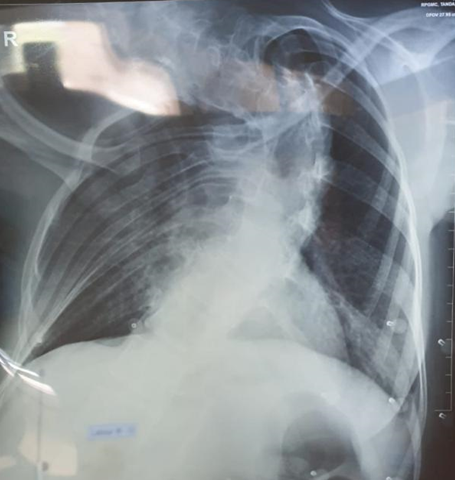
Figure 1: Chest X-Ray Showing Severe Thoracolumbar Scoliosis with Lateral Curvature from T4 To L5
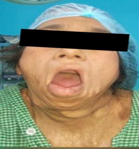
Figure 2: Pre-Operative Image Showing Restricted Neck Movements and Post-Burn Contractures of the Face and Neck in A Patient with Grade Iii Scoliosis
Preprocedural USG in PSO view done, starting from sacrum and then moving in cephalad direction. At L2-L3 where both posterior and anterior complex was visible the probe was oriented in the transverse plane and centred in the midline to get the clear view of the structures, (USG visibility score – 3). Depth of AC, PC and intrathecal space was measured in the midline. 18G Tuohy’s needle inserted at point of intersection of vertical and horizontal lines drawn from the centre of the probe in the longitudinal and transverse axis with Loss of Resistance technique. Epidural catheter inserted and fixed at 10 cm. Test dose of 3cc of Lignocaine (60mg) + Adrenaline (15 mcg) given. No tachycardia, no motor blockage seen. Sub arachnoid block given via 26 G Quincke’s needle with 2 ml of 0.5% heavy Bupivacaine.
Sensory and motor blockade was observed by loss of sensation to pinprick below T6 dermatome and Modified Bromage Scale of 4. Intraoperative period was uneventful.
DISCUSSION
The technical difficulty of neuraxial blockage is measured using 2 main parameters: The number of needle manipulations required for success and the time taken to perform the block. Of the two, the former is more important because multiple needle insertions are an independent predictor of complications. In our case, the feasibility and choice of anesthesia was a challenge [4]. In avoidance of airway manipulation in a setting of the difficult airway. Availability of USG for accessing spine, identifying point of insertion, depth of intrathecal space & needle trajectory is vital in such cases.Disadvantages include possibility of partial or incomplete block, which was taken care with USG guided bilateral rescue Transverse Abdominal Plane block with systemic analgesic for mitigation of pain.
CONCLUSION
Based on the clinical assessment, full stomach status and availability of Ultrasound, administration of spinal anesthesia was the best option for this patient. Ultrasound is boonfor accessing spine in patient with difficult spinal anatomy and pregnancy for deciding correct spinal interspace and successful outcome with fewer attempts and providing guided block and accessing airway.
REFERENCE
Kulkarni, A.H., Ambareesha, M. "Scoliosis and anesthetic considerations." Indian J. Anaesth., 2007, pp. 486–95.
Chin, K.J., Chan, V. "Ultrasonography as a preoperative assessment tool: predicting the feasibility of central neuraxial blockade." Anesth. Analg., 2010, pp. 252–3.
Bajwa, S.J. et al "Admixture of clonidine and fentanyl to ropivacaine in epidural anesthesia for lower abdominal surgery." Anesth. Essays Res., 2010, pp. 9–14.
de Filho, G.R. et al "Predictors of successful neuraxial block: a prospective study." Eur. J. Anaesthesiol., vol. 19, 2002, pp. 447–51.
Advertisement
Recommended Articles
Research Article
Clinicopathological Profile and Disease Presentation Patterns in Colorectal Cancer: A Prospective Observational Study from a Tertiary Care Center in North India
Rahul Rai,
...
Tushar
Published: 05/04/2025
Download PDF
Cite
x
APA
Rai, R., Paul, D. & None, T. (2025). Clinicopathological Profile and Disease Presentation Patterns in Colorectal Cancer: A Prospective Observational Study from a Tertiary Care Center in North India. Himalayan Journal of Medicine and Surgery, 6(1), 1-3.
MLA
Rai, Rahul, Dharam Paul and Tushar . "Clinicopathological Profile and Disease Presentation Patterns in Colorectal Cancer: A Prospective Observational Study from a Tertiary Care Center in North India." Himalayan Journal of Medicine and Surgery 6.1 (2025): 1-3.
Chicago
Rai, Rahul, Dharam Paul and Tushar . "Clinicopathological Profile and Disease Presentation Patterns in Colorectal Cancer: A Prospective Observational Study from a Tertiary Care Center in North India." Himalayan Journal of Medicine and Surgery 6, no. 1 (2025): 1-3.
Harvard
Rai, R., Paul, D. and None, T. (2025) 'Clinicopathological Profile and Disease Presentation Patterns in Colorectal Cancer: A Prospective Observational Study from a Tertiary Care Center in North India' Himalayan Journal of Medicine and Surgery 6(1), pp. 1-3.
Vancouver
Rai R, Paul D, Tushar T. Clinicopathological Profile and Disease Presentation Patterns in Colorectal Cancer: A Prospective Observational Study from a Tertiary Care Center in North India. Himalayan Journal of Medicine and Surgery. 2025 Jan;6(1):1-3.
Download PDF
Review Article
Clinicopathological Profile of Colorectal Cancer Patients: A Contemporary Review
Dharam Pau,
Tushar
Published: 05/04/2025
Download PDF
Cite
x
APA
Pau, D. & None, T. (2025). Clinicopathological Profile of Colorectal Cancer Patients: A Contemporary Review. Himalayan Journal of Medicine and Surgery, 6(1), 1-3.
MLA
Pau, Dharam and Tushar . "Clinicopathological Profile of Colorectal Cancer Patients: A Contemporary Review." Himalayan Journal of Medicine and Surgery 6.1 (2025): 1-3.
Chicago
Pau, Dharam and Tushar . "Clinicopathological Profile of Colorectal Cancer Patients: A Contemporary Review." Himalayan Journal of Medicine and Surgery 6, no. 1 (2025): 1-3.
Harvard
Pau, D. and None, T. (2025) 'Clinicopathological Profile of Colorectal Cancer Patients: A Contemporary Review' Himalayan Journal of Medicine and Surgery 6(1), pp. 1-3.
Vancouver
Pau D, Tushar T. Clinicopathological Profile of Colorectal Cancer Patients: A Contemporary Review. Himalayan Journal of Medicine and Surgery. 2025 Jan;6(1):1-3.
Download PDF
Research Article
Hernia and Its Surgical Management: A Cross-Sectional Study on Awareness Among the General Public of Kangra
Shallabh Sharma,
Ashish Guleria
Published: 05/04/2025
Download PDF
Cite
x
APA
Sharma, S. & Guleria, A. (2024). Hernia and Its Surgical Management: A Cross-Sectional Study on Awareness Among the General Public of Kangra. Himalayan Journal of Medicine and Surgery, 5(1), 1-5.
MLA
Sharma, Shallabh and Ashish Guleria. "Hernia and Its Surgical Management: A Cross-Sectional Study on Awareness Among the General Public of Kangra." Himalayan Journal of Medicine and Surgery 5.1 (2024): 1-5.
Chicago
Sharma, Shallabh and Ashish Guleria. "Hernia and Its Surgical Management: A Cross-Sectional Study on Awareness Among the General Public of Kangra." Himalayan Journal of Medicine and Surgery 5, no. 1 (2024): 1-5.
Harvard
Sharma, S. and Guleria, A. (2024) 'Hernia and Its Surgical Management: A Cross-Sectional Study on Awareness Among the General Public of Kangra' Himalayan Journal of Medicine and Surgery 5(1), pp. 1-5.
Vancouver
Sharma S, Guleria A. Hernia and Its Surgical Management: A Cross-Sectional Study on Awareness Among the General Public of Kangra. Himalayan Journal of Medicine and Surgery. 2024 Jan;5(1):1-5.
Download PDF
Research Article
A Silent Threat: Cervical Cancer Awareness and Prevention Among Women in Shimla
Shivali Ranote,
...
Ashutosh
Published: 05/04/2025
Download PDF
Cite
x
APA
Ranote, S., Sharma, A. & None, A. (2024). A Silent Threat: Cervical Cancer Awareness and Prevention Among Women in Shimla. Himalayan Journal of Medicine and Surgery, 5(1), 1-6.
MLA
Ranote, Shivali, Ajay Sharma and Ashutosh . "A Silent Threat: Cervical Cancer Awareness and Prevention Among Women in Shimla." Himalayan Journal of Medicine and Surgery 5.1 (2024): 1-6.
Chicago
Ranote, Shivali, Ajay Sharma and Ashutosh . "A Silent Threat: Cervical Cancer Awareness and Prevention Among Women in Shimla." Himalayan Journal of Medicine and Surgery 5, no. 1 (2024): 1-6.
Harvard
Ranote, S., Sharma, A. and None, A. (2024) 'A Silent Threat: Cervical Cancer Awareness and Prevention Among Women in Shimla' Himalayan Journal of Medicine and Surgery 5(1), pp. 1-6.
Vancouver
Ranote S, Sharma A, Ashutosh A. A Silent Threat: Cervical Cancer Awareness and Prevention Among Women in Shimla. Himalayan Journal of Medicine and Surgery. 2024 Jan;5(1):1-6.
Saphiya, L. & Singh Raizada, M. (2022). Pre-Procedural Usg Spine for Hystrectomy in A Patient with Thoracolumbar Scoliosis with Post Burn Contracture Neck. Himalayan Journal of Medicine and Surgery, 3(1), 1-2.
MLA
Saphiya, Latesh and Maninder Singh Raizada. "Pre-Procedural Usg Spine for Hystrectomy in A Patient with Thoracolumbar Scoliosis with Post Burn Contracture Neck." Himalayan Journal of Medicine and Surgery 3.1 (2022): 1-2.
Chicago
Saphiya, Latesh and Maninder Singh Raizada. "Pre-Procedural Usg Spine for Hystrectomy in A Patient with Thoracolumbar Scoliosis with Post Burn Contracture Neck." Himalayan Journal of Medicine and Surgery 3, no. 1 (2022): 1-2.
Harvard
Saphiya, L. and Singh Raizada, M. (2022) 'Pre-Procedural Usg Spine for Hystrectomy in A Patient with Thoracolumbar Scoliosis with Post Burn Contracture Neck' Himalayan Journal of Medicine and Surgery 3(1), pp. 1-2.
Vancouver
Saphiya L, Singh Raizada M. Pre-Procedural Usg Spine for Hystrectomy in A Patient with Thoracolumbar Scoliosis with Post Burn Contracture Neck. Himalayan Journal of Medicine and Surgery. 2022 Jan;3(1):1-2.