Incidence of Caecal volvulus is 2.8-7.1 persons per million per year and it is a rare cause of intestinal obstruction which may result in delay in the diagnosis and appropriate intervention. Patients with this condition may present with highly variable clinical presentations ranging from intermittent, self-limiting abdominal pain to acute abdominal pain associated with intestinal strangulation and sepsis.
A volvulus is a twisting or axial rotation of a portion of bowel about its mesentery. The rotation causes obstruction to the lumen (>180° torsion) and if tight enough also causes vascular occlusion in the mesentery (>360° torsion). Bacterial fermentation adds to the distension and increasing intraluminal pressure impairs capillary perfusion. Mesenteric veins become obstructed as a result of the mechanical twisting and thrombosis results and contributes to the ischaemia. Volvuli may be primary or secondary. The primary form occurs secondary to congenital malrotation of the gut, abnormal mesenteric attachments or congenital bands. Examples include volvulus neonatorum, caecal volvulus and sigmoid volvulus. A secondary volvulus, which is the more common variety, is due to rotation of a segment of bowel around an acquired adhesion or stoma.
Caecal volvulus is an uncommon cause of intestinal obstruction, this may occur as part of volvulus neonatorum or de novo and is usually a clockwise twist. It is estimated that it occurs in 2.8–7.1 per million people per year [1]. It is an infrequent cause of intestinal obstruction, accounting for 1−5% of all adult intestinal obstructions [2].It is more common in females in the 4th and 5th decades and usually presents acutely with the classic features of obstruction. Ischaemia is common. A mobile/non-fixed cecum is mentioned as one significant associated finding in patients with caecal volvulus [3].Its variable clinical symptoms make it difficult to diagnose early. At first the obstruction may be partial, with the passage of flatus and faeces. In 25% of cases, examination may reveal a palpable tympanic swelling in the midline or left side of the abdomen. The volvulus typically results in the caecum lying in the left upper quadrant. The diagnosis is not usually made preoperatively.
Case Report
A 70 year-old female presented in surgical department with Pain Abdomen for 1 Week and Non-Passage of flatus and stool for 3 Days.
H/o abdominal distention was present
H/o vomiting was present, bilious
Otherwise she did not have a previous history of similar complaints or constipation, no weight loss or bleeding from the rectum, no groin swelling, no prior abdominal surgery, or endoscopic procedures. On examination she was dehydrated, with a dry tongue and buccal mucosa. Her pulse rate was 110 beats per minute, blood pressure 110/60 mmHg and SPO2- 80-82% on Room Air.
On abdominal examination : Abdomen was uniformly distended, umblicus central, healed burn scar of size 5 cm was present in Rt iliac fossa region, visible intestinal loops in the central part of her abdomen
Palpation: Overlying temperature was normal, generalised tenderness and guard was present, Rebound tenderness was present. No lump/ organomegaly felt
Percussion: Hypertympanic note was present, Liver dullness was obliterated
BS: Absent
Digital rectal examination revealed an empty ballooned rectum.
Investigations
Hb: 14.3 g/dl
TLC: 5.5 th/ul
PC: 129th/ul
Urea: 82 mg/dl
Creat: 1.3 mg/dl
Na/k/Cl-132/4.2/97meq/l
RBS: 131mg/dl
Lipase/Amylase:-88/38U/L
Bilirubin-total 0.98/direct-0.20mg/dl
ALT/AST/ALP:-84/55/72U/L
PT/INR 15/1.16
A plain abdominal X-ray showed a markedly distended large bowel
USG Abdomen
Multiple dilated gut loops with inter loop fluid
F/S/O acute intestinal obstruction
CECT Abdomen: F/S/O Acute Intestinal Obstruction (transition point in transverse colon) with minimal free fluid in pelvis with pneumoperitonium with few air foci in the wall of ascending colon Pneumatosis intestinalis
Cause Midgut volvulus
Adhesion Band

Figure 1: Dilated Large Bowel Loops


Figure 2: Cect Showing Features of Intestinal Obstruction

Figure 3: Intraoperative Finding Showing a Distended, Mobile and Volvulated Cecum

Figure 4 Intraoperative Finding Showing a Distended, Mobile and Volvulated Cecum

Figure 5: Intraoperative Finding Depicting a Thinned Out and Perforated Cecum
She was admitted, resuscitated with intravenous fluid administration. Preoperative preparations were made; informed consent was obtained for the procedure. On opening abdomen, gush of air was present. Proximal gut was dilated, there was volvulus of caecum and caecum was mobile with perforation and pre-gangrenous changes and distally collapsed colon, right hemi-colectomy with end to side Ile transverse anastomosis done.
The patient had an uneventful postoperative course, she was started on a fluid diet on the 3rd post-operative day and advanced to a solid diet on the 5th day and was discharged with improvement on the 7th postoperative day. She had a follow-up at 6 months in the surgical referral clinic with no complications.
Caecal volvulus is the axial rotation of the ascending colon, cecum, and terminal ileum. It was first described by Rokitansky in 1837 [4]. Caecal volvulus could be divided into two major groups of loop axial ileocolic, the commonest, accounting for 90% of cases, which can appear as a clockwise or counter clockwise pattern in which the latter one is the most commonly seen variety [3]. Caecal bascule is where there is an upward folding of the cecum either anteriorly or posteriorly [5].
Caecal volvulus is said to be associated with inadequate right colon fixation or anomalies in which the right colon does not properly fuse to the lateral peritoneum [6]. Other well documented predisposing factors for CV are long-standing constipation, distal obstruction, previous abdominal surgeries, intraabdominal masses, being bedridden, and undergoing colonoscopy procedures [5,7].When it occurs in the younger populations its more commonly caused by post-OP adhesions, pregnancy, or aganganglionic megacolon [7-9] Reports have shown that there is no sex predilection and the mean age of incidence is 61.8 years6 .
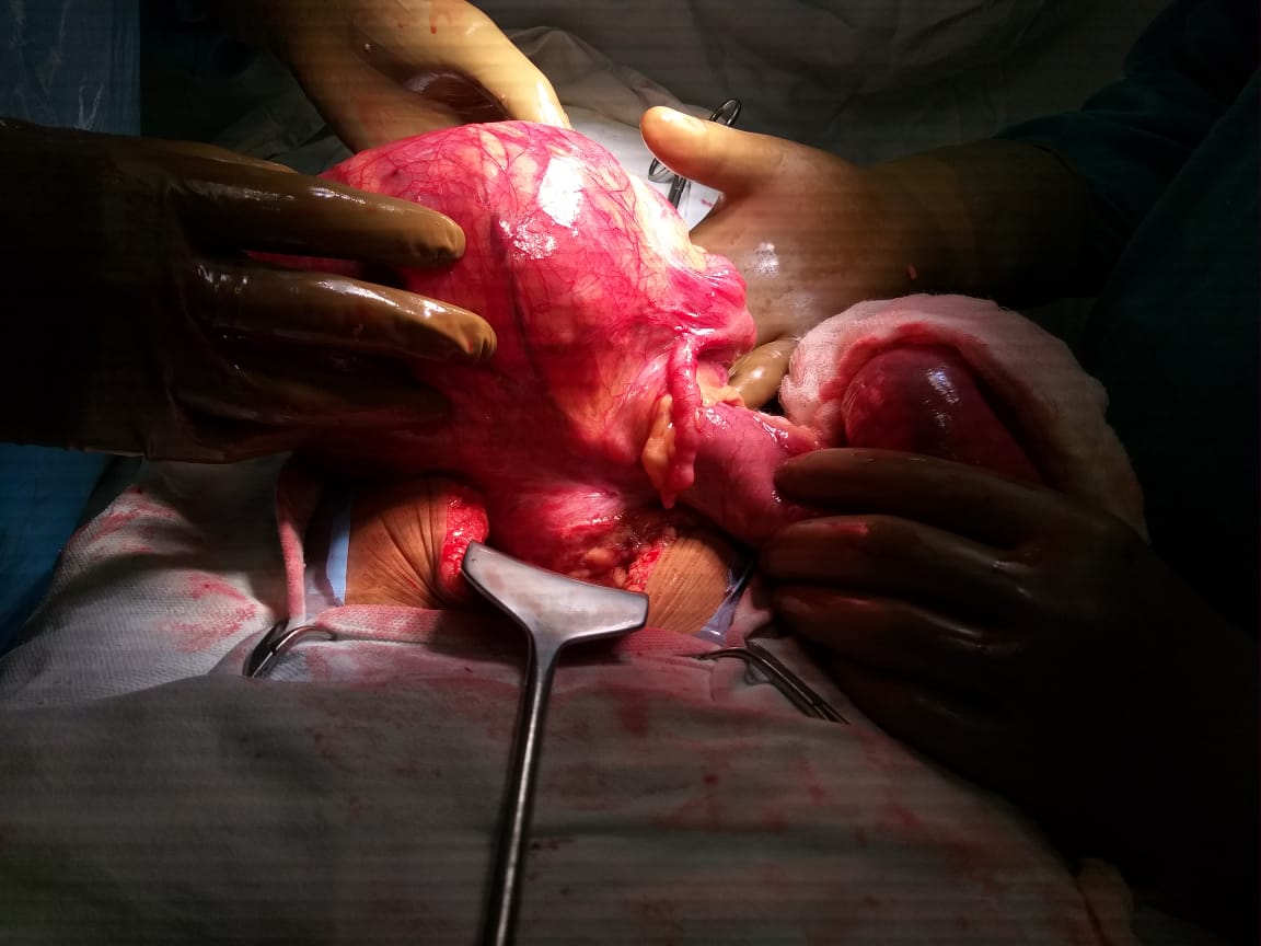
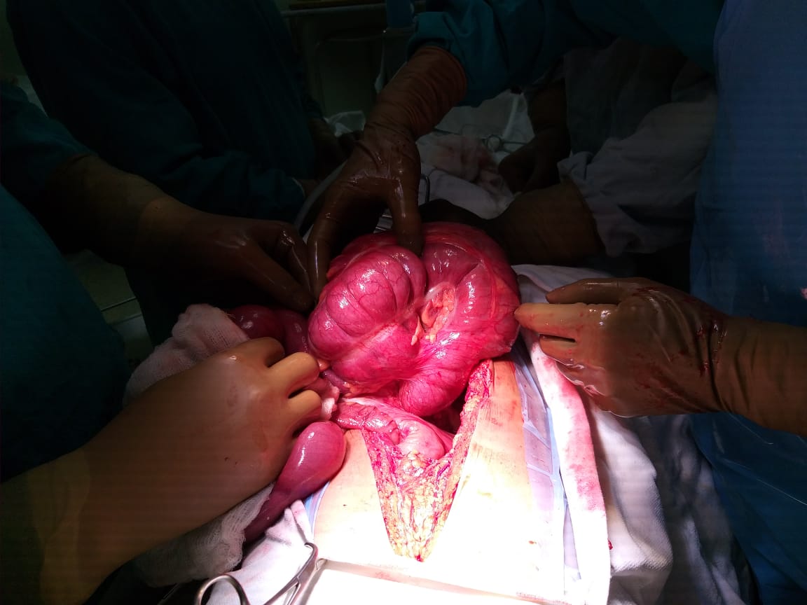
The clinical presentation depends on the duration of the complaints and the presence of complications. Symptoms of abdominal pain, associated with vomiting and abdominal distension, are reported as the commonest presentations [5]. Preoperative diagnosis of CV poses a significant challenge because of its rarity and nonspecific symptoms, mostly diagnosed intraoperatively.We were also able to reach the diagnosis intraoperatively [10]. Laboratory investigations are neither specific nor sensitive for the diagnosis of CV but may suggest the degree of obstruction and presence of complications [11].
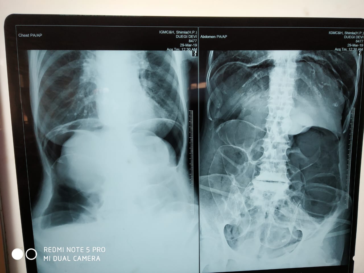
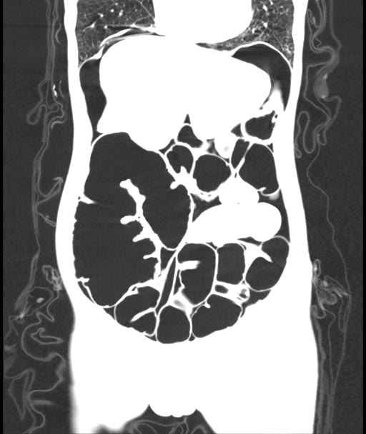
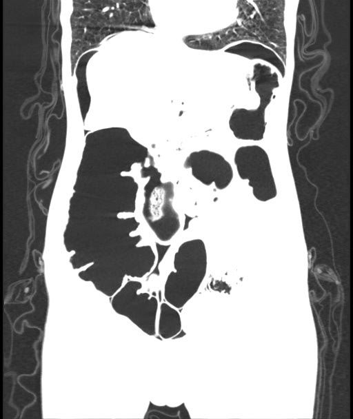
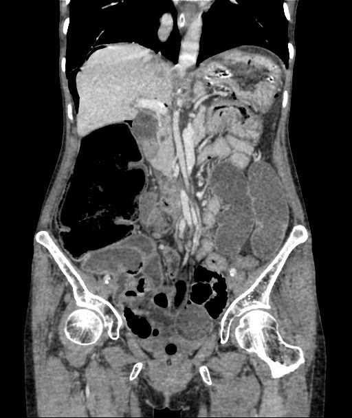
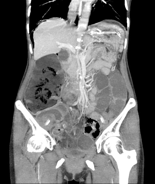
Radiologic imaging may be abnormal and detect CV in 45–56% of the cases [5] .Plain abdominal x-ray is highly sensitive for the diagnosis of CV with the characteristic “coffee bean“ sign deformity, its apex pointing to the left upper quadrant [9]. Other findings commonly seen are cecal dilatation (98−100%), single air–fluid level (72–88%), small bowel dilatation (42–55%), and absence of gas in the distal colon (82%). CT is more sensitive and specific for diagnosing CV and detecting complications [10].
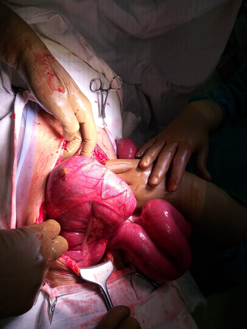
Once the diagnosis of CV was made patients should undergo urgent laparotomy or laparoscopic procedures to untwist the segment early to avoid the risk of strangulatio [5].
Once exploration has been done and if the intraoperative finding is a volvulated cecum; its viability should be assessed. Resection (right hemicolectomy) for all gangrenous and perforated or grossly distended thinned out cecal volvulus is the treatment of choice with primary anastomosis, ileostomy with mucous fistula is depending on the patient’s intraoperative physiology and bowel condition, resection has the absolute advantage of eliminating the risk of recurrence [5].
Manual detorsion without resection options is not advisable since it is associated with an unacceptable risk of recurrence (40%) [6]. Cecopexy or cecostomy are also available options depending on the overall status of the patient. Possible endoscopic detorsion in cecal volvulus is an emerging option for non-strangulated volvulus but associated with high risk of recurrence [12].
Caecal volvulus is a rare cause of adult intestinal obstruction with non-specific symptoms and laboratory findings. Its rarity makes its preoperative diagnosis challenging and hampers early intervention, resulting in a high incidence of complications and mortality, hence a high index of suspicion and urgent intervention is needed. Surgery is the mainstay of management for CV. Resection with hemicolectomy is the accepted method of treatment for gangrenous or perforated caecal volvulus. High quality studies including case series studies might be more helpful in the understanding of CV.
Mahruqi, G.A. et al. "Cecal volvulus case report and literature review." International Journal of Innovative Research in Medical Science, vol. 4, no. 6, 2019, pp. 391–394. doi:10.23958/ijirms/vol04-i06/680.
Mwita, C. et al. "Cecal volvulus in an adolescent African male: a case report and brief review of the literature." Pan African Medical Journal, vol. 17, 2014. doi:10.11604/ pamj.2014.17.92.3946.
Fenzl, N., DeSilva, S.G. and Scott, J. "Cecal volvulus case report." Imaging Medicine, vol. 9, no. 2, 2017.
Rojas, C.S. et al. "Cecal volvulus: a rare cause of intestinal obstruction." Case Reports in Gastroenterology, vol. 14, 2020, pp. 206–211. doi:10.1159/000506933.
Hasbahceci, M., Basak, F. and Alimoglu, O. "Cecal volvulus." Indian Journal of Surgery, vol. 74, no. 6, 2012, pp. 476–479. doi:10.1007/s12262-012-0432-9.
Zabeirou, A.A. et al. "Acute cecal volvulus: a diagnostic and therapeutic challenge in emergency: a case report." Annals of Medicine and Surgery, vol. 48, 2019, pp. 69–72. doi:10. 1016/j.amsu.2019.10.021.
Katoh, T. et al. "Cecal volvulus: report of a case and review of Japanese literature." World Journal of Gastroenterology, vol. 15, no. 20, 2009, pp. 2547–2549. doi:10.3748/ wjg.15.2547.
Basendowah, M. et al. "Cecal volvulus post cesarean section: a case report." Cureus, vol. 12, no. 1, 2020, e6644. doi:10.7759/cureus.6644.
Sedik, A., Bar, E.A. and Ismail, M. "Cecal volvulus: a case report and review of literature." Saudi Surgery Journal, vol. 3, 2015, p. 47. doi:10.4103/2320-3846.175211.
Sun, G. and Walsh, B. "Cecal volvulus diagnosed with a whirl sign case report." Journal of Education and Teaching in Emergency Medicine, vol. 5, no. 4, 2020. doi:10.5070 /M554050328.
Sharma, C. et al. "Gangrenous cecal volvulus complicating puerperium: is the delay in diagnosis really inevitable?" Case Reports in Obstetrics and Gynecology, vol. 2012, 2012. doi:10.1155/2012/236109.
Hasbahçeci, M. et al. "Cecal volvulus: report of two cases." Kolon Rektum Hastaliklari Dergisi, vol. 22, 2012, pp. 21–24.