Formation of the face and oral cavity is complex in nature and involves the development of multiple tissue processes that must merge and fuse in a highly orchestrated fashion. There are many developmental disorders affecting the head and neck region. Anomalies in the primary dentition are important because of their effect on the underlying permanent dentition. Early identification of these anomalies and intervention at the appropriate time would minimize complicated treatment in future. Careful observation and appropriate investigations are required to diagnose the condition and institute appropriate treatment. Frequently, more complex cases require multidisciplinary planning and treatment.
Keywords
Amelogenesis
Dentin
Dentinogenesis
Disturbance
Dysplasia.
INTRODUCTION
A series of factors influence the normal development of the occlusion, which further interfere with the correct alignment of teeth and its harmonic relationship. The most spectacular period of development of the human body takes place in utero and during this period various disturbances may occur, producing changes which are congenital but not always inherited [1].
Developmental disturbances of teeth may manifest by variations in number, position, size, shape, eruption or structure. It may occur in association with generalized disorder or may occur independently [2]. General or local factors may also be responsible for it. Both the teeth either primary or permanent may be involved and these disturbances may begin before or after birth.1Anomaly may be considered as irregular disturbance in epithelial mesenchymal interaction which may lead to alteration in normal odontogenesis which may further lead to developmental anomaly of teeth [3,4].
For diagnosis of disturbances, thorough evaluation of patient is required which involves medical, dental, familial, and clinical history. Clinical examination, radiographic evaluation and in some of the cases, specific laboratory tests are also needed for the patients. Early diagnosis of dental anomalies should allow for more comprehensive treatment planning, more proper prognosis and in certain instances, less extensive interception [4].
The etiology of each dental anomaly is important not only in identification but also to determine the course of the treatment [3]. In this article, we will discuss the developmental anomalies of tooth structure.
Anomalies of Tooth Structure
Abnormalities of enamel and dentine are caused by a variety of interacting factors ranging from genetic to environmental defects. Developmental enamel defects may present as enamel hypoplasia or hypomineralization while dentine defects demonstrate calcification and abnormalities of the dentine pulp complex. The main goal of managing the developmental abnormalities of enamel and dentine are early diagnosis of improvement of appearance and function by preserving the dentition and preventing complications [5]. Various anomalies have been listed below
Amelogenesis Imperfecta
Enamel Hypoplasia
Dentinogenesis Imperfecta
4.Dentin Dysplasia
Regional Odontodysplasia
Cemental Hypoplasia
Hypercementosis
Interglobular Dentin
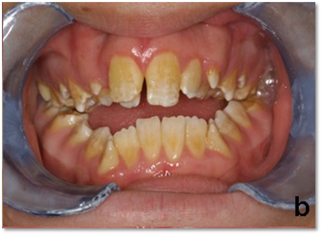
Amelogenesis Imperfecta (Figure 1)
Amelogenesis imperfecta (AI), a group of hereditary diseases affecting the tooth enamel in either quality or quantity, is associated with crown malformation and abnormal enamel density [6,7].
Classification and General Features
Weinmann et al 1945, subdivided amelogenesis imperfect into hypoplastic and hypocalcified types. Various classifications have been proposed but most commonly accepted classification is the one being proposed by Witkop 1988 which classified it into mainly 4 types, i.e [8].
IVA: Hypomaturation- Hypoplastic with Taurodontism, autosomal dominant
IVB: Hypoplastic- Hypomaturation with Taurodontism, autosomal dominant
Hypoplastic
Hypoplastic Amelogenesis Imperfecta occurs due to defect in enamel matrix deposition, i.e., the first stage of enamel formation.Patients arepresents with thin enamel with yellowish-brown, rough or smooth, flat occlusal surfaces of the posterior teeth due to attrition, and with/without grooves and/pitting. The enamel will be thin, well mineralized and do not chip. Radiographically thin enamel but normal radiodensity will be seen. Defects in matrix formation with a disturbance in the differentiation or viability of ameloblasts will be seen in histology section [6,9].
Hypomaturation Amelogenesis Imperfecta
In this type, Qualitative defect of the enamel is seen where the enamel is not sufficiently mineralized. The teeth are normal morphologically at the time of eruption, but eventually chip away posteruptively, especially in the occlusal areas. Clinically, the colour of teeth here varies from creamy opaque to marked yellow/brown. The surface of the teeth appear soft and rough leading to sensitivity due to dentinal exposure. Open bite malocclusion is a common feature. The enamel thickness is normal but often chips off and abrades away easily. Radiographically, there appears to be reduced differentiation between enamel and dentin which may be difficult to verify. Enamel has contrast similar to or greater than dentin, unerupted crowns have normal morphology radiographically.6,10
Hypocalcified Amelogenesis Imperfecta
Qualitative defect occurs when the enamel is insufficiently mineralized and soft. In comparison with hypo maturation type, the mineralization in this type is markedly reduced. Clinically, the crowns of the teeth appears to be opaque white to yellow-brown, soft rough enamel surface, dental sensitivity and very poor aesthetics. Due to severe hypomineralization, there may be early loss of enamel. The thickness of enamel appears to be normal at eruption that often chips and but, tends to abrade easily post eruptively. There may be delayed eruption of teeth. An anterior open bite of skeletal origin may be seen. Accumulation of a large amount of supragingival calculus is evident. There are two types, i.e., diffuse autosomal dominant (AD) and diffuse autosomal recessive (AR), with more severity in AR. Radiographically enamel is less radiopaque then dentin [9,10].
Hypomaturation-Hypoplastic with Taurodontism
Clinically, the crown appears to be white/yellow brown mottled. The teeth appear smaller than normal and they lack proximal contacts. In these cases, the enamel thickness is drastically reduced. The crowns show pitting and tend to have hypo mineralized areas. Radiographically, the enamel contrast is normal to slightly greater than dentin, and shows large or bulbous pulp chambers which appear taurodontic. In hypoplastic-hypomaturation with taurodontism, the enamel is thin, mottled yellow to brown, and pitted. Molar teeth exhibit taurodontism, and other teeth have enlarged pulp chambers [6,10].
Etiology2
Genetic, Febrile illness or Vitamin Deficiency, Local infection or Trauma, Fluoride Ingestion, Congenital syphilis, Birth defects or idiopathic factors responsible for Amelogenesis imperfecta.
Mutation or alteration in any of the genes encoding specific enamel proteins such as Enamelin gene (ENAM), Amelogenin gene (AMELX), Kallikrein 4 gene (KLK4), Matrix Metalloproteinase 20 gene (MMP‑20), and Distal‑less homeobox 3 gene (DLX3) have been linked with Amelogenesis Imperfecta.
Clinical Feature [6,9,10]
In the hypoplastic form, females show vertical ridging of the enamel, whereas, in males there is uniform hypoplasia.
In the hypomaturation form, males have teeth of normal size and shape, but with irregular, pigmented mottling. Females display vertical bands of mottling, often inconspicuous under normal lighting conditions.
Dentin and roots appear normal.
Amelogenesis Imperfecta with taurodontism is found to be associated with Trichodentoosseous (TDO) syndrome.
Histologically [9]
A ground section of the teeth involved showed very thin enamel, composed of laminations of irregularly arranged enamel prisms. The SEM studies of the extracted deciduous teeth, in a case of autosomal recessive rough hypoplastic amelogenesis imperfecta, showed an exposed outer enamel surface, with irregularly shaped globular protrusions.
Other Features Associated with Amelogenesis Imperfecta10
Amelogenesis Imperfecta may be associated with some other dental and skeletal developmental defects or abnormalities, such as crown and root resorption, attrition, microdontia, taurodontism, delayed eruption and tooth impaction, dens in dente, pulp stones, anterior open bite, and agenesis of teeth.
Diagnosis [10,11]
Accurate diagnosis requires a proper clinical history, clinical examination so that the presence of certain systemic diseases that may show generalized enamel hypoplasia can be excluded, identification of modes of inheritance determined by family pedigrees chart and proper radiographic interpretation. Accurate diagnosis enables genetic counseling in an early phase, and precautionary steps can be taken as an early step to prevent further dental complications for the patient and even for upcoming siblings in the future.
Management [7,10]
Management directed at three aspects of treatment includes prevention, restoration, and esthetics. Most of the time patient report to the dentist when the dental complication like dentinal sensitivity or dental caries would have been started, so restoration should be undertaken as a first step. The age and socioeconomic status of the patient, the type and severity of the disorder, and the intraoral situation at the time the treatment is planned. It includes removal of surface stains, reducing sensitivity, maintaining vertical dimension of occlusion and the esthetics.
Preventive aspects include dietary advice, regular use of fluoride mouthwashes, topical fluoride applications, and oral hygiene instructions. Oral hygiene can be difficult for these patients due to the sensitivity while brushing so warm water for tooth brushing can be advised. Along with preventive measures, long-term clinical follow-up term is mandatory.
Restorative aspects can be divided at the time of dentition, i.e., during primary dentition, direct composite veneers for anterior teeth, GIC and stainless steel crowns for primary molars can be advised while in the mixed dentition stainless steel crowns, onlays for permanent molars, composite or GICs for primary and permanent teeth and for permanent incisors, direct or indirect composite veneers should be advised. The minimal intervention GIC restorations should be advised in the primary dentition, direct and indirect composite resin veneers in the mixed dentition while porcelain veneers, full crowns, extractions of excessive defected teeth followed by fixed, or removable prosthesis should be advocated in permanent dentition based on number of teeth affected, patient age, and economical status. The accurate treatment should be planned based on the type of AI, severity and oral health habits of the patient. In cases of hypocalcified AI where the enamel is very weak and bonding of the restoration is questionable, full coverage is required. Anterior open bite can be treated with different treatment modalities ranging from orthodontic banding or orthognathic surgery.Orthodontic treatment can be performed only after once all restorative treatment is finished.
The esthetic appearance is the prime consideration. In some cases a lack of good enamel bonding of veneers occurs and does not result in a durable restoration. The use of glass ionomer cements with dentinal adhesives often overcomes this weakness.
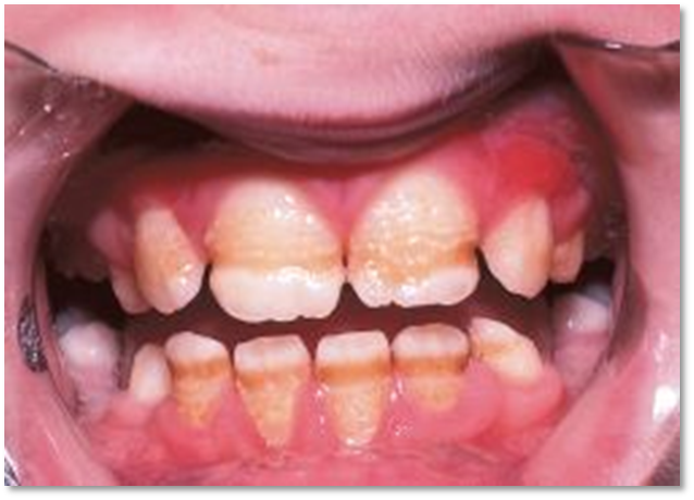
Enamel Hypoplasia (Figure 2)
Enamel Hypoplasia is defined as an incomplete or defective formation of the organic enamel matrix of teeth.12 Different types of enamel hypoplasia have been identified such as pit type, plane type and linear enamel type [13].
Etiology13
Enamel hypoplasia or hypo mineralization may be caused by hereditary factors and environmental factors that include systemic factors such as nutritional factors, exanthematous diseases like measles and chicken pox, congenital syphilis, hypocalcemia, birth injury or premature birth, fluoride ingestion or idiopathic causes, and local factors such as infection or trauma from a deciduous tooth.
Figure 1: Amelogenesis Imperfecta
Figure 2: Enamel Hypoplasia
Clinical Features
PindborgJJ, (1970)14 showed that 3 to 15% of young adults had enamel hypoplasia in the permanent teeth and Pedersen (1944) reported that 14% of 2- to 4-year-old children had mild enamel hypoplasia in the primary teeth.15
The spots may be localized or generalized and may involve only enamel or enamel and dentin.
Enamel hypoplasia due to trauma may vary from white to mild brownish discoloration of the enamel to severe pitting and irregularity of the crown of the tooth.16
Enamel hypoplasia presents with unfavourable aesthetics, increased dentin sensitivity, increased dental caries susceptibility, increased wear and malocclusion.12
It can occur in any permanent tooth, the most commonly involved sites of hypoplasia are the permanent first molars and incisors with specific areas of defect and well demarcated areas of hypomineralization.
When a deciduous tooth has been driven into the alveolus and has disturbed the permanent bud, it can manifest as a yellowish or brownish stain or pigmentation of the enamel usually on the labial surface or a true hypoplastic pitting defect or deformity.15
Environmental enamel hypoplasia/hypomineralization due to systemic factors are commonly manifested in the first permanent molars and incisors. It involves those teeth that form within the 1st year of birth. So, most frequently incisors and first permanent molars are affected. Hence, this condition is termed as “molar incisor hypomineralization.”
Environmental enamel hypoplasia / hypomineralization due to local factors is also called as “turners hypoplasia/ hypomineralization” seen most commonly in permanent maxillary incisors or upper lower premolar [15]
Similarly, when a deciduous tooth has been driven into the alveolus and has disturbed the permanent bud, it can manifest as a yellowish or brownish stain or pigmentation of the enamel usually on the labial surface or a true hypoplastic pitting defect or deformity [13].
Management [ 17,18,19]
A dental professional must be prepared to make the right diagnosis of these changes to determine the appropriate treatment. A differential diagnosis with other lesions that cause staining should be performed, including whether the white spots came from caries lesions, trauma, or posteruption wear. Adequate planning may ensure a conservative, effective and durable treatment.
Sometimes dental bleaching may be a conservative alternative, able to achieve a good result, even though many times the spots do not disappear completely.
In lesions involving enamel and dentin, either with or without the loss of structure, direct restorations may be indicated.
Invasive treatments should be avoided, especially in childhood. However, when the esthetic damages compromise the child’s social life, restorative treatment is indicated and should be as conservative as possible.
Composite resin is a restorative material alternative that may restore esthetics with high quality, minimal wear and durability. This procedure should be performed before restorative treatments using a low-concentration bleaching gel.
In pediatric patients, it is known that a bleaching treatment should follow the policy determined by the American Association of Pediatric Dentistry (AAPD), which has defined the judicious use of bleaching for vital and nonvital teeth in children. The procedure must be strictly controlled by the dentist, who should determine the appropriate method and timing of the treatment within the context of an individualized, comprehensive, and sequenced planning, while also considering the side effects of tooth whitening for children and adolescents.
By using the stratification technique, the opaque shades restored the affected dentin and reconstructed anatomical structures, such as dentin mamelons. When applied on the dentin composite, the incisal edge shade generates a counter-opalescent effect, reflecting an orange hue. Thicker layers of enamel resin generate more gray restorations because of the difference of the refractive index between the resin and the natural tooth. Therefore, each step of the treatment is essential for a successful outcome: diagnosis, planning, and execution.
Dentinogenesis Imperfecta (Figure 3)
It comprises of a group of autosomal dominant genetic conditions characterised by abnormal dentine structure affecting either the primary or both the primary and secondary dentitions [20]. It was recognized by WC Barrett (1882). The defect is primarily due to abnormal dentin was first recognized by Fargin-Foyelle and Malassez in 1908 [21].
Classification
The most familiar classification system is that formulated by Shields22 in 1973.
This categorisation discriminates three types of dentinogenesis imperfecta (types I, II and II).
The Shields' classification is summarised below:20,23
Dentinogenesis Imperfecta type I: Individuals with DGI-I have osteogenesis imperfecta. The teeth of both dentitions are typically amber and translucent and show significant attrition. Radiographically, the teeth have short, constricted roots and dentine hypertrophy leading to pulpal obliteration either before or just after eruption. Some teeth shows total pulpal obliteration while in others the dentine appears normal.
Dentinogenesis Imperfecta type II: The dental features of DGI-II are similar to those of DGI-I but penetrance is virtually complete and osteogenesis imperfecta is not a feature. Bulbous crowns are a typical feature with marked cervical constriction. Normal teeth are never found in DGI-II. Sensorineural hearing loss has also been reported as a rare feature of the condition.
Dentinogenesis Imperfecta type III: This is a form of DGI found in a tri-racial population from Maryland and Washington DC known as the Brandywine isolate. The clinical features are variable and resemble those seen in DGI-I and -II but the primary teeth show multiple pulp exposures and radiographically, they often manifest "shell" teeth i.e. teeth which appear hollow due to hypotrophy of the dentine.
The most current classification adopted by the Mendelian Inheritance is based on that of Shields but excludes DGI with osteogenesis imperfecta. Thus, the entity once termed DGI-II has now become DGI-I (MIM 125490), while the classification of DGI-III (MIM 125500) is unchanged.
Etiology
Mutations in the genes encoding the major protein constituents of dentine seem to underlie most hereditary dentine defects.
Figure 3: Dentinogenesis Imperfecta
Figure 4: Dentine Dysplasia
Dentinogenesis Imperfecta type I24: DGI-I is a syndromic form of DGI associated with osteogenesis imperfecta. Osteogenesis imperfecta is an autosomal dominant condition usually resulting from mis-sense mutations affecting either of the two genes encoding type I collagen (COL1A1 and COL1A2)
Dentinogenesis Imperfecta type II, Dentinogenesis Imperfecta type III25: The only mutations causative of DGI are found in the Dentine Sialophosphoproteingene (DSPP), suggesting that these conditionsare indeed allelic. DSPPis located within human chromosome 4q22.1 and consists of 5 exons spanning approximately 8343 bp.DSPP is expressed in a number of tissues including bone, kidney, salivary gland and lung but its expression in dentine is hundreds of times higher than in other tissues. Three distinct protein products are formed from the initially translated polypeptide: dentine sialoprotein (DSP) results from the cleavage of amino acids 16–374 of the nascent polypeptide, dentine glycoprotein (DGP) is constituted from amino acids 375– 462 and dentine phosphoprotein (DPP) is composed of the remaining amino acids of the nascent polypeptide
Clinical Features [21,23].
The affected teeth show a peculiar color deviation from the natural dentition that ranges from gray to brownish violet or yellowish brown, with a characteristic unusual translucent or opalescent hue
These teeth are prone to excessive wear and fracture due to the primary abnormality in the structure and composition of dentin and presumably abnormal dentin enamel junction that lacks normal scalloping
The caries incidence is low in these patients due to early wear of the fissures and contact points. There is an early attrition of dentin in deciduous teeth with hyperplasia of the residual ridges
Radiographically [21,26].
The teeth affected have partial or complete precocious obliteration of pulpal space. This is attributed to the continual dentin formation
Teeth have short and blunt or spike-like roots, and bulbous crowns with cervical constrictions, giving them a typical tulip appearance
The cementum, periodontal ligament and supporting bone are normal
Histologically [21,26].
The appearance of dentin in affected teeth shows sparse and irregular tubules, which may be irregular in shape, size, number, and course
Characteristically, a layer of mantle dentin is succeeded by irregularly formeddentin, often with large areas of uncalcified matrix (globular dentin)
The increased water content (as much as 60% of the normal) and decreased inorganic mineral content of dentin takes its hardness close to that of cementum
Diagnostic Methods
Clinical History20: A medical history should aim to establish if the dental condition is a 'syndromic' form of DGI as this is a variable feature of a number of heritable conditionsincluding Osteogenesis imperfecta, Ehlers Danlos sydrome, Goldblatt syndrome, Schimke immunoosseous dysplasia, Brachio-skeleto-genital syndrome, and osteodysplastic and primordial short stature with severe microdontia, opalescent teeth, and rootless molars. Since DGI may be the most penetrant clinical finding in individuals with DGI-I, it is very important to ask patients with DGI about histories of bone fracture with minimal trauma, joint hyperextensibility, short stature, hearing loss and scleral hue. Details such as colour, tooth wear, abscess formation, tooth mobility and early loss of primary teeth may help to establish what type of DGI the patient has. Dental history, experience and age often influence treatment options and mode of treatment
Clinical Examination20: Extraoral features such as short stature and blue sclera may be consistent with osteogenesis imperfecta. Intraorally, in both dentitions, it is important to consider tooth colour (which may vary from normal to amber, grey or purple to bluish translucent discolouration), tooth wear, abscess formation, tooth mobility and early loss of teeth. The tooth enamel may have sheared off leaving dentine exposed; in such cases the exposed dentine often has a hard glassy appearance due to sclerosis. For this reason, patients rarely complain of sensitivity
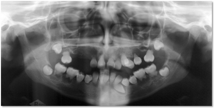
Radiographs [22]: Radiographs should reveal normal enamel and dentine radiodensity; however, the enamel may already be lost with only dentine remaining. Crowns may appear bulbous with marked cervical constriction. Pulp chambers and canals may be normal, contain pulp stones or, more often, be partially or totally obliterated. Roots are often short but may be of normal length or absent. There may be numerous periapical radiolucencies in non-carious teeth
Differential Diagnosis
Included in the differential diagnosis are conditions that have similar clinical or radiographic features to DGI.
Clinical [20,22].
Exposure of underlying dentine: Hypocalcified forms of amelogenesis imperfecta initially develop normal enamel thickness but the poorly calcified enamel is soft and friable and is rapidly lost by attrition leaving dentine cores. Unlike DGI the teeth are usually sensitive and on radiographs enamel is less radio-dense than dentine. Pulp chamber and root canals are usually not sclerosed.
Intrinsic discolouration: Congenital erythropoietic porphyria is a rare condition resulting from an inborn error of porphyrin metabolism. This deficiency leads to haemolytic anaemia, photosensitivity, blistering of the skin, and deposition of red-brown pigments in the bones and teeth. A number of prenatal and neonatal enamel discolourations and hypoplasias are due to neonatal haemolytic anaemias. Most cases are due to Rhesus incompatibility. The discolouration which ranges from yellow through to green, brown and grey to black is usually found at the necks of teeth and the enamel hypoplasias are usually located in the coronal third of the teeth. Tetracyclines have the ability to chelate calcium ions and to be incorporated into developing teeth, cartilage and bone, resulting in discolouration of both the primary and permanent dentitions. This permanent discolouration varies from yellow or grey to brown depending on the dose or the type of the drug received in relation to body weight.
Mobility leading to early tooth loss: Other causes of early loss of teeth as in DGI-III and DD-I include: Hypophosphatasia, Immunological deficiencies e.g. severe Congenital Neutropenia (Kostmann's disease), Cyclic neutropenia, Chediak-Hegashi syndrome, Neutropenias, Histiocytosis X, Papillon-Lefevre syndrome and leucocyte adhesion deficiency syndrome. With the exception of hypophosphatasia, all of these conditions have an underlying immunological defect which makes those with these conditions susceptible to periodontal breakdown. Mobility of teeth in those with hypophosphatasia however is due to aplasia or marked hypoplasia of cementum
Radiographic [22].
Regional odontodysplasia is of unknown aetiology. Radiographically, roots are short with wide open apices and very wide pulp canals, and often become infected as in DGI-III . Primary and permanent teeth are affected.
Clinical and radiographic [20].
Vitamin D-dependent rickets and vitamin D-resistant rickets have clinical and radiographic features of DGI. It is characterised by yellowish to brown enamel, chronic periodontal disease, large quadrangular pulp chambers and short roots
Vitamin D-resistant rickets include attrition and exposure of abnormally formed dentine of primary teeth and abscessed non-carious primary or permanent teeth. As previously mentioned, DGI-I is a variable feature of Ehlers Danlos syndrome, Goldblatt syndrome, Schimke immuno-osseous dysplasia and Brachioskeleto genital syndrome and osteodysplastic and primordial short stature with severe microdontia, opalescent teeth, and rootless molars
Management [20,23,26].
The aims of treatment are to remove sources of infection or pain, restore aesthetics and protect posterior teeth from wear. Treatment varies according to the age of the patient, severity of the problem and the presenting complaint.
In the primary dentition, stainless steel crowns on the molars may be used to prevent tooth wear and maintain the occlusal vertical dimension. The aesthetics may be improved using composite facings or composite strip crowns. If, however the child presents late, the teeth may have undergone attrition to the level of the gingivae and the only treatment option then is to provide over-dentures. If abscesses develop, pulp therapy is not successful and removal of the affected teeth is required
In younger children, where co-operation is limited, or the level of treatment required is extensive, a general anaesthetic may be required to facilitate treatment. In some cases, the parents or child may not be concerned with aesthetics and may request removal of sources of pain or infection only
As the permanent dentition erupts, it should be closely monitored in relation to the rate of tooth wear with intervention only if necessary. Cast occlusal onlays on the first permanent molars and eventually the premolars, help to minimise tooth wear and maintain the occlusal vertical dimension. Obliteration of the pulp chambers and root canals in teeth that develop abscesses makes endodontic therapy difficult if not impossible. If conventional therapy is not an option, periapical curettage and retrograde root filling is another possible alternative, but is not recommended for teeth with short roots
Dental implants may be considered when growth is complete at about 18 years of age. Maxillo-mandibular atrophy is a consequence of no or rudimentary root development and early tooth loss. Ridge augmentation prior to implants is often required
Exposed dentine is more susceptible to tooth decay than enamel. For all patients, regular dental checkups and prevention of tooth decay in the form of oral hygiene instruction, dietary advice and appropriate use of fluoride is essential.
Prognosis [20].
The outcome of a diagnosis of DGI/DD largely depends upon the age at which the diagnosis was given and the speed and quality of the treatment provided. Where diagnosis occurs early in the life good aesthetics and function can be obtained thereby minimizing nutritional deficits and psychosocial distress.
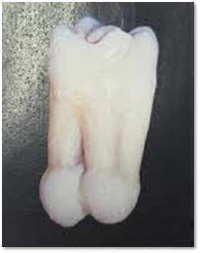
Dentine Dysplasia (Figure 4)
Dentin dysplasia (DD) is an autosomal dominant hereditary disturbance in dentin formation, which may present with either mobile teeth or pain associated with spontaneous dental abscesses or cysts. It was Ballschmiede in 1920 who first reported such a condition as ‘rootless teeth’ and in 1939 Rushton termed this condition as Dentin dysplasia.27,28
Classification
In 1988, Witkop8 classified Dentin Dysplasia into two types, radicular DD as type I and coronal DD as type II.
In type I, both the deciduous and permanent dentitions are affected. The crowns of the teeth appear clinically normal in morphology but defects in dentin formation and pulp obliteration are present. Radiographic examination is important for the identification of DD type I. There are four subtypes for this abnormality
In type 1a, there is no pulp chamber and root formation, and there are frequent periradicular radiolucencies
In type 1b has a single small horizontally oriented and crescent shaped pulp, and roots are only a few millimeters in length and there are frequent periapical radiolucencies
In type 1c, there are two horizontal or vertical and crescent shaped pulpal remnants surrounding a central island of dentine and with significant but shortened root length and variable periapical radiolucencies
In type 1d, there is a visible pulp chamber and canal with near-normal root length, and large pulp stones that are located in the coronal portion of the canal and create a localized bulging in the canal, as well as root constriction of the pulp canal apical to the stone and few peri-apical radiolucencies [27]
Histologically28,29
The enamel and the immediately subjacent dentin appear normal. Deeper layers of dentin show an atypical tubular pattern with an amorphous, atubular area and irregular organization. Pulpally to normal appearing mantle dentin, and globular or nodular masses of abnormal dentin are seen.
DD type II is characterized by yellow, brown, grey amber, translucent primary teeth with complete pulpal obliteration. The permanent teeth have a normal appearance or might be slightly amber colored. The radiological evidence of enlarged pulp chamber extending into roots, thistle tube appearance of pulp chamber, and root canal in multiple teeth along with presence of pulp stones and normal root length were typical of DD type II. The histopathological evidence of irregularly arranged dentinal tubules along with presence of entrapped lacunae suggestive of osteodentin confirmed our diagnosis of DD type II.
Pathogenesis
It is still unknown in the dental literature.
Sauk et al in a scanning electron microscope study, postulated that dentin dysplasia is a defect in the epithelial component of the developing tooth germ in which the invagination of the root sheath occurs too soon and, in a sequence of futile attempts to correct itself, results in a stunted root form with an unusual whorl like pattern of dentin obliterating the pulp chambers [30]
Witkop suggested that the dysplasia results from epithelial cells from the sheath of Hertwig breaking off and migrating into dental papilla, where they induce odontoblast differentiation and dentin formation [8]
Melnick et al suggested that the abnormal root morphology is caused by abnormal differentiation and/or function of the odontoblasts. Clearly, the exact etiology of dentin dysplasia has yet to be explored [30]
Systemic Diseases [28,30]
It is correlated with dentin dysplasia like alterations:
Calcinosis Universalis: Deposition of calcium salts in skin, subcutaneoustissue, tendons and muscles. Clinically, may have arthralgia to movement limitation
Rheumatoid Arthritis and Vitaminosis D: It is an autoimmune disease characterized by symmetric, inflammatory arthritis of small and large joints with constitutional symptoms including fatigue, weight loss, morning stiffness, low grade fever and anemia.
Sclerotic Bone and Skeletal Anomalies: Patients with teeth showing all features ofradicular dentine dysplasia (type I) have been found to have dense sclerotic bone and skeletal anomalies of the wrists and hand bones.
Tumoral Calcinosis: Calcium deposition in the soft tissue in periarticular location that is around joints. Frequently seen in patients undergoing renal dialysis.
Management [28,30,31]
The patients with dentinal dysplasia have presented dentists with many problems. Extraction has been suggested as a treatment alternative for teeth with pulp necrosis and periapical abscess. Follow-up and routine conservative treatment is another choice of treatment plan.
Another approach for the treatment of teeth included periapical surgery and retrograde filling, which is recommended in teeth with long roots.
Premature loss of teeth and loose teeth as a result of rudimentary or absence of roots has presented dentists with many problems in management of patients with Dentin dysplasia I.
Tooth wear seen in primary dentition can be maintained with stainless steel crowns to prevent tooth wear and maintain occlusal dimension. Composite facings or composite strip crowns can be added for esthetic reasons on demand.
In permanent dentition, conserving existing health of teeth may demand endodontic intervention. Obliteration of the pulp chambers and root canals in teeth that develop abscesses makes endodontic therapy difficult. If conventional endodontic therapy is not possible, periapical curettage and retrograde filling is another alternative but not recommended in teeth with too short roots. Teeth with short thin roots and marked cervical constrictions, however, are often unfavorable for crowns. If abscess develops and pulp therapy is not successful, removal of the affected teeth is required and full mouth rehabilitation is considered with dentures or over dentures until growth is complete. In case of few teeth having enough root development, cast partial dentures can be a treatment option.
Dental implants should be considered when growth is complete. Ridge augmentation procedures need to be carried out prior to implant placement in maxillo‑mandibular alveolar atrophy due to early loss of teeth. A combination of onlay bone grafting and a sinus lift technique to accomplish implant placement can be done.
Regional Odontodysplasia (Figure 5)
Regional odontodysplasia (ROD) is uncommon developmental anomaly, which tends to be localised and involves the ectodermal and mesodermal tooth components [32]. The first report of this condition was published by McCall et al., in 1947, but the term “odontodysplasia” was introduced by Zegarelli et al in 1963.33,34
Etiology [32,33]
The etiology remains uncertain. This included somatic mutation of the dental lamina, viral infection, medication taken during pregnancy, local circulating disorder like vascular nevi on the skin of the affected side of the face, failure of migration, and differentiation of neural crest cells.
Figure 5: Regional Odontodysplasia
Figure 6: Hpercementosis
Clinical Features [32,33,34]
Pain and dental abscess formation which can be seen even in the absence of dental caries.
Delay or failure of eruption, gingival swelling, and abnormal clinical appearance of the teeth.
Teeth are usually smaller than normal with a rough surface texture and extensive pits and grooves.
The enamel is hypocalcified and/or hypoplastic with a yellow or brown discolouration and can be soft on exploration with a dental probe.
Affected teeth are more susceptible to dental caries due to defective mineralisation.
Clinically, affected teeth have an abnormal morphology and a rough surface with defective mineralization.
It occurs in both deciduous and permanent dentitions, and it has a marked preference for the maxilla.
Female is slightly more affected than male at ratio of 1.4:1.
Affects one quadrant of the jaw, although it does occasionally cross the midline.
Affected teeth are usually in a continuous series, although it may skip a tooth or a group of teeth.
Primary teeth affected by this anomaly are usually followed by affected permanent successors; however it is very rare to find normal permanent teeth to follow affected primary ones.
Radiographically [32,34]
A marked reduction in the radiodensity of the enamel and dentine can be seen, which leads to a poor contrast between them. Classically, a “ghost-like appearance”(thus the name ghost teeth) of the teeth is observed. These teeth present with a shorter appearance, wide open apices, and a large pulp chamber.
Histologically [34]
The dental hard tissues are characterized as hypocalcified. Enamel prisms are irregularly distributed, with aprismatic and degenerative hypocalcifiedareas. There is a marked reduction in the dentinal tubules, and areas of clefts can be seen within them. The pulp and dental follicle may contain calcifications of varying degrees.
Management
Treatment of Regional Odontodysplasia remains a clinical dilemma as it is controversial with lack of consensuses in managing this anomaly. The management involves interventional dental care in both dentitions and it requires multidisciplinary care. The treatment is aimed at improving function and esthetics, reducing the psychological impact of early tooth loss and facilitating normal jaw growth.
Early extraction of the affected teeth has been proposed by many authors (M.A. Hamdan,2004)35 as these teeth might develop dental pathology even in the absence of dental caries due to the thin enamel layer and the presence of enamel and dentinal cleft which allow ingress of microorganism to the dental pulp
The defective mineralisation of the involved teeth results in undesirable appearance and poor dental aesthetics. The extraction was followed in some cases by prosthetic replacement. Some authors( A.Cahuana, 36 have argued for maintaining the noninfected affected teeth to allow normal jaw development and reduce the risk of psychological trauma associated with premature tooth loss. Other treatment approaches in ROD include coverage restorations and autotransplantation of teeth in the permanent dentition [32]
Cahuna et al 36 proposed autotransplantation as a treatment option in RO patients; however it is limited because of availability of an appropriate donor
Bulut et al suggested the use of endosseous implant for oral rehabilitation in patients with odontodysplasia, however, this treatment can be done at a later stage of life.The treatment should be considered based upon the patient’s age, compliance, attitude towards the dental treatment, and the degree of odontodysplasia [34]
Hypercementosis (Figure 6)
Hypercementosis is a non-neoplastic condition characterised by excessive deposition of cementum on the roots of teeth.37 Although some cases of hypercementosis are idiopathic, certain circumstances favor the association with hypercementosis, including the following:
Supra-eruption of a tooth because of the loss of an antagonist tooth
Inflammation at the apex of a tooth
Traumatic occlusion
Systemic diseases such as Paget’s disease, toxic goiter, acromegaly, and gigantism [38]
Classification [39]
It is a form of cemental hyperplasia which can be generalized or localized.
Generalized hypercementosis is characterized by increased thickness of cementum involving all teeth and is a classical feature of Paget’s disease. Other systemic disturbances associated with hypercementosis include acromegaly, arthritis, calcinosis, rheumatic fever and thyroid goiter
Localized hypercementosis affects single tooth and usually presents as generalized thickening of cementum with nodular thickening of apical third of root
Etiology
The aetiopathogenesis of hypercementosis is ambiguous. This condition is idiopathic in most cases, but may also be associated with several local or systemic factors.
The local factors implicated to cause hypercementosis are occlusal trauma, inflammation secondary to pulpal or periodontal disease, tooth mobility repair of root fracture and transplantation of teeth [37].
Supra-eruption of a tooth
Periapical inflammation resulting from pulpal infection sometimes stimulates excessive formation of cementum
Rushton and Cooke stated that mild traumatic occlusion may cause hypercementosis [38]
As with resorption, a direct causal relationship with periodontal diseases is not proven, but hypercementosis is seen occasionally on teeth with bone loss [38]
Systemic conditions associated with hypercementosis are acromegaly, goitre, arthritis, rheumatic fever, calcinosis, Gardner’s syndrome, Paget’s disease and vitamin A deficiency [37].
Osteitis deformans or Paget’s disease of bone is a generalized skeletal disease characterized by deposition of excessive amounts of secondary cementum on the roots of the teeth and by the apparent disappearance of the lamina dura of the teeth, as well as by other features related to the bone itself [38]
Clinical Features [37,38]
It may affect a single tooth or multiple teeth.
The condition is asymptomatic and is detected on radiographic examination
The most frequently affected teeth are mandibular molar, followed by mandibular and maxillary second premolars and mandibular first premolars. However, some authors have found premolars to be the most commonly affected teeth
Frequency of about 4.9% in population, hypercementosis may be considered as a relatively common finding
Radiographically38
The excess cementum may be of two types
The secondary cementum is of the same density as the primary cementum and dentin.
ii)The secondary cementum appears less dense and is clearly differentiated from the primary cementum and dentin.
Root areas affected by hypercementosis are separated from the periapical bone by a normal-appearing periodontal ligament space; the surrounding lamina dura appears normal as well.
Differential Diagnosis [38]
It include any radiopaque structure that is seen in the vicinity of the root, such as
A dense bone island or mature cemento-osseous dysplasia
There may be a resemblance to a small cementoblastoma
Occasionally, a severely dilacerated root may have the appearance of hypercementosis
Management37
Hypercementosis generally requires no active treatment. However, any interventional dental therapy, be it extraction of teeth, endodontic therapyor orthodontic therapy,will require considerable precautionary measures to avoid complications owing to this condition.
The only practical clinical significance of hypercementosis is the difficulties that may be encountered in extracting such teeth. Sectioning of the tooth may be necessary in certain cases to aid in removal
During endodontic treatment various difficulties may be encountered so the clinician needs to know the limits for root canal shaping and filling to obtain endodontic treatment success
CONCLUSION
Developmental anomalies of teeth are clinically evident abnormalities. Some of the developmental disorders develop during intrauterine development and are manifested at the time of birth. Careful observation and appropriate investigations are required to diagnose the condition and institute appropriate treatment. Clinical and radiographic evaluation of these conditions is essential so as to provide the best treatment modality for these patients and to prevent further complications in head and neck region due to the same. These developmental disorders also have impact on esthetics thereby affecting psychosocial behavior of the patient; hence the patients should also be managed on psychological basis.
REFERENCE
Rohilla, M. "Etiology of Various Dental Developmental Anomalies: Review of Literature." Journal of Dental Problems and Solutions, vol. 4, no. 2, 2017, pp. 19–25.
Kerr, Ash. An Introduction to General and Oral Pathology for Hygienist. 5th ed., Lea and Febiger, 1986. Link.
Shrestha, A., et al. "Developmental Anomalies Affecting the Morphology of Teeth: A Review." RSBO, vol. 12, no. 1, 2015, pp. 68–78.
Jahanimoghadam, F. "Dental Anomalies: An Update." Advances in Human Biology, vol. 6, 2016, pp. 112–18.
Seow, W.K. "Developmental Defects of Enamel and Dentine: Challenges for Basic Science Research and Clinical Management." Australian Dental Journal, vol. 59, no. 1, 2014, pp. 143–54.
Roma, M., and D.S. Hegde. "Amelogenesis Imperfecta: A Review of the Literature." Journal of Pharmaceutical Science and Research, vol. 8, no. 9, 2016, pp. 1042–1044.
Mehta, D.N., et al. "Amelogenesis Imperfecta: Four Case Reports." Journal of Natural Science, Biology and Medicine, vol. 4, no. 2, 2013, pp. 462–465.
Witkop, C.J., Jr. "Amelogenesis Imperfecta, Dentinogenesis Imperfecta, and Dentin Dysplasia Revisited: Problems in Classification." Journal of Oral Pathology, vol. 17, 1988, pp. 547–53.
Chaudhary, M., et al. "Amelogenesis Imperfecta: Report of a Case and Review of Literature." Journal of Oral and Maxillofacial Pathology, vol. 13, no. 2, 2009, pp. 70–77.
Shivhare, P., et al. "Amelogenesis Imperfecta: A Review." Journal of Advanced Oral Research, vol. 7, no. 1, 2016.
Gadhia, K., et al. "Amelogenesis Imperfecta: An Introduction." British Dental Journal, vol. 212, no. 8, 2012, pp. 377–79.
Sandhu, M., et al. "Circular Enamel Hypoplasia: A Rare Enamel Developmental Disturbance in Permanent Teeth." Journal of Clinical and Diagnostic Research, vol. 8, no. 8, 2014, pp. 39–40.
Kanchan, T., et al. "Enamel Hypoplasia and Its Role in Identification of Individuals: A Review of Literature." Indian Journal of Dentistry, vol. 6, no. 2, 2015, pp. 99–102.
Pindborg, J.J. Pathology of the Dental Hard Tissues. 1970, pp. 88–89.
Nikiforuk, G., and D. Fraser. "The Etiology of Enamel Hypoplasia: A Unifying Concept." Journal of Pediatrics, vol. 98, no. 6, 1981, pp. 888–93.Andreasen JO, Andreasen FM. Textbook and color atlas of traumatic injuries to the teeth.1994.
Nahsan, F.P., et al. "Conservative Approach for a Clinical Resolution of Enamel White Spot Lesions." Quintessence International, vol. 42, no. 5, 2011, pp. 423–26.
Carvalho, L.D., et al. "Hypoplastic Enamel Treatment in Permanent Anterior Teeth of a Child." Operative Dentistry, vol. 38, no. 4, 2013, pp. 363–68.
Ribas, A.O., and G.D. Czlusniak. "Anomalies in Dental Enamel: Etiology, Diagnostic, and Treatment." Publicações da UEPG Ciências Biológicas e da Saúde, vol. 10, no. 1, 2004, pp. 23–36.
Barron, M.J., et al. "Hereditary Dentine Disorders: Dentinogenesis Imperfecta and Dentine Dysplasia." Orphanet Journal of Rare Diseases, vol. 3, 2008, p. 31.
Bhandari, S., and K. Pannu. "Dentinogenesis Imperfecta: A Review and Case Report of a Family Over Four Generations." Indian Journal of Dental Research, vol. 19, no. 4, 2008, pp. 357–61.
Shields, et al. "A Proposed Classification for Heritable Human Dentine Defects with a Description of a New Entity." Archives of Oral Biology, vol. 18, 1973, pp. 543–53.
Sapir, S., and J. Shapira. "Dentinogenesis Imperfecta: An Early Treatment Strategy." Pediatric Dentistry, vol. 23, no. 3, 2001, pp. 232–37.
Martin, E., and J.R. Shapiro. "Osteogenesis Imperfecta: Epidemiology and Pathophysiology." Current Osteoporosis Reports, vol. 5, 2007, pp. 91–97.
MacDougall, M., et al. "Assignment of Dentin Sialophosphoprotein (DSPP) to the Critical DGI2 Locus on Human Chromosome 4 Band q21.3 by In Situ Hybridization." Cytogenetics and Cell Genetics, vol. 79, 1997, pp. 121–22.
Subramaniam, P., et al. "Dentinogenesis Imperfecta: A Case Report." Journal of the Indian Society of Pedodontics and Preventive Dentistry, vol. 26, no. 2, 2008, pp. 85–87.
Toomarian, L., et al. "Dentin Dysplasia Type I: A Case Report and Review of the Literature." Journal of Medical Case Reports, vol. 4, 2010, p. 1.
Fulari, S.G., and D.P. Tambake. "Rootless Teeth: Dentin Dysplasia Type I." Contemporary Clinical Dentistry, vol. 4, no. 4, 2013, pp. 520–22.
Daryani, D., et al. "Dentin Dysplasia Type II: An Exclusive Report of Two Cases in Siblings." Journal of the Indian Academy of Oral Medicine and Radiology, vol. 29, 2017, pp. 132–34.
Malik, S., et al. "Dentin Dysplasia Type I: A Rare Entity." Journal of Oral and Maxillofacial Pathology, vol. 19, no. 1, 2015, p. 110.
Logan, J., et al. "Dentinal Dysplasia." Oral Surgery, vol. 15, 1962, pp. 317–33.Al- Mullahi AM, Toumba KJ. Regional Odontodysplasia with Generalised Enamel Defect. Case Rep Dent 2016(2):1-5.
Desai, V.D., et al. "Regional Odontodysplasia with Supernumerary Teeth in Pediatric Patients: Coincident/New Finding?" Chrismed Journal of Health Research, vol. 2, no. 3, 2015, p. 272.
Malhotra, R., et al. "Regional Odontodysplasia: A Classical Case Report." SRM Journal of Research in Dental Sciences, vol. 4, no. 2, 2013, p. 86.
Hamdan, M.A., et al. "Regional Odontodysplasia: A Review of the Literature and Report of a Case." International Journal of Paediatric Dentistry, vol. 14, no. 5, 2004, pp. 363–70.
Cahuana, A., Y. Gonzalez, and C. Palma. "Clinical Management of Regional Odontodysplasia." Pediatric Dentistry, vol. 27, no. 1, 2005, pp. 34–39.
Shoor, H., et al. "Hypercementosis: A Rare Finding in a Patient with Systemic Lupus Erythematosus." BMJ Case Reports, 2014, pp. 1–3.
Raghavan, V., and C. Singh. "Hypercementosis: Review of Literature and Report of a Case of Mammoth, Dumbbell-Shaped Hypercementosis." Journal of the Indian Academy of Oral Medicine and Radiology, vol. 27, 2015, pp. 160–63.
Jeddy, N., et al. "Localized Multiple Cemental Excrescences: A Rare Presentation of Hypercementosis." Journal of Clinical and Diagnostic Research, vol. 8, no. 5, 2014, pp. 16–17.
License
Creative Commons Attribution-NonCommercial-NoDerivatives 4.0 International License
All papers should be submitted electronically. All submitted manuscripts must be original work that is not under submission at another journal or under consideration for publication in another form, such as a monograph or chapter of a book. Authors of submitted papers are obligated not to submit their paper for publication elsewhere until an editorial decision is rendered on their submission. Further, authors of accepted papers are prohibited from publishing the results in other publications that appear before the paper is published in the Journal unless they receive approval for doing so from the Editor-In-Chief.
Himalayan Journal of Applied Medical Sciences and Research open access articles are licensed under a Creative Commons Attribution-Share A like 4.0 International License. This license lets the audience to give appropriate credit, provide a link to the license, and indicate if changes were made and if they remix, transform, or build upon the material, they must distribute contributions under the same license as the original.
Recommended Articles
Research Article
Study of the Role of Serum Interleukin-6 in Inflammation among Pregnant Women with COVID-19
Aynar Talib Samad,
Yossra Saleh Khudhur
Published: 27/07/2021
Download PDF
Cite
x
APA
Talib Samad, A. & Khudhur, Y. S. (2021). Study of the Role of Serum Interleukin-6 in Inflammation among Pregnant Women with COVID-19. Himalayan Journal of Applied Medical Sciences and Research, 2(2), 1-6.
MLA
Talib Samad, Aynar and Yossra Saleh Khudhur. "Study of the Role of Serum Interleukin-6 in Inflammation among Pregnant Women with COVID-19." Himalayan Journal of Applied Medical Sciences and Research 2.2 (2021): 1-6.
Chicago
Talib Samad, Aynar and Yossra Saleh Khudhur. "Study of the Role of Serum Interleukin-6 in Inflammation among Pregnant Women with COVID-19." Himalayan Journal of Applied Medical Sciences and Research 2, no. 2 (2021): 1-6.
Harvard
Talib Samad, A. and Khudhur, Y. S. (2021) 'Study of the Role of Serum Interleukin-6 in Inflammation among Pregnant Women with COVID-19' Himalayan Journal of Applied Medical Sciences and Research 2(2), pp. 1-6.
Vancouver
Talib Samad A, Khudhur YS. Study of the Role of Serum Interleukin-6 in Inflammation among Pregnant Women with COVID-19. Himalayan Journal of Applied Medical Sciences and Research. 2021 Jul;2(2):1-6.
Download PDF
Research Article
Bones in Balance: Awareness of Rickets and Nutritional Deficiencies Among the Hamirpur Community
Vishal Dhatwalia,
Swati Chandel
Published: 10/12/2024
Download PDF
Cite
x
APA
Dhatwalia, V. & Chandel, S. (2024). Bones in Balance: Awareness of Rickets and Nutritional Deficiencies Among the Hamirpur Community. Himalayan Journal of Applied Medical Sciences and Research, 5(2), 1-5.
MLA
Dhatwalia, Vishal and Swati Chandel. "Bones in Balance: Awareness of Rickets and Nutritional Deficiencies Among the Hamirpur Community." Himalayan Journal of Applied Medical Sciences and Research 5.2 (2024): 1-5.
Chicago
Dhatwalia, Vishal and Swati Chandel. "Bones in Balance: Awareness of Rickets and Nutritional Deficiencies Among the Hamirpur Community." Himalayan Journal of Applied Medical Sciences and Research 5, no. 2 (2024): 1-5.
Harvard
Dhatwalia, V. and Chandel, S. (2024) 'Bones in Balance: Awareness of Rickets and Nutritional Deficiencies Among the Hamirpur Community' Himalayan Journal of Applied Medical Sciences and Research 5(2), pp. 1-5.
Vancouver
Dhatwalia V, Chandel S. Bones in Balance: Awareness of Rickets and Nutritional Deficiencies Among the Hamirpur Community. Himalayan Journal of Applied Medical Sciences and Research. 2024 Jul;5(2):1-5.
Download PDF
Research Article
Assessing First Trimester Maternal Serum Pentraxin-3 Levels in Primary Unexplained Recurrent Pregnancy Loss: A Case-Control Study
Raniah Ibrahim Khaleel Yaseen,
Israa Hashim Abdalkareem
Published: 28/02/2026
Download PDF
Cite
x
APA
Yaseen, R. I. K. & Abdalkareem, I. H. (2026). Assessing First Trimester Maternal Serum Pentraxin-3 Levels in Primary Unexplained Recurrent Pregnancy Loss: A Case-Control Study. Himalayan Journal of Applied Medical Sciences and Research, 7(1), 1-6.
MLA
Yaseen, Raniah I. K. and Israa H. Abdalkareem. "Assessing First Trimester Maternal Serum Pentraxin-3 Levels in Primary Unexplained Recurrent Pregnancy Loss: A Case-Control Study." Himalayan Journal of Applied Medical Sciences and Research 7.1 (2026): 1-6.
Chicago
Yaseen, Raniah I. K. and Israa H. Abdalkareem. "Assessing First Trimester Maternal Serum Pentraxin-3 Levels in Primary Unexplained Recurrent Pregnancy Loss: A Case-Control Study." Himalayan Journal of Applied Medical Sciences and Research 7, no. 1 (2026): 1-6.
Harvard
Yaseen, R. I. K. and Abdalkareem, I. H. (2026) 'Assessing First Trimester Maternal Serum Pentraxin-3 Levels in Primary Unexplained Recurrent Pregnancy Loss: A Case-Control Study' Himalayan Journal of Applied Medical Sciences and Research 7(1), pp. 1-6.
Vancouver
Yaseen RIK, Abdalkareem IH. Assessing First Trimester Maternal Serum Pentraxin-3 Levels in Primary Unexplained Recurrent Pregnancy Loss: A Case-Control Study. Himalayan Journal of Applied Medical Sciences and Research. 2026 Jan;7(1):1-6.
Download PDF
Research Article
Evaluation of Serum Heat Shock Protein 70 as a Diagnostic Biomarker for Ectopic Pregnancy: A Comparative Case-Control Study
Hala Ahmed Atiya,
Ayla Khedher Ghalib
Published: 28/02/2026
Download PDF
Cite
x
APA
Atiya, H. A. & Ghalib, A. K. (2026). Evaluation of Serum Heat Shock Protein 70 as a Diagnostic Biomarker for Ectopic Pregnancy: A Comparative Case-Control Study. Himalayan Journal of Applied Medical Sciences and Research, 7(1), 1-5.
MLA
Atiya, Hala A. and Ayla K. Ghalib. "Evaluation of Serum Heat Shock Protein 70 as a Diagnostic Biomarker for Ectopic Pregnancy: A Comparative Case-Control Study." Himalayan Journal of Applied Medical Sciences and Research 7.1 (2026): 1-5.
Chicago
Atiya, Hala A. and Ayla K. Ghalib. "Evaluation of Serum Heat Shock Protein 70 as a Diagnostic Biomarker for Ectopic Pregnancy: A Comparative Case-Control Study." Himalayan Journal of Applied Medical Sciences and Research 7, no. 1 (2026): 1-5.
Harvard
Atiya, H. A. and Ghalib, A. K. (2026) 'Evaluation of Serum Heat Shock Protein 70 as a Diagnostic Biomarker for Ectopic Pregnancy: A Comparative Case-Control Study' Himalayan Journal of Applied Medical Sciences and Research 7(1), pp. 1-5.
Vancouver
Atiya HA, Ghalib AK. Evaluation of Serum Heat Shock Protein 70 as a Diagnostic Biomarker for Ectopic Pregnancy: A Comparative Case-Control Study. Himalayan Journal of Applied Medical Sciences and Research. 2026 Jan;7(1):1-5.
Kour, P., Kaur, A., Singh, N., Singh, R., Kumar, S. & Chauhan, M. (2022). Developmental Anomalies of Tooth Structure: A Comprehensive Review. Himalayan Journal of Applied Medical Sciences and Research, 3(1), 1-13.
MLA
Kour, Puneet, et al. "Developmental Anomalies of Tooth Structure: A Comprehensive Review." Himalayan Journal of Applied Medical Sciences and Research 3.1 (2022): 1-13.
Chicago
Kour, Puneet, Avninder Kaur, Neetika Singh, Reetu Singh, Sanchit Kumar and Madhvi Chauhan. "Developmental Anomalies of Tooth Structure: A Comprehensive Review." Himalayan Journal of Applied Medical Sciences and Research 3, no. 1 (2022): 1-13.
Harvard
Kour, P., Kaur, A., Singh, N., Singh, R., Kumar, S. and Chauhan, M. (2022) 'Developmental Anomalies of Tooth Structure: A Comprehensive Review' Himalayan Journal of Applied Medical Sciences and Research 3(1), pp. 1-13.
Vancouver
Kour P, Kaur A, Singh N, Singh R, Kumar S, Chauhan M. Developmental Anomalies of Tooth Structure: A Comprehensive Review. Himalayan Journal of Applied Medical Sciences and Research. 2022 Jan;3(1):1-13.