Contents
Download PDF
pdf Download XML
1750 Views
54 Downloads
Share this article
Research Article | Volume 3 Issue 2 (July-Dec, 2022) | Pages 1 - 6
Mycetoma of the Foot Leading to Amputation - A Case Report
 ,
 ,
 ,
 ,
1
Assistant Professor, Department of Pathology, AMU, Aligarh, Uttar Pradesh, India, 202001.
2
Chairman and Professor, Department of Pathology, AMU, Aligarh, Uttar Pradesh, India, 202001.
3
Junior Resident, Department of Pathology, AMU, Aligarh, Uttar Pradesh, India, 202001.
4
Senior Resident, Department of Pathology, AMU, Aligarh, Uttar Pradesh, India, 202001.
Under a Creative Commons license
Open Access
Received
April 20, 2022
Revised
May 10, 2022
Accepted
July 30, 2022
Published
Aug. 10, 2022
Abstract

Introduction: Mycetoma is an infective process leading to numerous draining sinuses with exudates containing coloured grains. It can be caused both by bacteria as well as fungi. In this case report we present a 24-year female who complained of multiple discharging sinuses from the left foot. She was examined and was being conservatively treated but increased pain for the last one month. Her left leg was amputated as she was not responding to any treatment. Clinical history, radiology and histopathology were used to diagnose the condition. Conclusion: Madura foot must be kept in mind in patients presenting with discharging sinuses as it is notorious for its chronicity.

Keywords
IMPORTANT

Key findings:

Key findings from this case report on mycetoma include: A 24-year-old female presented with multiple discharging sinuses from her left foot, leading to amputation due to non-responsiveness to conservative treatment. The diagnosis was confirmed using clinical history, radiology, and histopathology, highlighting the chronic nature and severity of Madura foot infections.


What is known and what is new?

The known aspect in this abstract is the existence of mycetoma, a chronic infectious disease characterized by discharging sinuses and grains. The new contribution is the presentation of a specific case of a 24-year-old female with Madura foot, emphasizing the importance of considering this condition in patients with chronic discharging sinuses and the potential need for amputation in severe cases.


What is the implication, and what should change now?

The implication of this case report on mycetoma highlights the importance of considering Madura foot in patients with chronic discharging sinuses, emphasizing the need for early diagnosis and appropriate management to prevent severe complications like amputation. Changes needed include raising awareness among healthcare providers about the diagnostic challenges and potential consequences of delayed treatment for mycetoma.

INTRODUCTION

Mycetoma, also called Madura foot, is a chronic, slow growing infectious disease which involves skin, subcutaneous tissues and even bones in severe cases [1]. It presents as a classic triad of tumefaction, numerous draining sinuses and exudates containing coloured grains [2]. About 40% of cases are caused by true fungi, hence known as eumycetoma and remaining 60% cases are due to filamentous bacteria of order actinomycetes, called actinomycetoma [3]. The most common causative organism in the world is Madurella mycetomatis [4]. The causative organism is commonly introduced in the body by traumatic inoculation. Mycetoma usually involves the foot, though involvement of other body parts has been reported [5]. Mycetoma is most prevalent in tropical and subtropical regions, expanding between latitude 15°S and 30°N, known as ‘mycetoma belt’ [6]. In India, eumycetoma is common in North India and central parts of Rajasthan whereas actinomycetoma is more common in south India, south-east Rajasthan and Chandigarh [7].

CASE STUDY

A 24-year-old female patient presented to the outpatient Department of Surgery with a history of swelling, pain and discharging sinus involving left foot for the last 4 years. The swelling began as a single nodule, which was accompanied by numerous nodules in successive years. The lesions involved dorsum of the left foot. She was currently working as a farmer and she worked barefoot on the farm.  She was non hypertensive, non-diabetic. A thorough examination of all the systems were performed, which were within normal limits.


A clinical diagnosis of Madura Foot was made on the basis of history and examination. The patient was treated conservatively for 3 months as per the guidelines. However, failure to respond adequately was seen. She complained of severe pain which increased last month.


The patient underwent an MRI of left foot. MRI showed multiple nodular lesions with typical ‘central dot ‘sign with surrounding inflammatory tissue involving subcutaneous and intermuscular plane of dorsal and palmar aspect of left foot, with associated involvement of 2nd, 3rd and 4th metatarsals. She underwent soft tissue resection of the lesion. (Fig. 1)


Left leg amputation was done as there was no remedy in sight. (Fig. 1)


Grossly: Received a single partially skin covered white firm tissue piece measuring (7.5x 6.5x 1) cm, along with a fragmented creamish white nodular tissue piece measuring (7.5x6x1 cm), largest tissue piece measured 2x1x0.4 cm. (Fig. 2)


The specimens were photographed and representative sections taken from all the specimens, which were sent for histopathological examination. Extensive sampling was also done. (Fig. 3)


Microscopically, haematoxylin & eosin-stained section showed a tissue piece focally lined by fragmented keratinised stratified squamous epithelium with underlying subcutis showing dense suppurative granulomas surrounding the actinomyces colony and radiating filamentous organisms, showing Splendore-Hoeppli phenomenon. Dense mixed inflammatory infiltrate along with areas of necrosis were seen. (Fig. 4)


PAS staining was positive for the above-described filamentous colonies. (Fig. 5)


A final diagnosis of Madura Foot was made on the basis of clinical, radiological and histopathological findings.

DISCUSSION

Mycetoma was first described by Gill in the Madurai district of South India, in the year of 1842, hence the term Madura Foot [8]. It commonly affects agricultural workers or people walking barefoot in dry, dusty conditions. Traumatic inoculation of organisms is the major route of introduction of disease into the body [3]. 


Mycetoma is caused by two main groups of organisms; actinomyces (bacteria) and true fungi. Actinomycetoma is caused by a group of filamentous bacteria including N. brasiliensis. Streptomyces madurae, Actinomyces israelli, N. asteroids [9]. Eumycetoma is caused by true fungi possessing thick, septate hyphae which includes Allescheria boydiiMadurella griesia and Madurella mycetomi [10]


The discharges from sinuses contain grains of variable size, colour and consistency which help in providing povisional diagnosis of the causative agent. However, culture is required for its accurate diagnosis [3]. Eumycetoma is associated with dark coloured grains due to presence of the pigment, melanoprotein or related substance [11].  Larger grains are associated with Actinomadura madurae and A. pelletieri [12]. 


The incubation period ranges from a few weeks to several months. Sinuses extend beyond underlying fascia, involving muscles and bones. Lymphatic dissemination is rare.  Actinomycetoma is more invasive, with a larger number of sinuses as compared to eumycetoma [13]. 


H & E staining from actinomycetes shows grains of varied sizes. The grains may be rounded and multilobulated having basophilic outer border and paler center (Actinomadura madurae), or may show fractures (A. pelletieri), or may be rounded having homogenous eosinophilic appearance along with longitudinal cracks (Streptomyces madurae) [14-16]. They are negative on PAS staining. Nocardia are partially acid fast on ZN staining [1].


Eumycetomas show septate hyphae which are PAS positive. Presence of amorphous matrix can help in providing provisional diagnosis of eumycotic agent [17]. 


Differential diagnosis of discharging sinuses are bacterial osteomyelitis, tuberculosis, syphilis, yaws, coccidiomycosis, sporotrichosis, botryomycosis and neoplasia [18].


It is important to differentiate between actinomycetoma and eumycetoma, as they have different treatment and prognosis. Actinomycetoma may require debridement surgery with multidrug therapy, particularly the modified Welsh regimen. Eumycetoma usually responds to antifungal therapy with surgery [19].

CONCLUSION

The diagnosis of Madura foot must be considered when patients come with discharging sinuses and with a history of walking barefooted. A high index of suspicion must be kept to diagnose the disease as it leads to significant morbidity.


Funding: No funding sources.

Conflict of interest: None declared.

Ethical approval: The study was approved by the Institutional Ethics Committee of Aligarh Muslim University.

REFERENCES
  1. Alam, Kiran, et al. "Histological Diagnosis of Madura Foot (Mycetoma): A Must for Definitive Treatment." Journal of Global Infectious Diseases, vol. 1, no. 1, 2009, pp. 64-67. DOI: 10.4103/0974-777X.52985.

  2. Davis, James D., Paul A. Stone, and John J. McGarry. "Recurrent Mycetoma of the Foot." The Journal of Foot and Ankle Surgery, vol. 38, no. 1, 1999, pp. 55-60. DOI: 10.1016/S1067-2516(99)80089-1.

  3. Magana, Mario. "Mycetoma." International Journal of Dermatology, vol. 23, no. 4, 1984, pp. 299-308. DOI: 10.1111/j.1365-4362.1984.tb01238.x.

  4. Bitan, Ohad, et al. "Mycetoma (Madura Foot) in Israel: Recent Cases and a Systematic Review of the Literature." The American Journal of Tropical Medicine and Hygiene, vol. 96, no. 6, 2017, pp. 1355-1361. DOI: 10.4269/ajtmh.16-0710.

  5. Zijlstra, Eduard E., et al. "Mycetoma: A Unique Neglected Tropical Disease." The Lancet Infectious Diseases, vol. 16, no. 1, 2016, pp. 100-112. DOI: 10.1016/S1473-3099(15)00359-X.

  6. Karrakchou, Basma, et al. "Madurella Mycetomatis Infection of the Foot: A Case Report of a Neglected Tropical Disease in a Non-Endemic Region." BMC Dermatology, vol. 20, no. 1, 2020, pp. 1-5. DOI: 10.1186/s12895-019-0097-1.

  7. Dubey, N., et al. "Epidemiological Profile and Spectrum of Neglected Tropical Disease Eumycetoma from Delhi, North India." Epidemiology & Infection, vol. 147, 2019, e294. DOI: 10.1017/S0950268819001822.

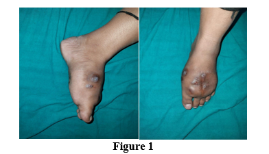
  8. Venkatswami, Sandhya, Anandan Sankarasubramanian, and Shobana Subramanyam. "The Madura Foot: Looking Deep." The International Journal of Lower Extremity Wounds, vol. 11, no. 1, 2012, pp. 31-42. DOI: 10.1177/1534734612438549.

  9. Gosselink, Carrie, et al. "Nocardiosis Causing Pedal Actinomycetoma: A Case Report and Review of the Literature." The Journal of Foot and Ankle Surgery, vol. 47, no. 5, 2008, pp. 457-462. DOI: 10.1053/j.jfas.2008.04.009.

  10. Barnetson, R. StC, and L. J. R. Milne. "Mycetoma." British Journal of Dermatology, vol. 99, no. 2, 1978, pp. 227-231. DOI: 10.1111/j.1365-2133.1978.tb00064.x.

  11. van de Sande, Wendy W.J., et al. "Melanin Biosynthesis in Madurella Mycetomatis and Its Effect on Susceptibility to Itraconazole and Ketoconazole." Microbes and Infection, vol. 9, no. 9, 2007, pp. 1114-1123. DOI: 10.1016/j.micinf.2007.05.015.

  12. Pilsczek, Florian H., and Michael Augenbraun. "Mycetoma Fungal Infection: Multiple Organisms as Colonizers or Pathogens?" Revista da Sociedade Brasileira de Medicina Tropical, vol. 40, no. 4, 2007, pp. 463-465. DOI: 10.1590/S0037-86822007000400007.

  13. Mahgoub, E. S. "Mycetoma." Seminars in Dermatology, vol. 99, 1978, pp. 227-231.

  14. Yousif, B. M., A. H. Fahal, and M. Y. Shakir. "A New Technique for the Diagnosis of Mycetoma Using Fixed Blocks of Aspirated Material." Transactions of the Royal Society of Tropical Medicine and Hygiene, vol. 104, no. 1, 2010, pp. 6-9. DOI: 10.1016/j.trstmh.2009.06.015.

  15. Padhi, Somanath, et al. "Mycetoma in South India: Retrospective Analysis of 13 Cases and Description of Two Cases Caused by Unusual Pathogens: Neoscytalidium Dimidiatum and Aspergillus Flavus." International Journal of Dermatology, vol. 49, no. 11, 2010, pp. 1289-1296. DOI: 10.1111/j.1365-4632.2010.04610.x.

  16. Weedon, David, Strutton, Gillian, and Rubin, Alan I. Weedon’s Skin Pathology. 3rd ed., Churchill Livingstone, London, n.d.

  17. Chufal, Sanjay Singh, Naveen Chandra Thapliyal, and Manoj Kumar Gupta. "An Approach to Histology-Based Diagnosis and Treatment of Madura Foot." The Journal of Infection in Developing Countries, vol. 6, no. 9, 2012, pp. 684-688. DOI: 10.3855/jidc.2387.

  18. Sharma, Nand Lal, et al. "Nocardial Mycetoma: Diverse Clinical Presentations." Indian Journal of Dermatology, Venereology and Leprology, vol. 74, no. 6, 2008, pp. 635-637. DOI: 10.4103/0378-6323.45110.

  19. Ahmed, Abdalla A.O., et al. "Management of Mycetoma: Major Challenge in Tropical Mycoses with Limited International Recognition." Current Opinion in Infectious Diseases, vol. 20, no. 2, 2007, pp. 146-151. DOI: 10.1097/QCO.0b013e328045fc92.

License
CC BY-NC-ND
Creative Commons Attribution-NonCommercial-NoDerivatives 4.0 International License
Mycetoma of the Foot Leading to Amputation - A Case Report © 2026 by Dr. Ruquiya Afrose, Dr. Sayeedul Hasan Arif, Dr. Dipanjan Sinha, Dr. Zeeshan Nahid, Dr. Avadh Bihari Lal Sharma licensed under CC BY-NC-ND 4.0
All papers should be submitted electronically. All submitted manuscripts must be original work that is not under submission at another journal or under consideration for publication in another form, such as a monograph or chapter of a book. Authors of submitted papers are obligated not to submit their paper for publication elsewhere until an editorial decision is rendered on their submission. Further, authors of accepted papers are prohibited from publishing the results in other publications that appear before the paper is published in the Journal unless they receive approval for doing so from the Editor-In-Chief.
Himalayan Journal of Applied Medical Sciences and Research open access articles are licensed under a Creative Commons Attribution-Share A like 4.0 International License. This license lets the audience to give appropriate credit, provide a link to the license, and indicate if changes were made and if they remix, transform, or build upon the material, they must distribute contributions under the same license as the original.
Advertisement
Recommended Articles
Research Article
Evaluation of Serum Heat Shock Protein 70 as a Diagnostic Biomarker for Ectopic Pregnancy: A Comparative Case-Control Study
Published: 28/02/2026
Download PDF
Research Article
Assessing First Trimester Maternal Serum Pentraxin-3 Levels in Primary Unexplained Recurrent Pregnancy Loss: A Case-Control Study
Published: 28/02/2026
Download PDF
Research Article
Patent Ductus Arteriosus Medical Closure and Associated Co-morbidities in Premature Infants in Sulaymaniyah Neonatal Intensive Care Unit
...
Published: 28/02/2026
Download PDF
Research Article
Bones in Balance: Awareness of Rickets and Nutritional Deficiencies Among the Hamirpur Community
Published: 10/12/2024
Download PDF
Chat on WhatsApp
Flowbite Logo
Najmal Complex,
Opposite Farwaniya,
Kuwait.
Email: kuwait@iarcon.org

Editorial Office:
J.L Bhavan, Near Radison Blu Hotel,
Jalukbari, Guwahati-India
Useful Links
Order Hard Copy
Privacy policy
Terms and Conditions
Refund Policy
Others
About Us
Team Members
Contact Us
Online Payments
Join as Editor
Join as Reviewer
Subscribe to our Newsletter
Follow us
MOST SEARCHED KEYWORDS
scientific journal
 | 
business journal
 | 
medical journals
 | 
Scientific Journals
 | 
Academic Publisher
 | 
Peer-reviewed Journals
 | 
Open Access Journals
 | 
Impact Factor
 | 
Indexing Services
 | 
Journal Citation Reports
 | 
Publication Process
 | 
Impact factor of journals
 | 
Finding reputable journals for publication
 | 
Submitting a manuscript for publication
 | 
Copyright and licensing of published papers
 | 
Writing an abstract for a research paper
 | 
Manuscript formatting guidelines
 | 
Promoting published research
 | 
Publication in high-impact journals
Copyright © iARCON Internaltional LLP . All Rights Reserved.