Contents
Download PDF
pdf Download XML
1282 Views
406 Downloads
Share this article
Research Article | Volume 3 Issue 2 (July-Dec, 2022) | Pages 1 - 7
Role of Computed Axial Tomography in Determination of Bone Depth Density in Dental Implants
 ,
1
College of Dentistry, University of Wasit, Wasit, Iraq
Under a Creative Commons license
Open Access
Received
Aug. 13, 2022
Revised
Sept. 16, 2022
Accepted
Oct. 20, 2022
Published
Nov. 10, 2022
Abstract

Introduction: Primary stability is a key factor in implant success. This study used computed axial tomography to determine the bone depth in implant candidate sites and its correlation with radio frequency findings. Materials and Methods: This study was included a total of 85 dental implants in 24 patients; 11 males and 13 females. Surgical guides were made from stereolithography 3D designing. Electronic surgery was performed on patient-selected implant sites based on computed axial tomography data entered into surgical guide software. Bone depth value determined bone depth. After osteotomy, the manufacturer-recommended implant was placed. Primary implant stability was determined by installing relevant stability. P. value was significant (<0.05). Results: In this particular research, the mean value of the scale was found to be 433.3 with a standard deviation of 198.4, and the implant stability was measured at 55.7 with a standard deviation of 5.6. The Pearson correlation test was used to evaluate the correlation between the dental stability and bone depth density. Respectively the results indicated that there is a significant relation between the dental stability and bone depth density. Conclusion: Both the dental stability followed by a density, and they were significantly correlated. In other words, the bone depth density is determined by the tomography methods of dental implants.

Keywords
INTRODUCTION

Dental implants can restore both the appearance and function of a person's missing teeth. They're popular for replacing destroyed tooth [1]. Supporting methods of dental replacement are effectively related with new methods of screening. Dental implants are one of the most successful treatments [2]. A dental implant is inserted into the jawbone to support a prosthetic [3]. With proper care, dental implants can last a lifetime and properly cared for implants can last a lifetime and replace missing teeth [4]. After osseointegration, the implant becomes microscopically connected to the living bone [5]. This follows osseointegration and this step follows implant insertion and dental implant success depends on the bone contact surface, the implant's ability to transfer forces to the depth density of bone, and dental stability of new teeth [6]. After the implant is placed, its initial stability can be assessed to accomplish this, consider the bone structure, implant shape, and surgical technique. Initial implant stability depends on several factors, most importantly looseness [7]. Primary implant stability is required for osseointegration and immediate implant treatments [8]. Bone and implant tissue fuse during osseointegration. Osseointegration describes this process [9]. Several studies have suggested a variety of methods for evaluating dental implant initial stability [10]. These methods have been suggested for evaluating dental implant initial stability [11]. Torque analysis and radio frequency ablation analysis are still used during dental implantations [12]. The insertion torque method measures implant osseointegration [13]. This method has the potential to be destructive due to the application of additive steps to the implant and the miss of degree on the levels of each stage [14]. Lack of quantity and quality grading makes it difficult to evaluate staging's effectiveness of staging cannot be graded of staging cannot be graded and this simple and noninvasive method of radio frequency ablation measures implant stability at various time intervals [15]. Insertion torque is used during surgery to place implants. This is its only application. This method measures bone implant complex rigidity  and  pliability   simultaneously  [16]. Implant stability will be the unit of measurement for this method and if the number is between 60 and 85, the implant should be successful while, if the score was below 49, the implant may not be suitable and several researches have investigated the correlations between bone density depth and dental stability [16]. After clinical exams to determine if a sample selected for tooth implantation, radiographic exams are performed to evaluate the bone at implant sites and,this step follows clinical exams to determine if a patient is a dental implant candidate [17]. After clinical exams to determine a patient's candidacy for dental implant treatment, the next step is to determine if the patient is a good candidate [18]. Competed axial tomography scans are commonly performed before surgery and the bone's quality and quantity are evaluated by depth [19]. Using the Hounsfield unit to determine bone depth is one of its main benefits, other benefits: This quality is directly proportional to X-ray attenuation and bone depth [20]. Using computed axial tomography to determine which regions need implants exposes patients to a high dose of radiation, and this is a major drawback of using this method to determine implant areas [21]. This is one of the major drawbacks of this approach. Competed axial tomography scans produce higher-quality images while requiring less radiation [22]. The conclusive findings can't be trusted for technical reasons, including calibration issues with several computed axial tomography devices and this is why the findings are unreliable [22-25]. This study examined the bone depth at implant candidate sites using on demand 3-D CT and its relationship with radio frequency ablation values after implant insertion. The aim of this study was to detect the role of competed axial tomography in the determination of bone depth for the 12 dental implants.

MATERIALS AND METHODS

Study Design 

For the purpose of this descriptive-analytic study, a total of 24 patients provided data on the implant treatment sites for which they were being evaluated, and 12 of those patients' reports were selected (including 11 males and 13 females). The range age of the samples was 49.21 years old, and there was a standard deviation of 2.44 years among them. Potential participants had to be between the ages of 32 and 48 and in good health in order to take part in the study. Additionally, they needed to be in the appropriate age range. In addition to this, they needed to have implantation performed in the mandibular and maxillary sites of their mouths. Conditions such as a systemic bone disease as well as other diseases that could potentially affected the immunity, have been determined to be exclusion criteria for this study. In addition to this, having a history of receiving antibiotic treatment within the past four months was also considered to be a criterion for exclusion.


Computed Axial Tomography 

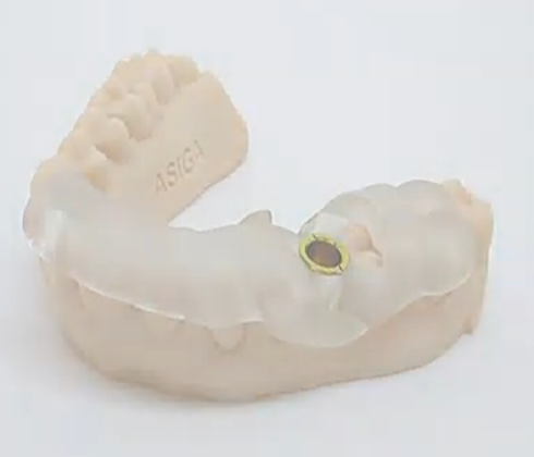
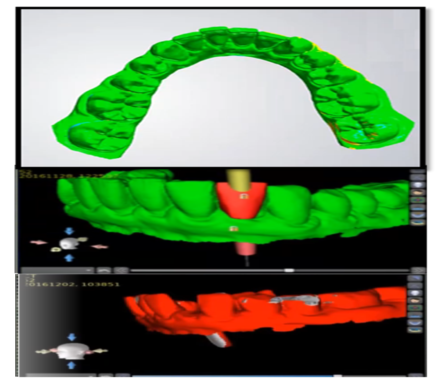
Every patient was provided with information regarding the processes that would be followed during the study, and their participation in the research was contingent on their providing informed consent. After the clinical examinations of patients who fulfilled the requirements were finished, impressions were taken in order to create a surgical guide for each patient using a 3D printer, which is one of the quickest methods for guide methods Figure 1. During this time period, patients were referred to a center for orally and maxillary of facial tomography so that computed axial tomography of implantation areas could be obtained. All of the computed axial tomography was obtained by utilizing 3D Dental Software and Cranex 3D, both of which had a field of view of 9 x 7, and high-resolution mode was utilized for both of these applications. After that, the information regarding each patient's computed axial tomography images as well as the images concerning the 3D scanning of the templates into the surgical software guide. This software was developed by a company called 3D (Iraq). After that, 3D shapes of implant treatment designed forsites were prepared using surgical software guide for patient's computed axial tomography images and scanning the patient's templates. This was done before moving on to the next step. Before continuing on to the subsequent step, this was completed.


Surgical Assessment of Implants 

The digital implants placement before surgery in the areas was performed that were selected in the 3D shape methods in the platform that was used for the surgical guide software. After completing the prosthetic reconstruction of the implants, the designed data of the implant site was transferred to the implant virtual placement so that the bone density could be evaluated there. In the software platform, 3D dental software was used to determine the bone density of the region surrounding each implant site, and the results of this calculation were recorded for each individual implant site. On the day of surgery, in addition to the appropriate analgesic medication, three grams of amoxicillin was given to each patient an hour before the procedure. To induce a state of anesthesia in the parts of the body that required implant surgery, an articaine anesthetic carpule that included epinephrine and contained articaine at a concentration of 3.5% was administered. Following the administration of local anesthesia, the toothless ridge underwent crestal incision, and then vertical incisions were made. After that, a periosteal elevator reflected the flaps in the opposite direction. Following that, the surgical guide was positioned atop the ridge that lacked teeth. A pilot drill was utilized during the initial osteotomy procedure, which was performed in order to properly position the implant. After the process of drilling the implants to their final diameter in accordance with the guidelines provided by the manufacturer was finished, the implants with the optimal predetermined diameters were positioned at the location that was desired. This allowed for the best possible outcome. Following completion of all of the procedures, the surgical guide was extracted from the patient. The implants that were utilized were those that had been purchased prior to the procedure. Two of the implants had a diameter of 4.5 mm, 14 of the other implants had a diameter of 5 mm, and the remaining three implants had a diameter of 5 mm. Every implant measured 12 millimeters in length, with the exception of one that was only 10 millimeters in length.


Figure 1: Guide for Surgical Implantation (3d Printed)


Analysis Steps 

After that, the intelligent device that was going to be installed on the body of the implant was chosen after taking into consideration the diameter of the implant that was going to be used, and it was then installed. After being activated, the device started recording the frequency values it had received in the form of numbers ranging from 0 to 100, as indicated by the dental stability unit. It was possible to accomplish this by causing a vibration in the body of the device that was connected to the implants and recording the amount of response that was received from the body of the implant. Following that, the component was extracted from the body of the implant by utilizing the appropriate instruments. After either a cover screw or a healing abutment had been installed on the body of the implant, a region was stitched using a three-needle suture to close the gap left by the previous procedure. The particular conditions were the primary consideration used in making a decision regarding the kind of abutment that was put into place. We utilized the Kolmogorov–Smirnov test and the Pearson correlation statistical test in order to investigate the normality of the distribution in both depth and stability. Both of these tests are statistical investigations. In addition, the statistical technique known as the Pearson correlation was used in order to conduct an investigation into the extent of the connection that exists between the level of stability and the gray scale. Patients had their sutures removed after a period of three weeks, and then they were given instructions on how to take care of themselves at home before being discharged. The prosthetic phase of the treatment was started after a period of time equaling four months had passed during which time the wound was allowed to heal.

RESULTS

The surgery was performed on three patients, and a total of 12 implants were inserted into their bodies. The maxilla of the first patient received all of the implants, while the mandibles of the other two patients each received implants. All of the implants were placed in the maxilla of the first patient. In each of the patients' individual tables, the precise location of each implant was presented in its own column. This was done so that the information could be compared between patients. During the course of the investigation, it was found that the initial stability values of the implant as well as the bone density varied from one patient to the next as well as from one implant area to the next. In addition, the initial stability values also differed depending on which implant area they were measured in. In the first patient, for instance, the initial stability of the implant was calculated to be 65, and it was positioned in the right canine region of the maxilla. In addition to this, the bone density was calculated to be 433.3. The values of the gray scale and the stability were reported in Table 1 in a fashion that was unique to each individual patient as well as the implant site. This was done in order to keep the information as accurate as possible. It was necessary to do this in order to guarantee that the data was correct.


Correlation between Depth Density of Bone and Dental Stability of Implants

The procedure was performed on a total of three patients, and during the course of the operation, a combined total of twelve implants were inserted into the patients' bodies. It was decided to place all of the implants in the patient's maxilla, while the implants for the other two patients were placed in their respective mandibles. This decision was made after some discussion. One implant at a time was inserted into the maxilla of the first patient, and that patient was given each and every implant that was placed there. In each of the patients' individual tables, the specific location of each implant was presented in its own column. These columns were labeled according to the patient's name. 


Table 1: Dental stability of implants and the depth of bone density of patient number 1

Implanted tooth

StabilityDepth density

Mandibular left central incisor

66302.23

Mandibular left third molar

68299.45

Mandibular left second molar

85456.21

Mandibular left premolar

71342.21

Maxillary right third molar

63443.61

Maxillary right second molar

86543.78

Maxillary right premolar

75521.89

Maxillary right canine

83433.32

Maxillary right first molar

73323.45


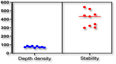
Figure 2: Correlation of Depth Density of Bone with Stability of Plants


Figure 3: The 3D Printed For Dental Guide


This was done in order to facilitate the comparison of the information gathered from the various patients. According to the labels, the patient's name was written in each of these columns after they were labeled. This was done in order to facilitate a more straightforward comparison of the data obtained from each of the various patients. During the course of the investigation, it was found that the initial stability values of the implant as well as the bone density varied from one patient to the next as well as from one implant area to the next. In addition, the winitial stability values also differed depending on which implant area they were measured in. The initial stability values also differed from one implant area to the next, which was another factor that contributed to this phenomenon. In addition, the values for the implant's initial stability varied from one part of the implant to another, depending on the location at which the measurements were taken within the implant. It was determined that the implant would have an initial stability of 65 for the first patient, and the canine region of the right maxilla would be the location of its placement. This is just one example among many. In addition, the calculations showed that the bone density was 433.3. This was determined by the bone density test. The values of the gray scale and the stability were reported in Table 1 in a fashion that was unique to each individual patient as well as the implant site. This was done in order to keep the information as accurate as possible. This was done in order to maintain the highest possible level of precision in the information. This was done in order to keep the information as accurate as possible and maintain the highest possible level of precision. It was necessary to carry out these procedures in order to guarantee that the data were correct. The researchers discovered that there is a significant correlation between the two variables, and that there is a normal distribution for both the gray scale and the initial stability of the implant. Both of these variables were found to have a normal distribution. The following is a condensed version of what was discovered in this study: The findings also demonstrated that the gray scale followed a normal distribution, which was yet another fascinating finding to emerge from the investigation. One is able to make predictions regarding changes in the level of stability by utilizing the correlation coefficient of the grayscale values.


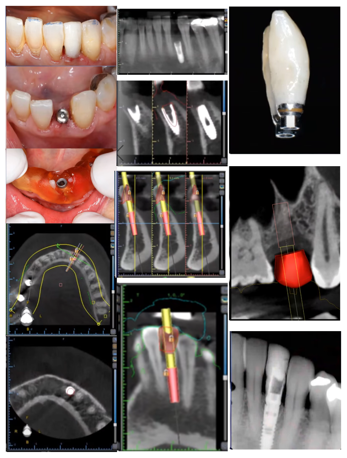
Figure 4: Computed Tomography View of the Dental Implants and Bone Depth Density

DISCUSSION

Osseointegration cannot occur without the capacity for the patient to keep their stability [1,26]. Dental implants' rates of stability and survival can be affected by a wide range of factors, including the patient's bone density, implant geometry, surgical procedures, and general health [2,27]. In this investigation, the distribution of bone density and bone stability followed a normal curve and displayed a high degree of correlation [4,28]. This was demonstrated by the fact that the distributions were highly correlated and before implant surgery, CT scans are typically done as a precautionary measure [5]. There were elements of the method that contributed to both its benefits and its drawbacks [5,29-30]. The most significant benefit that resulted from applying this method was the enhancement of one's capacity to evaluate bone quality and density on the basis of bone depth density [31-33]. In accordance with the findings of the current study, it was discovered that the bone density, which was measured using computed axial tomography in HU units, had a significant association with the primary implant stability, which was measured using stability units [34-36]. Frequency ablation values is a simple intraoral procedure that does not involve any kind of invasive tissue removal and can be performed in just a few minutes [37-39]. Measurement of implant stability and an analysis of the implant interface are the goals of this procedure. RF ablation analysis has been shown to result in improved implant stability in multiple studies and, there is no method that can absolutely guarantee accuracy when measuring stability [40-42]. The concepts of repeatability and reproducibility can be utilized in order to establish a reliable standard of measurement [43   -44]. The findings of a number of separate studies regarding the typical bone density of the maxilla and mandible have been found to be in agreement with one another [45-46]. The numbers for stability and bone density at the beginning of the process of implant placement were, respectively, 67.5 5.8 and 683.48 78.63. The findings of this study have yielded numbers that are lower than those presented here. In line with the results of the present investigation, the authors also found that there is a positive correlation between the grayscale and the stability values [47-48]. Several studies looked into the capability of the stability device to determine the degree to which an implant is stable. The program carried out three separate measurements in their entirety. The gap between the two earlier assessments of implant stability was found to be significant after the third evaluation of implant stability [42,49-50]. It is possible to determine the stability of an implant in a reliable manner by using a method that is both repeatable and based on the stability of the frequency ablation system. In the course of this particular piece of research, the steadiness of the initial implant was analyzed through the utilization of frequency ablation values [7-9]. According to the findings of the study, the stability of the mandible was found to be superior to that of the maxilla [51]. It's possible that this is because the mandible has an above-average bone density, which contributes to an increase in implant primary stability [10-12]. Previous clinical research has found what the current study has found to be true, namely that the stability and longevity of dental implants in the mandible are superior to those in the maxilla [10-14]. This finding is in line with the findings of the current study [25]. The density of the cortical bone has an effect on how securely dental implants can be anchored into place [24]. In contrast to the maxillary bones, which have a substantial layer of trabecular bone covered by a thin layer of superficial cortical bone, the cortical bone of the mandible is thicker and more substantial [52]. The maxillary bones have a substantial layer of trabecular bone [23]. The findings of a few studies have shown that decreased stability is associated with loss of crestal bone [53]. Radiographs need to be taken from a preapical position in order to obtain the most accurate results. In primary implant stability, the degree to which bone density, surgical procedures, and implant surface parameters all play a role is determined by a number of different factors [54]. These factors include: Increasing primary stability and preventing preimplant bone resorption are two goals that have been the focus of a number of studies, and each of these studies has suggested a different strategy for accomplishing these goals [55]. These are two objectives that have been the focal point of a number of different research projects [56]. As a result of this, a low bone density can sometimes be compensated for by other factors in order to achieve primary implant stability [56]. This is because of the fact that this is a consequence of this [57]. The radio frequency ablation technique makes it possible to perform an examination of the bone tissue that is surrounding the implant at any point during the treatment process [58]. This allows for greater precision and accuracy in the procedure [59]. Implants that have a low level of stability going into the initial healing period have a greater chance of failing after the initial healing period has passed [60].

CONCLUSION

Both the initial stability of the implant and the gray scale were found to have a normal distribution, and the researchers found that there is a significant correlation between the two variables. The findings of this study can be summarized as follows: The findings also showed that the gray scale had a normal distribution, which was another interesting discovery. With the help of the bone depth density values, it is possible to make a prediction about the correlation coefficient of the changes in the level of stability.

REFERENCE
  1. Shabana Azmin. et al. "Applications of cone beam computed tomography in the field of prosthodontics: A review." 2022.

  2. Ouldyerou, A. et al. "Biomechanical performance of Ti-PEEK dental implants in bone: An in-silico analysis." Journal of the Mechanical Behavior of Biomedical Materials, vol. 2022, 2022, p. 105422.

  3. Brockmeyer, P. et al. "Informational content of two-dimensional panoramic radiographs and lateral cephalometric radiographs with respect to the bone volume of intraoral donor regions considering CBCT imaging." BMC Oral Health, vol. 22, no. 1, 2022, pp. 1–11.

  4. Chakraborty, A. et al. "Probing the influence of hybrid thread design on biomechanical response of dental implants: Finite element study and experimental validation." Journal of Biomechanical Engineering, vol. 145, no. 1, 2022, p. 11011.

  5. Gelețu, G.L. et al. "Customized 3D-printed titanium mesh developed for an aesthetic zone to regenerate a complex bone defect resulting after a deficient odontectomy: A case report." Medicina (Buenos Aires), vol. 58, no. 9, 2022, p. 1192.

  6. Akers, J.A. et al. "Dental implants at sites of focal high and mixed density osseous lesions: Clinical practice guidelines." Clinical Advances in Periodontics, 2022.

  7. Arango, E. et al. "Age differences in relation to bone thickness and length of the zygomatic process of the maxilla, infrazygomatic crest, and buccal shelf area." American Journal of Orthodontics and Dentofacial Orthopedics, vol. 161, no. 4, 2022, pp. 510–18.

  8. Szabó, Á.L. et al. "Distally tilted implants according to the All-on-Four® treatment concept for the rehabilitation of complete edentulism: A 3.5-year retrospective radiographic study of clinical outcomes and marginal bone level changes." Dentistry Journal, vol. 10, no. 5, 2022, p. 82.

  9. AlSamak, S. et al. "Evaluation of potential mini-implant insertion sites for maxillary skeletal expander: A computerized tomography study." Clinical Investigations in Orthodontics, 2022, pp. 1–9.

  10. Houshmand, B. et al. "Effect of implant design on stress distribution: A finite element study." Journal of Long-Term Effects of Medical Implants, vol. 32, no. 4, 2022.

  11. Omaish, H.H.M. et al. "Comparison of the strain developed around implants with angled abutments with two reinforced polymeric CAD-CAM superstructure materials: An in vitro comparative study." Journal of Prosthetic Dentistry, 2022.

  12. Sayed, A.M., and Abdelazim, I.A. "Effect of different framework materials on stresses induced at the implant/bone interface in all-on-four implant treatment concept: Three-dimensional finite element analysis." Tanta Dental Journal, vol. 19, no. 3, 2022, p. 105.

  13. Seo, D.J. et al. "The effect of under-drilling and osseodensification drilling on low-density bone: A comparative ex vivo study." Applied Sciences, vol. 12, no. 3, 2022, p. 1163.

  14. Büyük, F.N. et al. "Review on finite element analysis of dental implants." Journal of Dental Implant Research, vol. 41, no. 3, 2022, pp. 50–63.

  15. Karami, N. and Razeghi, M. "Structural evaluation of the effect of bone quality on the mechanical interaction between dental implants and bone." Shock and Vibration, vol. 2022, 2022.

  16. Datta, M. et al. "Importance of CBCT as a diagnostic tool for evaluating the position of inferior alveolar canal during dental implant placement: A systemic review." Dental Research, 2022.

  17. Hsu, Y.Y. et al. "Insertion speed affects the initial stability of dental implants." Journal of Medical and Biological Engineering, vol. 42, no. 4, 2022, pp. 516–25.

  18. Jha, S. et al. "Marginal bone level and bone quality evaluation using CBCT after functional loading around dental implant in the population of Bhubaneswar, Odisha: A longitudinal study." Dental Research, 2022.

  19. Tinfer, S. et al. "Method to support dental implant process based on image processing." Personalized Orthopedics, Springer, 2022, pp. 221–50.

  20. Leblebicioğlu Kurtuluş, I. et al. "Finite element analysis of the stress distribution associated with different implant designs for different bone densities." Journal of Prosthodontics, vol. 31, no. 7, 2022, pp. 614–22.

  21. Li, Q. et al. "Biomechanical evaluation of customized root implants in alveolar bone: A comparative study with traditional implants and natural teeth." Journal of Prosthodontics, 2022.

  22. Nakra, P. et al. "Skeletal changes after miniscrew-assisted rapid palatal expansion in young adults: A cone-beam computed tomography technique study." World Journal of Dentistry, vol. 13, no. 6, 2022, pp. 617–22.

  23. Satpathy, M. et al. "Effect of bone remodeling on dental implant fatigue limit predicted using 3D finite element analysis." Journal of Dental and Oral Epidemiology, vol. 2, no. 1, 2022.

  24. Sakamaki, T. et al. "Thread shape, cortical bone thickness, and magnitude and distribution of stress caused by the loading of orthodontic miniscrews: Finite element analysis." Scientific Reports, vol. 12, no. 1, 2022, pp. 1–11.

  25. Gil, J. et al. "Influence of bone-level dental implants placement and of cortical thickness on osseointegration: In silico and in vivo analyses." Journal of Clinical Medicine, vol. 11, no. 4, 2022, p. 1027.

  26. Mohammed Alwan, A. et al. "Significance of the estrogen hormone and single nucleotide polymorphisms in the progression of breast cancer among female." Archives of Razi Institute, vol. 77, no. 3, 2022, pp. 943–58, https://archrazi .areeo.ac.ir/artic le_126343.html.

  27. Alwan, A.M. et al. "The impact of CYP19A1 variants and haplotypes on breast cancer risk, clinicopathological features and prognosis." Molecular Genetics & Genomic Medicine, vol. 9, no. 7, 2021, p. e1705.

  28. Alwan, A.M., and Afshari, J.T. "In vivo growth inhibition of human Caucasian prostate adenocarcinoma in nude mice induced by amygdalin with metabolic enzyme combinations." BioMed Research International, vol. 2022, 2022.

  29. Rahi, M. et al. "Polymerase chain reaction-based malaria diagnosis can be increasingly adopted during current phase of malaria elimination in India." American Journal of Tropical Medicine and Hygiene, 2022.

  30. Deo, D.A. et al. "Difference between microscopic and PCR examination result for malaria diagnosis and treatment evaluation in Sumba Barat Daya, Indonesia." Tropical Medicine and Infectious Disease, vol. 7, no. 8, 2022, p. 153.

  31. Kann, S. et al. "Comparison of the influence of different nucleic acid extraction assays on the sensitivity of Trypanosoma cruzi-specific real-time PCR." Microorganisms, vol. 10, no. 8, 2022, p. 1554.

  32. Troll, H. et al. "Simple differential detection of Entamoeba histolytica and Entamoeba dispar in fresh stool specimens by sodium acetate-acetic acid-formalin concentration and PCR." Journal of Clinical Microbiology, vol. 35, no. 7, 1997, pp. 1701–5.

  33. Pian, H. et al. "Sandwich hybridization-based loop-mediated isothermal amplification (SHB-LAMP) for high-throughput detection of malaria RNA from asymptomatic infections." Sensors and Actuators B: Chemical, vol. 365, 2022, p. 131973.

  34. Freitas, M.A.R. et al. "A single step duplex PCR to distinguish Entamoeba histolytica from Entamoeba dispar." Parasitology, vol. 128, no. 6, 2004, pp. 625–8.

  35. Khairnar, K. and Parija, S.C. "A novel nested multiplex polymerase chain reaction (PCR) assay for differential detection of Entamoeba histolytica, E. moshkovskii and E. dispar DNA in stool samples." BMC Microbiology, vol. 7, no. 1, 2007, pp. 1–9.

  36. Aryal, M. et al. "First report on the molecular detection of Entamoeba bovis from the endangered wild water buffalo (Bubalus arnee) in Nepal." Veterinary Medicine and Science, vol. 8, no. 2, 2022, pp. 799–807.

  37. Verweij, J.J. et al. "Simultaneous detection of Entamoeba histolytica, Giardia lamblia, and Cryptosporidium parvum in fecal samples by using multiplex real-time PCR." Journal of Clinical Microbiology, vol. 42, no. 3, 2004, pp. 1220–3.

  38. Shankar, H. and Kumar, G. "Truenat: An affordable and user-friendly option for screening of sub-microscopic Plasmodium infections in low resource countries." Lancet Regional Health – Asia, vol. 5, 2022.

  39. Belachew, M. et al. "Evaluating performance of multiplex real time PCR for the diagnosis of malaria at elimination targeted low transmission settings of Ethiopia." Malaria Journal, vol. 21, no. 1, 2022, pp. 1–9.

  40. Bhullar, S. and Mishra, N. "Molecular assays for determining sulphadoxine-pyrimethamine drug resistance in India: A systematic review." Parasitology Research, 2022, pp. 1–10.

  41. Nema, S. et al. "Unreported mixed Plasmodium species infection may increase vivax malaria in India: A challenge for malaria elimination." Transactions of the Royal Society of Tropical Medicine and Hygiene, 2022.

  42. Haque, R. et al. "Comparison of PCR, isoenzyme analysis, and antigen detection for diagnosis of Entamoeba histolytica infection." Journal of Clinical Microbiology, vol. 36, no. 2, 1998, pp. 449–52.

  43. Deora, N. et al. "Multiplexing for Plasmodium spp.? Think again! Comment on Bhowmick et al. Dry post wintertime mass surveillance unearths a huge burden of P. vivax, and mixed infection with P. vivax P. falciparum, a threat to malaria elimination, in Dhalai, Tripura, India." Pathogens, vol. 11, no. 7, 2022, p. 737.

  44. Santos, H.L.C. et al. "Comparison of multiplex-PCR and antigen detection for differential diagnosis of Entamoeba histolytica." Brazilian Journal of Infectious Diseases, vol. 11, 2007, pp. 365–70.

  45. Kumari, P. et al. "Comparative assessment of diagnostic performance of cytochrome oxidase multiplex PCR and 18S rRNA nested PCR." Korean Journal of Parasitology, vol. 60, no. 4, 2022, pp. 295–9.

  46. Rivera, W.L. et al. "Field study on the distribution of Entamoeba histolytica and Entamoeba dispar in the northern Philippines as detected by the polymerase chain reaction." American Journal of Tropical Medicine and Hygiene, vol. 59, no. 6, 1998, pp. 916–21.

  47. Gonin, P. and Trudel, L. "Detection and differentiation of Entamoeba histolytica and Entamoeba dispar isolates in clinical samples by PCR and enzyme-linked immunosorbent assay." Journal of Clinical Microbiology, vol. 41, no. 1, 2003, pp. 237–41.

  48. Ramírez, A.M. et al. "Usefulness of Malachite-Green LAMP for diagnosis of Plasmodium and five human malaria species in a nonendemic setting." American Journal of Tropical Medicine and Hygiene, 2022.

  49. Leiva, B. et al. "Overdiagnosis of Entamoeba histolytica and Entamoeba dispar in Nicaragua: A microscopic, triage parasite panel and PCR study." Archives of Medical Research, vol. 37, no. 4, 2006, pp. 529–34.

  50. Kamau, E. et al. "Safety and tolerability of mosquito bite-induced controlled human infection with Plasmodium vivax in malaria-naive study participants—Clinical profile and utility of molecular diagnostic methods." Journal of Infectious Diseases, vol. 225, no. 1, 2022, pp. 146–56.

  51. Van Baar, G.J.C. et al. "The Amsterdam UMC protocol for computer-assisted mandibular and maxillary reconstruction: A cadaveric study." Oral Oncology, vol. 133, 2022, p. 106050.

  52. Gu, Y. et al. "Evaluation of the clinical efficacy and safety of modified alveolar cleft bone graft with cone-beam CT digital imaging in children." Translational Pediatrics, vol. 11, no. 7, 2022, p. 1140.

  53. Naeem, D.M. and Al-Jumaily, H.A. "Can the immediate implantation with immediate loading achieve an acceptable esthetic outcome? A prospective observational clinical study." Journal of Craniofacial Surgery, 2022, pp. 10–1097.

  54. Yu, W. et al. "Biomechanical analysis of different framework design, framework material and bone density in the edentulous mandible with fixed implant-supported prostheses: A three-dimensional finite element study." Journal of Prosthodontics, 2022.

  55. Park, J. et al. "Biomechanical effects of bone quality and design features in dental implants in long-term bone stability." Journal of Computational Design and Engineering, vol. 9, no. 5, 2022, pp. 1538–48.

  56. Chen, Z. et al. "Influence of bone density on the accuracy of artificial intelligence–guided implant surgery: An in vitro study." Journal of Prosthetic Dentistry, 2022.

  57. AlMugeiren, O.M. et al. "Anterior mandibular alveolar bone measurements between diabetic and non-diabetic individuals using cone-beam computed tomography." European Review for Medical and Pharmacological Sciences, vol. 26, no. 15, 2022, pp. 5476–84.

  58. Issrani, R. et al. "Cone-beam computed tomography: A new tool on the horizon for forensic dentistry." International Journal of Environmental Research and Public Health, vol. 19, no. 9, 2022, p. 5352.

  59. Tawfik, B.E.A. et al. "Tissue expander followed by autogenous bone graft versus autogenous bone graft alone for mandibular reconstruction: Quantitative assessment." Journal of Craniofacial Surgery, 2022, pp. 10–1097.

  60. Mao, C. et al. "Mechanobiologically optimized Ti–35Nb–2Ta–3Zr improves load transduction and enhances bone remodeling in tilted dental implant therapy." Bioactive Materials, vol. 16, 2022, pp. 15–26.


License
CC BY-NC-ND
Creative Commons Attribution-NonCommercial-NoDerivatives 4.0 International License
Role of Computed Axial Tomography in Determination of Bone Depth Density in Dental Implants © 2026 by Zainab A.H. Al-Tamemi, Hani M. Khlaif licensed under CC BY-NC-ND 4.0
All papers should be submitted electronically. All submitted manuscripts must be original work that is not under submission at another journal or under consideration for publication in another form, such as a monograph or chapter of a book. Authors of submitted papers are obligated not to submit their paper for publication elsewhere until an editorial decision is rendered on their submission. Further, authors of accepted papers are prohibited from publishing the results in other publications that appear before the paper is published in the Journal unless they receive approval for doing so from the Editor-In-Chief.
Himalayan Journal of Medicine and Surgery open access articles are licensed under a Creative Commons Attribution-Share A like 4.0 International License. This license lets the audience to give appropriate credit, provide a link to the license, and indicate if changes were made and if they remix, transform, or build upon the material, they must distribute contributions under the same license as the original.
Recommended Articles
Research Article
Effect of Land Degradation on Livelihood
Published: 04/01/2024
Download PDF
Research Article
Impact of Gut-Liver Axis: Hepatic Biochemical and Metabolic Changes Associated with Chronic Gastritis in Iraqi Patients with Helicobacter Pylori
Published: 20/02/2026
Download PDF
Research Article
Amniotic Fluid and Umbilical Cord Serum Erythropoietin as Biomarkers of Fetal Hypoxia in Term and Prolonged Pregnancies
Published: 15/01/2026
Download PDF
Research Article
Awareness and Practices among Professional Healthcare Workers towards COVID-19 in Iraq
...
Published: 30/03/2024
Download PDF
Flowbite Logo
Najmal Complex,
Opposite Farwaniya,
Kuwait.
Email: support@himjournals.com

Useful Links
Order Hard Copy
Privacy policy
Terms and Conditions
Refund Policy
Others
About Us
Team Members
Contact Us
Online Payments
Join as Editor
Join as Reviewer
Subscribe to our Newsletter
Follow us
MOST SEARCHED KEYWORDS
scientific journal
 | 
business journal
 | 
medical journals
 | 
Scientific Journals
 | 
Academic Publisher
 | 
Peer-reviewed Journals
 | 
Open Access Journals
 | 
Impact Factor
 | 
Indexing Services
 | 
Journal Citation Reports
 | 
Publication Process
 | 
Impact factor of journals
 | 
Finding reputable journals for publication
 | 
Submitting a manuscript for publication
 | 
Copyright and licensing of published papers
 | 
Writing an abstract for a research paper
 | 
Manuscript formatting guidelines
 | 
Promoting published research
 | 
Publication in high-impact journals
Copyright © Himalayan Journals . All Rights Reserved.