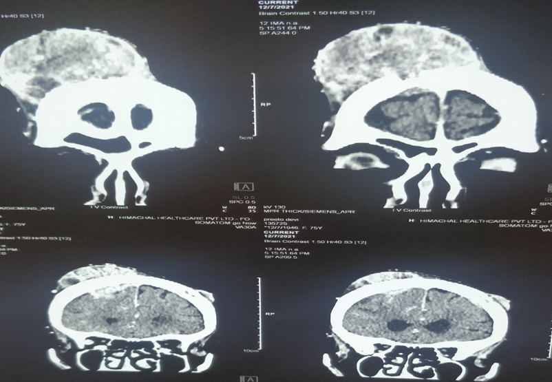
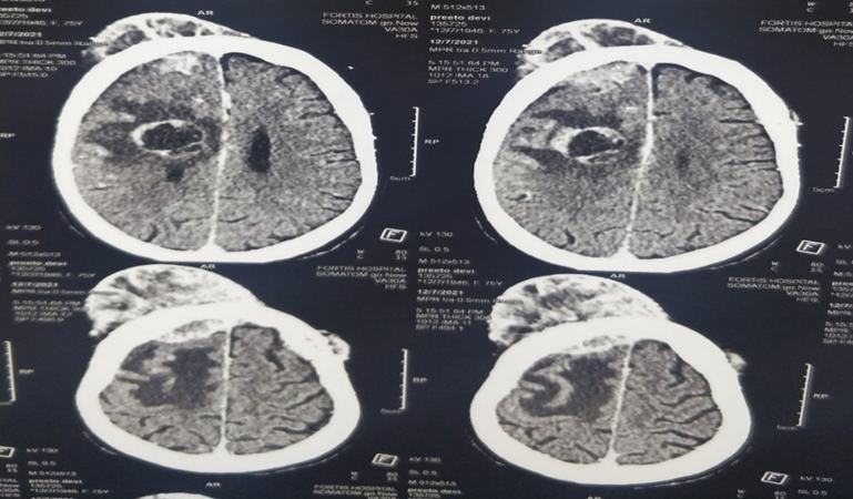
We present the case of a 75-year-old female with an unusual presentation of metastatic carcinoma of the lung. The case emonstrated a small cell carcinoma of the lung with an intracranial metastatic lesion destroying the bilateral frontal bones and extending into the extracranial soft tissue.
Lung cancer contributes significantly to the burden of disease [1,2]. The brain is a frequent site of metastases for carcinoma of the lung, and lung cancer is responsible for approximately 50% of all brain metastases. Most of brain tumours are the result of metastatic disease [3].


Figure 1

Figure 2
Of brain metastases, 80% originate from the hemispheres of the cerebrum and most are well demarcated with a capsule. A minority of lesions may demonstrate infiltrative growth [4]. Metastatic brain lesions are responsible for significant morbidity and mortality and have a dismal prognosis (Figure
1) [4,5]. The clinical features of brain metastases vary depending on the location of the lesion [6]. The most common complaint of brain metastases is headache, found in maximum patients. Other common symptoms include altered mental status, focal weakness, seizures and ataxia [7].
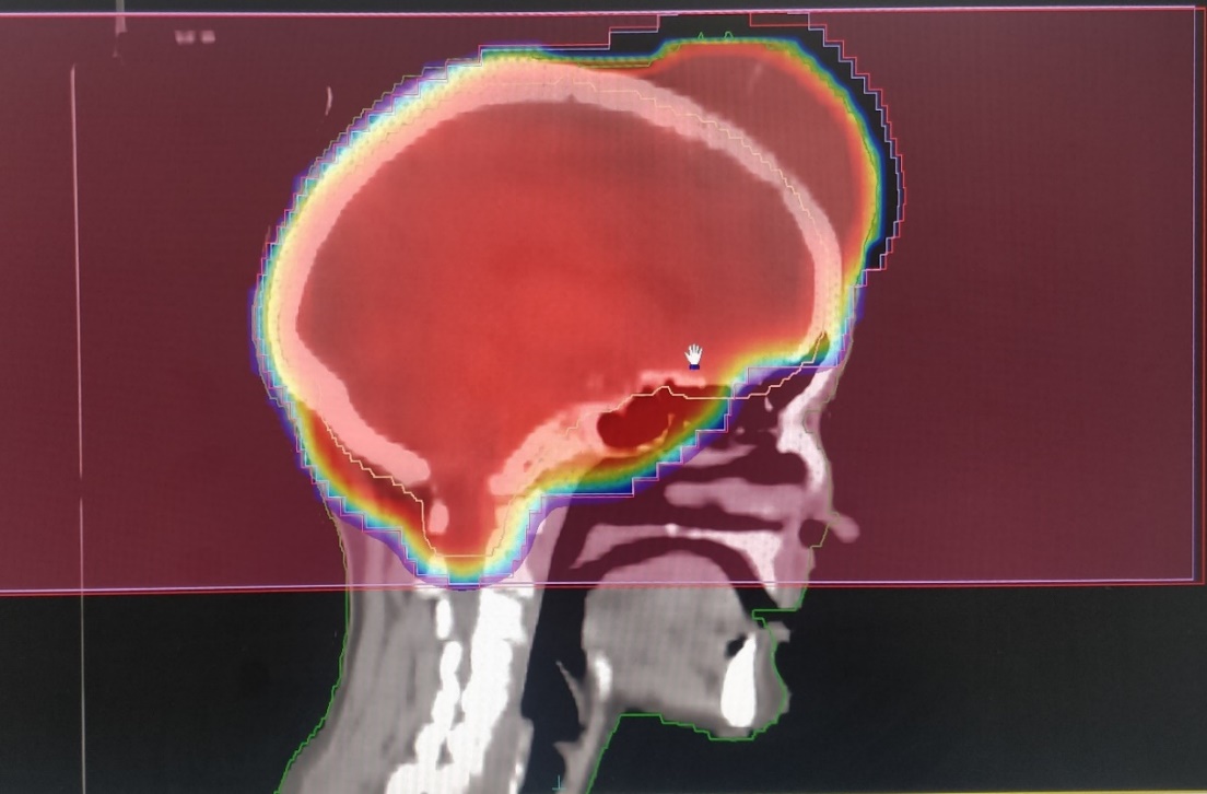
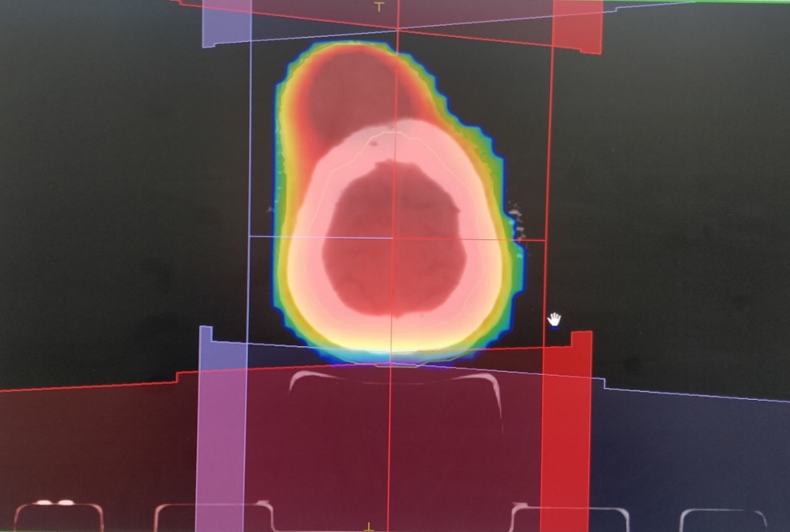
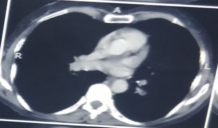
A 75-year-old female presented with complaint of a persistent headache and weakness bilateral lower limb with abnormal behaviour and history of forgetfulness for two months. On physical examination, swelling on scalp overlying the frontal bone was seen. He had no history of trauma to the head. CECT Head revealed lytic destructive lesions in bilateral frontal lobes with extracranial soft tissue component (right- 5cm, left- 1cm). Multiple necrotic peripherally enhancing lesions noted in right frontal lobe (23x24mm), right occipital lobe (9x10mm), two in left cerebellar hemispheres (10mm and 25mm) with significant surrounding oedema (fig. 1). The CECT chest demonstrated multilobulated area of size 4x2.3cm in superior segment of left lower lobe with adjacent interstitial septal thickening. No lymphadenopathy was noted (fig. 2). FNAC from lesion on scalp suggestive of metastatic small cell carcinoma. A bronchoscopy was done and a biopsy for histopathology was taken. Patient was then subjected to palliative whole brain radiotherapy (Fig. 3) and decongestive therapy for palliation of symptoms.


Figure 3
This case highlights an unusual presentation of lung cancer, an intracranial lesion producing destruction of frontal bone to invade beyond the skull and into the extracranial soft tissue. It is extremely rare for intracranial metastases from lung carcinomas to produce destruction of bone. It was previously reported by Foco et al. in the only published case of adenocarcinoma metastasis that produced destruction of the frontal bone [8]. Secondary metastases from Ewing’s sarcoma, carcinoma of the prostate, breast, thyroid and neuroblastoma have been reported to lead to destruction of the bone on rare occasions [9,10] The prognosis of patients with stage four small cell carcinoma lung is extremely poor with a median survival of four months [10]. If not treated patients with brain metastases from any primary source have a median survival of one month from diagnosis [4]. As a result, treatment should be focused primarily on palliation. Management of patients with intracranial metastatic lesions extending extracranially is poorly studied. There is a demonstrated role for surgical resection followed by whole brain radiation therapy with doses of 30Gy to 40Gy in patients with a single intracranial metastatic lesion, particularly if the lesion appears capsulated and there are no extracranial lesions. It shows improved neurocognitive function, elimination of mass effects and removal of the source of perifocal oedema [5]. Palliative radiotherapy without surgery has also been used along with steroids. This patient was subjected to palliative radiotherapy along with decongestive therapy for palliation of symptoms.
The authors find this case quite rare due to intracranial metastasis from a small cell carcinoma lung invading through the skull into the extracranial soft tissues and raises questions over the most appropriate course of management for these kind of patients.
Perez-Moreno P, et al "Squamous Cell Carcinoma of the Lung: Molecular Subtypes and Therapeutic Opportunities." Clinical Cancer Research, vol. 18, 2012, pp. 2443–2451. https://doi.org/10.1158 /1078-0432.CCR-11-2370.
Ruysscher D, et al. "Radical Treatment of Non-Small-Cell Lung Cancer Patients with Synchronous Oligometastases: Long-Term Results of a Prospective Phase II Trial." Journal of Thoracic Oncology, vol. 7, 2012, pp. 1547–1555. https://doi.org/10.1097/ JTO.0b013e318262caf6.
Platta C, et al "Current Treatment Strategies for Brain Metastasis and Complications from Therapeutic Techniques: A Review of Current Literature". American Journal of Clinical Oncology, vol. 33, 2010, pp. 398–407. https://doi.org/10.1097/COC.0b013e 318194f744.
Vogelbaum M, et al "Resectable Brain Metastases." Journal of Clinical Oncology, vol. 24, 2006, pp. 1289–1294. https://doi.org/10.1200/JC O.2005.04.6235.
Soffietti R, et al "Brain Metastases: Current Management and New Developments." Current Opinion in Oncology, vol. 20, 2008, pp. 676–684. https://doi.org/10.1097/CCO .0b013e32831186fe.
Meyers C. "Neurocognitive Dysfunction in Cancer Patients." Oncology (Huntington), vol. 14, 2000, pp. 75–79.
Schellinger P, et al. "Diagnostic Accuracy of MRI Compared to CCT in Patients with Brain Metastases." Journal of Neuro-Oncology, vol. 44, 1999, pp. 275–281. https://doi.org/10.1023/A:1 006308808769.