Thirty samples of endometriosis were collected from Alhusaina Hospital, Karbala, Iraq, and Fatimah Al-Zahraa Hospital For gynecology and obstetrics, Baghdad, Iraq. The aim of the research was to explore the generated relationship between chronic endometriosis in the endometriotic group. The samples were divided into the first group (20 patients) Endometriosis group and the second group consisting of 10 women (non-endometriosis groups) and concloude in this study, the occurrence of endometriotic foci is closely associated with pathological changes in the endometrium. However, the sequence of development of pathological changes in the uterine cavity and in the pelvic cavity is still not clear, and no statistically significant differences were found between endometriosis and non-endometriosis groups.
Endometriosis is an inflammation of the endometrium, the mucous layer that lines the lining of the endometrium [1].
Chronic endometritis and infertility is an association that was not well known until recently and was not considered and therefore untreated as it is characterized by endometrial distension, with a high density of stromal cells, intermittent maturation between the epithelium and stroma, and plasma cell infiltration into the endometrial stroma, and this The latter is the most important finding and this endometrial involvement has been known for a long time but is currently being given special attention in patients with infertility problems[2,3].
The prevalence of chronic endometriosis is 24.4% in premenopausal women; in women with fertility problems, it ranges from 2.8 to 56.8%, and from 14 to 67.5% in women with recurrent implantation failure, and pregnancy loss ranges Frequency between 9.3 and 67.6% [4,5].
As for the relationship generated with endometriosis, which is a condition in which tissue very similar to what lines the inside of the uterus (called the endometrium) grows outside the uterus in places where it should not be. When you have endometriosis, this tissue tends to grow on the ovaries, fallopian tubes, external uterus and abdominal organs [6,7].
The most common symptom of endometriosis is pain before and during menstruation. The colon causes pain that no one enjoys; This pain can be more severe if have this condition; in many cases, the patient also feels chronic (persistent) pain in the lower abdomen or back [8,9].
Other symptoms include bleeding between periods, heavy periods, and infertility, as endometriosis sometimes causes pain when urinating or defecating.
As for the accompanying symptoms, cyclical pelvic pain is usually in the midline, especially pain before or during menstruation (dysmenorrhea) and during intercourse (dyspareunia), which may develop into a chronic phase (lasting > six months). Also typical are endometrial tumors and infertility. Interstitial cystitis with suprapubic or pelvic pain, frequent urination and incontinence are common—the possibility of intermenstrual bleeding [10].
In some patients, even with extensive endometriosis, the disease may be asymptomatic, and in patients with minimal manifestations of the disease, there may be debilitating pain. Dysmenorrhea is an important diagnostic criterion, especially if its manifestations begin several years after a relatively painless menstruation. During pregnancy, symptoms improve or disappear [11].
Endometriosis of the cervix is characterized by the appearance of small (2-5 mm) reddish "eyes" on the vaginal part. Before menstruation, these foci change color and increase in size. The occurrence of endometriosis of the cervix is enhanced by erosion therapy by thermocoagulation, postpartum cervical trauma, abortion, and diagnostic curettage [12].
Endometriosis of the ovaries is manifested in the form of foci of endometrial tissue in the thickness or on the surface of the ovary. With a slight severity, the disease manifests itself in the form of infertility. As this process becomes more widespread, endometrial cysts (sometimes called "chocolate cysts") appear [13,14].
At the moment, a lot of attention is paid to the "small" forms of endometriosis. So-called small single areas of endometriosis. Their only manifestation, as a rule, is sterility. Before the introduction of laparoscopy, this form of the disease was not detected, and infertility in such cases was considered “unjustified”.
Endometriosis of the uterine body usually occurs at the age of 40-50 years, but in recent years it has become "younger". In its occurrence, pathological childbirth, abortion, diagnostic curettage of the uterine cavity and inflammatory processes play an important role. Endometriosis is often combined with uterine fibroids [15].
Patients sample
30 patients were collected from Alhusaina Hospital, Karbala, Iraq, and Fatimah Al-Zahraa Hospital For gynecology and obstetrics, Baghdad, Iraq. and divided into two groups of patients,
The first group (10 patients) suffering from endometriosis, and the second group consisting of 10 women (endometriosis, but not chronic)
Study design
This study was designed by relying on the evaluation found in endometrial obstruction; in addition, light microscopy was used to count the number of stem cells present and clinical information of patients was found.
As for the patients who were excluded from this study
Menstrual samples
Cervical cancer
Patients who have been treated with oral contraceptives
The diagnosis of endometriosis is based on typical symptoms. A common misdiagnosis is pelvic inflammatory disease, urinary tract infections, or irritable bowel syndrome. Negative cervical and/or urine cultures suggest a diagnosis of endometriosis.
The diagnosis of endometriosis should be confirmed by direct imaging and sometimes by laparoscopy, but sometimes by laparotomy, vaginal examination, sigmoidoscopy, or cystoscopy. A biopsy is not required, but the results may aid the diagnosis.
Study period
The duration of the study, which consisted of collecting samples and demographic information about patients, was for a full year, from 9-9-202 to 7-8-2021.
Aim of research
This research aims to Exploring the relationship generated between chronic endometriosis in the endometriotic group.

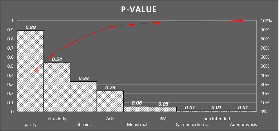
Figure 1- p-value of main results between endometriosis and non-endometriosis groups
Table 1- characteristics main of patients Endometriosis
| Statistics | ||||||
| Age patient | Gravidity | Parity | Menstrual period | BMI | ||
| N | Valid | 20 | 20 | 20 | 20 | 20 |
| Missing | 1 | 1 | 1 | 1 | 1 | |
| Mean | 41.8500 | 2.10 | 1.23 | 6.5000 | 25.5500 | |
| Std. Error of Mean | .44293 | .177 | .094 | .25649 | .81265 | |
| Median | 42.0000 | 2.11 | 1.10 | 6.5000 | 25.0000 | |
| Mode | 39.00a | 2a | 2 | 5.00a | 25.00 | |
| Std. Deviation | 1.98083 | .790 | .423 | 1.14708 | 3.63427 | |
| Range | 6.00 | 2 | 1 | 3.00 | 11.00 | |
| Sum | 837.00 | 42 | 25 | 130.00 | 511.00 | |
| a. Multiple modes exist. The smallest value is shown | ||||||
a. Multiple modes exist. The smallest value is shown

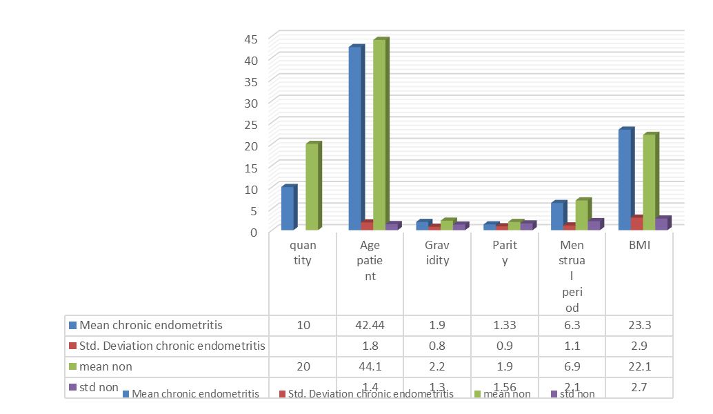
Figure 2- compression between Chronic Endometritis group and non-chronic Endometritis

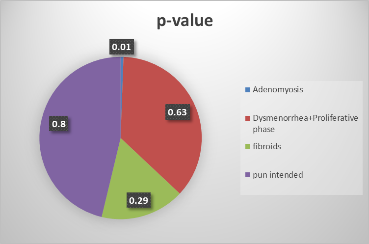
Figure 3- p-value of results between non-chronic endometritis and chronic endometritis
Table 2- Main characteristics of Group II (NON) non-endometriosis groups
| Statistics | ||||||
| AGE | parity | Menstrual | BMI | Gravidity | ||
| N | Valid | 10 | 10 | 10 | 10 | 10 |
| Missing | 0 | 0 | 0 | 0 | 0 | |
| Mean | 43.7000 | 1.8000 | 7.5000 | 22.7000 | 2.9000 | |
| Std. Error of Mean | .76085 | .13333 | .37268 | .42295 | .27689 | |
| Median | 43.5000 | 2.0000 | 7.0000 | 22.5000 | 3.0000 | |
| Mode | 43.00a | 2.00 | 7.00 | 22.00 | 2.00 | |
| Std. Deviation | 2.40601 | .42164 | 1.17851 | 1.33749 | .87560 | |
| Variance | 5.789 | .178 | 1.389 | 1.789 | .767 | |
| Range | 7.00 | 1.00 | 3.00 | 4.00 | 2.00 | |
| Percentiles | 25 | 41.7500 | 1.7500 | 6.7500 | 21.7500 | 2.0000 |
| 50 | 43.5000 | 2.0000 | 7.0000 | 22.5000 | 3.0000 | |
| 75 | 46.2500 | 2.0000 | 9.0000 | 24.0000 | 4.0000 | |
| a. Multiple modes exist. The smallest value is shown | ||||||
| age patient * BMI * Crosstabulation | ||||||||||||||
| Count | ||||||||||||||
| p | BMI | Total | ||||||||||||
| 20 | 21 | 23 | 24 | 25 | 26 | 28 | 29 | 30 | 31 | |||||
| Adenomyosis | age | 39 | 0 | 0 | 0 | 1 | 0 | 1 | ||||||
| 42 | 0 | 1 | 0 | 0 | 1 | 2 | ||||||||
| 43 | 1 | 0 | 0 | 0 | 0 | 1 | ||||||||
| 44 | 0 | 0 | 1 | 0 | 0 | 1 | ||||||||
| Total | 1 | 1 | 1 | 1 | 1 | 5 | ||||||||
| Dysmenorrhea+Proliferative phase | age | 39 | 1 | 0 | 0 | 0 | 0 | 1 | 0 | 2 | ||||
| 40 | 0 | 0 | 0 | 0 | 0 | 0 | 1 | 1 | ||||||
| 41 | 0 | 0 | 0 | 0 | 0 | 1 | 0 | 1 | ||||||
| 43 | 0 | 1 | 0 | 1 | 0 | 0 | 0 | 2 | ||||||
| 44 | 0 | 0 | 1 | 0 | 0 | 0 | 0 | 1 | ||||||
| 45 | 0 | 0 | 0 | 0 | 1 | 0 | 0 | 1 | ||||||
| Total | 1 | 1 | 1 | 1 | 1 | 2 | 1 | 8 | ||||||
| fibroids | age | 40 | 0 | 0 | 1 | 1 | ||||||||
| 41 | 1 | 0 | 0 | 1 | ||||||||||
| 44 | 0 | 1 | 0 | 1 | ||||||||||
| Total | 1 | 1 | 1 | 3 | ||||||||||
| pun intended | age | 40 | 0 | 1 | 0 | 1 | ||||||||
| 41 | 1 | 0 | 0 | 1 | ||||||||||
| 42 | 0 | 1 | 0 | 1 | ||||||||||
| 45 | 0 | 0 | 1 | 1 | ||||||||||
| Total | 1 | 2 | 1 | 4 | ||||||||||
| Total | age | 39 | 1 | 0 | 0 | 0 | 0 | 1 | 0 | 0 | 1 | 0 | 3 | |
| 40 | 0 | 1 | 0 | 0 | 0 | 0 | 0 | 1 | 0 | 1 | 3 | |||
| 41 | 1 | 0 | 1 | 0 | 0 | 0 | 0 | 0 | 1 | 0 | 3 | |||
| 42 | 0 | 1 | 0 | 1 | 0 | 0 | 0 | 0 | 0 | 1 | 3 | |||
| 43 | 0 | 0 | 1 | 0 | 1 | 0 | 1 | 0 | 0 | 0 | 3 | |||
| 44 | 0 | 0 | 0 | 1 | 1 | 1 | 0 | 0 | 0 | 0 | 3 | |||
| 45 | 0 | 0 | 0 | 0 | 1 | 0 | 0 | 1 | 0 | 0 | 2 | |||
| Total | 2 | 2 | 2 | 2 | 3 | 2 | 1 | 2 | 2 | 2 | 20 | |||
Table 3- demographic results of endometriosis groups

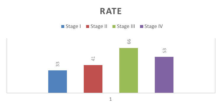
Figure 4 – results chronic endometritis in each stage of endometriosis
Table 4: Frequency Diseases of the female reproductive system (non-endometriosis groups)
| p | |||||
| Frequency | Percent | Valid Percent | Cumulative Percent | ||
| Valid | Adenomyosis | 2 | 20.0 | 20.0 | 20.0 |
| Dysmenorrhea+Proliferative phase | 3 | 30.0 | 30.0 | 50.0 | |
| fibroids | 3 | 30.0 | 30.0 | 80.0 | |
| pun intended | 2 | 20.0 | 20.0 | 100.0 | |
| Total | 10 | 100.0 | 100.0 | ||
Table 5- classification disease of non-endometriosis groups results according to age
| AGE * disease Crosstabulation | ||||||
| Count | ||||||
| p | Total | |||||
| Adenomyosis | Dysmenorrhea+Proliferative phase | fibroids | pun intended | |||
| AGE | 40.00 | 0 | 0 | 0 | 1 | 1 |
| 41.00 | 0 | 0 | 1 | 0 | 1 | |
| 42.00 | 0 | 1 | 0 | 0 | 1 | |
| 43.00 | 1 | 0 | 1 | 0 | 2 | |
| 44.00 | 0 | 1 | 0 | 1 | 2 | |
| 46.00 | 1 | 0 | 0 | 0 | 1 | |
| 47.00 | 0 | 1 | 1 | 0 | 2 | |
| Total | 2 | 3 | 3 | 2 | 10 | |
Table 6- characteristics of patient chronic endometritis according to disease
| chronic endometritis | |||||
| Frequency | Percent | Valid Percent | Cumulative Percent | ||
| Valid | 2 | 16.7 | 16.7 | 16.7 | |
| Adenomyosis | 2 | 16.7 | 16.7 | 33.3 | |
| Dysmenorrhea+Proliferative phase | 4 | 33.3 | 33.3 | 66.7 | |
| fibroids | 3 | 25.0 | 25.0 | 91.7 | |
| pun intended | 1 | 8.3 | 8.3 | 100.0 | |
| Total | 12 | 100.0 | 100.0 | ||
Table 7- characteristics of patient non-chronic endometritis groups according to disease
| non-chronic endometritis | |||||
| Frequency | Percent | Valid Percent | Cumulative Percent | ||
| Valid | Adenomyosis | 7 | 35.0 | 35.0 | 35.0 |
| Dysmenorrhea+Proliferative phase | 6 | 30.0 | 30.0 | 65.0 | |
| fibroids | 4 | 20.0 | 20.0 | 85.0 | |
| pun intended | 3 | 15.0 | 15.0 | 100.0 | |
| Total | 20 | 100.0 | 100.0 | ||
Samples were collected from Alhusaina Hospital, Karbala, Iraq, and Fatimah Al-Zahraa Hospital For gynecology and obstetrics, Baghdad, Iraq. where the research aimed to Exploring the relationship generated between chronic endometriosis in the endometriotic group Through the use of statistical analysis to analyze data and demographic information of patients, mean value and SD of the endometriosis group were (41.85 ± 1.98) and the ratio of gravidity (2.10 ± 0,790) And the proportion of the Menstrual period (6.5 ±1.14) and when comparing these ratios with a group non-endometriosis groups We find that these percentages are rather low in terms of age, parity, gravidity and Menstrual period
In Table 3, which looks to know the results of the demographic relationship between BMI and diseases for the group of Endometriosis, where we find that Adenomyosis was at a rate of 20%, and for the Dysmenorrhea + Proliferative phase, it was for 8 out of 20 patients, and it was associated with women who had a BMI that was Between 26 and 31 (kg/m2), as for the other diseases formed by fibroids and pun intended, it was present in 7 out of 20 patients.
In a previous study by AG Tylor, it was found that the level of pathological changes in the endometrium was significantly higher in the group of patients with endometriosis than in the women without this disease (50% vs 27.1%). Chronic endometriosis was diagnosed in 22.03% of cases (26/118) in the group of patients with endometriosis and in 11.02% of cases (10/118) in the group of patients without endometriosis. Endometriosis was detected in 17.8% of cases (21/118) among endometriotic patients and in 8.47% of cases (10/118) among patients from the comparison group.
There is little correlation between the severity of endometriotic lesions and the severity or duration of symptoms: symptoms are mild in some who have significantly large lesions, while symptoms are severe in others with few lesions. Symptoms often improve after menopause, but painful symptoms can persist in some cases. Chronic pain may be due to pain centers in the brain becoming over-responsive over time (central sensitization), which can occur at any stage of ectopic endometriosis
It is suggested that endometriosis also has immunological changes at the level of the endometrium. Thus, it appears that the immune system is significantly altered not only in endometrial lesions but also within the endometrium of women with endometriosis, affecting the viability and function of the endometrium.
The occurrence of endometriotic foci is closely associated with pathological changes in the endometrium. However, the sequence of development of pathological changes in the uterine cavity and in the pelvic cavity is still not clear, and no statistically significant differences were found between endometriosis and non-endometriosis groups.
Recommendation
Doctor recommends taking over-the-counter pain relievers such as nonsteroidal anti-inflammatory drugs (NSAIDs) that help relieve symptoms associated with endometriosis.
Hormonal therapy that helps reduce the amount of estrogen that the body produces, thus stopping the menstrual cycle and bleeding caused by endometriosis.
Abbott, J., et al. "Laparoscopic Excision of Endometriosis: A Randomized, Placebo-Controlled Trial." Fertility and Sterility, vol. 82, 2004, pp. 878–884. DOI: 10.1016/j.fertnstert.2004.03.046.
Adamson, G., et al. "Creating Solutions in Endometriosis: Global Collaboration Through the World Endometriosis Research Foundation." Journal of Endometriosis, vol. 2, 2010, pp. 3–6. DOI: 10.1177/228402651000200102.
Adamson, G.D. "Endometriosis Classification: An Update." Current Opinion in Obstetrics and Gynecology, vol. 23, 2011, pp. 213–220. DOI: 10.1097/gco.0b013e328348a3ba.
Alvarez, P., et al. "Role of Nociceptor Estrogen Receptor GPR30 in a Rat Model of Endometriosis Pain." PAIN®, vol. 155, no. 12, 2014, pp. 2680–2686.
Amaral, V.F., et al. "Positive Correlation Between Serum and Peritoneal Fluid CA-125 Levels in Women with Pelvic Endometriosis." Sao Paulo Medical Journal, vol. 124, 2006, pp. 223–227. DOI: 10.1590/s1516-31802006000400010.
Anaf, V., et al. "Pain, Mast Cells, and Nerves in Peritoneal, Ovarian, and Deep Infiltrating Endometriosis." Fertility and Sterility, vol. 86, 2006, pp. 1336–1343. DOI: 10.1016/j.fertnstert.2006.03.057.
Anaf, V., et al. "Relationship Between Endometriotic Foci and Nerves in Rectovaginal Endometriotic Nodules." Human Reproduction, vol. 15, 2000, pp. 1744–1750. DOI: 10.1093/humrep/15.8.1744.
Matalliotakis, I.M., et al. "Serum Concentrations of Growth Factors in Women with and Without Endometriosis: The Action of Anti-Endometriosis Medicines." International Immunopharmacology, vol. 3, 2003, pp. 81–90.
Banu, S.K., et al. "Cyclooxygenase-2 Regulates Survival, Migration, and Invasion of Human Endometriotic Cells Through Multiple Mechanisms." Endocrinology, vol. 149, 2008, pp. 1180–1189. DOI: 10.1210/en.2007-1168.
Gazavani, R., et al. "Effect of Interleukin-8 (IL-8), Anti-IL-8, and IL-12 on Endometrial Cell Survival in Combined Endometrial Gland and Stromal Cell Cultures Derived from Women with and Without Endometriosis." Fertility and Sterility, vol. 77, 2002, pp. 62–67.
Bedaiwy, M.A., et al. "Prediction of Endometriosis with Serum and Peritoneal Fluid Markers: A Prospective Controlled Trial." Human Reproduction, vol. 17, 2002, pp. 426–431. DOI: 10.1093/humrep/17.2.426.
Hever, A., et al. "Human Endometriosis is Associated with Plasma Cells and Overexpression of B Lymphocyte Stimulator." Proceedings of the National Academy of Sciences of the USA, vol. 104, 2007, pp. 12451–12456.
Bohonyi, N., et al. "Local Upregulation of Transient Receptor Potential Ankyrin One and Transient Receptor Potential Vanilloid One Ion Channels in Rectosigmoid Deep Infiltrating Endometriosis." Molecular Pain, vol. 13, 2017, 1744806917705564.
Brierley, S.M., and D.R. Linden. "Neuroplasticity and Dysfunction After Gastrointestinal Inflammation." Nature Reviews Gastroenterology and Hepatology, vol. 11, 2014, pp. 611–627. DOI: 10.1038/nrgastro.2014.103.
Castro, J., et al. "Pharmacological Modulation of Voltage-Gated Sodium (NaV) Channels Alters Nociception Arising from the Female Reproductive Tract." Pain, 2020. DOI: 10.1097/j.pain.0000000000002036.