Color Stability of Acrylic Teeth for Mobile Dentures Under the Influence of Coffee, Carbonated Dark Drink, Wine and Tea
Amra Hadzipasic
1
1
Public Institution Health Centar of Sarajevo Canton, Organisation Unit Specialty Consultative Health Care, Dental department, Alajbegovića 1, 71000 Sarajevo, Bosnia and Herzegovina
Introduction: Crucial property of esthetic restorative materials is their color stability. Aim: to evaluate color stability of Gnathostar Ivoclar Vivadent acrylic artificial teeth in contact with the dark carbonated soft drink, coffee, sage tea and red wine, compared to distilled water. Materials and methods: a total of 15 samples were monitored, the same type, color and position of the teeth. The samples were divided into 5 groups, 3 teeth in each (n = 3). The artificial teeth were placed in an appropriate liquid at the room temperature. Tooth color was registered after 60 days, using spectrophotometer. Data were analyzed by the Kruskal-Wallis test. Results: dark carbonated beverage had the greatest pigmenting effect (ΔE = 22.1). Conclusion: there was no significant difference in staining ability between the four different fluids (χ² (3) = 4.385, p = 0.223). Each fluid showed perceptible difference in tooth color, compared to the control group.
Keywords
Color Change
Acrylic Teeth
Coffee
Red Wine
Tea
INTRODUCTION
For many years, the comparative and the subjective way of determining the shade of the teeth, dental color guides, have been used. Later were invented spectrophotometers that were able to detect the color change more accurately than a human eye [1] Spectrophotometer data are in the form of quantitative values.
Clark was the first to try to objectify and measure tooth color using the Munsell color scale [2].
The CIE, the Commission International de l’Eclairage, an international organization authorized for light, lighting, and color, presented the CIELab system, a uniform color scale, where L represents brightness, and a and b are chromatic characteristics [3].
In this study, the CIE L * c * h coordinate system was used. In Lch coordinate space, L represents brightness, c represents color and h is the hue. The values of c and h are calculated from the a and b coordinates in L * a * b * system. The difference in color is represented by delta values: ΔL *, ΔC *, and Δh *, compared under standard conditions. The total color difference (ΔE * ch) shows the difference between the L *, c *, and h * of the sample and the control measurement. This difference is calculated by the following formula (https://sensing.konica minolta.us):
ΔE ∗ ch = [(ΔL ∗) 2 + (Δc ∗) 2 + (Δh ∗) 2] ½
Delta values for L, c and h can be positive or negative. From the aspect of clinical assessment, if ΔE is less than 3.3, changes in the color of the restoration are acceptable by the human eye [4]. Perceptiveness is a difference in tooth color that can be detected, and acceptability is a tolerable difference in color [5-9].
Many researchers investigated color changes of the artificial or natural teeth, under the influence of different conditions and solutions [10-14].
MATERIALS AND METHODS
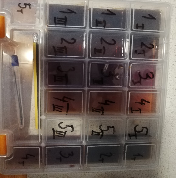
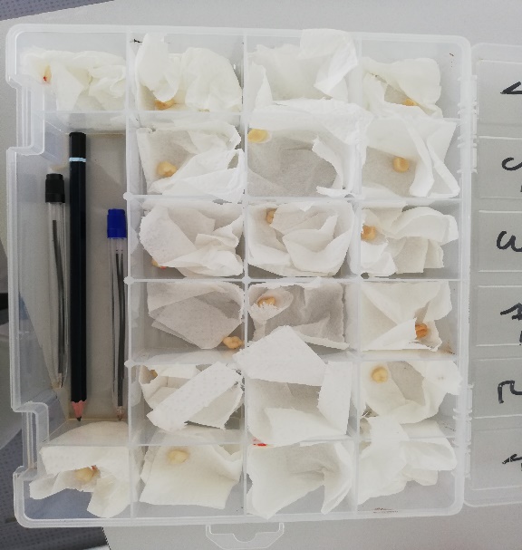
Fifteen upper left second acrylic molars, color shade 2B, size 84, type Gnathostar, produced by Ivoclar Vivadent, were divided into 5 groups, 3 in each series. Samples were immersed in Bosnian coffee, carbonated dark drink, red wine, sage tea, and distilled water, each tooth separately (Figure 1).
Figure 1: Artificial Teeth Immersed in Five Different Solutions
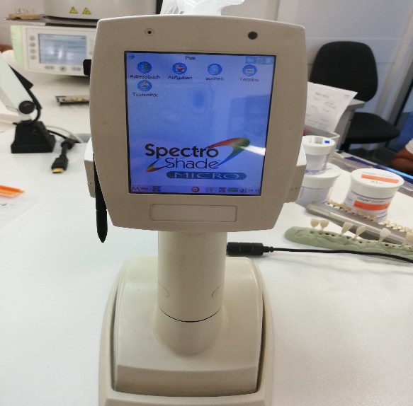
Figure 2: Apparatus On Which the Measurement was Performed (Spectroshade, Handy Dental, Mht S. P. A. Verona, Italy)
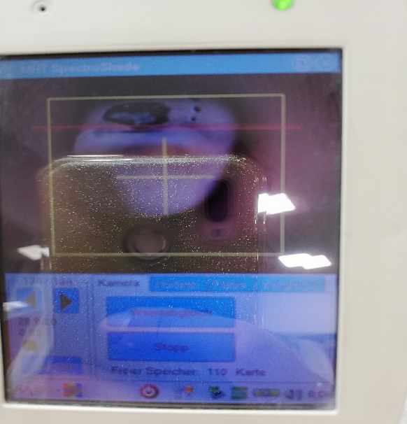
Figure 3: Tooth Scan
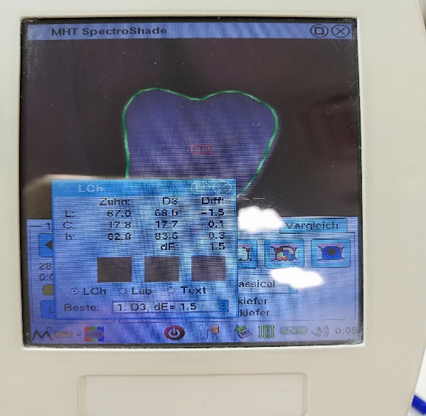
Figure 4: Tooth Color Data Obtained Using the Device
Figure 5: Teeth Cleaned and Air Dried Before Measuring
RESULTS
For each sample, L, c and h values were measured (Table 1).
ΔL, Δc and Δh were obtained by calculating the differences between L, c or h of the sample, and mean values of L, c and h from the control group of distilled water (Table 2).
Afterwards, ΔL, Δc and Δh were used in the formula ΔE ∗ ch = [(ΔL ∗) 2 + (Δc ∗) 2 + (Δh ∗) 2] ½, in order to calculate ΔE for each sample (Table 3).
Statistical analysis was performed by using the SPSS software version 26.0.
The absolute mean values of color change, ΔE, in each series, were much above the lower limit of human eye perception, which is 3.3 (5).
Carbonated dark drink caused the highest color change (ΔE = 22.1). Coffee and tea stained the teeth with similar intensity (ΔE 13.1, and 11.0, respectively, Table 4). The medians and standard deviations of ΔE are also presented (Table 4).
The Kruskal-Wallis H test was used to compare different solutions (Table 5).
Table 1: Measured Values For L, C and H.
Tooth sample
Coffee
Carbonated drink
Wine
Tea
Water
1
L
C
H
67.0
17.8
82.8
62.2
54.7
110.9
63.2, 42.1 100.5
70.1 27.4 90.6
70.1
31.5 102.6
2
L
C
H
70.4
37.4
105.2
67.7
16.9
85.0
72.7
28.9 97.8
73.6
22.4 88.1
69.1
36.4 103.4
3
L
C
H
71.5
28.2
100.1
72.6
16.9
85.5
72.8,
27.4 90.5
73.6
19.1 83.3
74.0
20.6
89.0
Mean values
L
C
H
71.06
29.5
98.33
Table 2. Values Δ L, Δ C and Δ H For Each Sample
Tooth
Difference
Coffee
Carbonated drink
Wine
Tea
I
Δ l
-4.96
-8.86
-7.86
-0.96
Δ c
-11.7
25.2
12.6
-2.1
Δ h
-16.1
12.57
2.17
-7.73
Ii
Δ l
-0.66
-3.36
1.64
2.54
Δ c
7.9
-12.6
-0.6
-7.1
Δ h
6.87
-13.33
-0.53
-10.23
Iii
Δ l
-0.44
1.54
1.74
2.54
Δc
-1.3
-12.6
-2.1
-10.4
Δ h
1.77
-12.83
-7.83
-15.03
Table 3. Δe Values for Every Sample
Δ E
Tooth Sample
Coffee
Carbonated Drink
Wine
Tea
1
20.31
29.52
15.01
8.07
2
10.49
18.68
1.83
12.70
3
2.29
18.05
8.29
18.45
Table 4. Mean Value for ΔE for Each Fluid
Parameters
Δe
Mean Value Δe
Standard Deviation
Median
Minimum
Maximum
Fluid
Coffee
11.0
9.0
10.5
2.3
20.3
Carbonated Drink
22.1
6.4
18.7
18.0
29.5
Wine
8.4
6.6
8.3
1.8
15.0
Tea
13.1
5.2
12.7
8.1
18.5
Table 5. ΔE Analysis Using Kruskal Wallis Test
Kruskal Walls test
ΔE
Chi-Square
4.385
df
3
Asymp. Sig.
.223
a. Kruskal Wallis Test, b. Grouping Variable: Fluid
p value less than 0.05 was considered as significant. There was no statistically significant difference in tooth color change between different fluids (χ² (3) = 4.385, p = 0.223, Table 5).
DISCUSSION
Goiato MC examined the coloring effect of coffee, wine, carbonated dark drink, two mouthwashes, and concluded that color changes occurred on acrylic dentures in all cases [10]. In a study by Indian authors Gupta R et al., it was shown that the composite is most discolored by coffee, tea and carbonated drink [11]. In another study, the discoloring effect of chlorhexidine, tannic acid and iron on plaque formed on acrylate was monitored. None of the solutions individually caused a statistically significant discoloration, but did cause it under the influence of iron after the use of chlorhexidine and tannic acid [12]. Guler AU et al. found that red wine and coffee and tea with sugar have the greatest coloring effect on acrylic provisional prosthetic material [13]. The color stability of acrylic and composite temporary materials was examined in a study conducted by Turker SB, et al., where coffee, tea, carbonated dark drink and orange juice led to discoloration of the material [14].
CONCLUSION
The study showed that the coloring ability of four different liquids, Bosnian coffee, tea, wine and carbonated drink is greatest in the case of carbonated dark drink and sage tea (acrylic Gnathostar Ivoclar Vivadent artificial teeth were used). There was no statistically significant difference in the coloring effect between four solutions.
REFERENCE
Browning, W.D. et al. "A comparison of human raters and an intra-oral spectrophotometer." Operative Dentistry, vol. 34, no. 3, 2009, pp. 337-343.
Clark, E.B. "Tooth color selection." Journal of the American Dental Association, vol. 20, 1933, pp. 1065-1073.
Vichi, A., M. Ferrari and C.L. Davidson. "Color and opacity variations in three different resin-based composite products after water aging." Dental Materials, vol. 20, no. 6, 2004, pp. 530-534.
Wee, A.G. et al. "Use of a porcelain color discrimination test to evaluate color difference formulas." The Journal of Prosthetic Dentistry, vol. 98, no. 2, 2007, pp. 101-109.
Douglas, R.D., T.J. Steinhauer and A.G. Wee. "Intraoral determination of the tolerance of dentists for perceptibility and acceptability of shade mismatch." The Journal of Prosthetic Dentistry, vol. 97, no. 4, 2007, pp. 200-208.
Lindsey, D.T. and A.G. Wee. "Perceptibility and acceptability of CIELAB color differences in computer-simulated teeth." Journal of Dentistry, vol. 35, no. 7, 2007, pp. 593-599.
Ghinea, R. et al. "Color difference thresholds in dental ceramics." Journal of Dentistry, vol. 38, 2010, pp. e57-e64.
Douglas, R.D. and J.D. Brewer. "Acceptability of shade differences in metal ceramic crowns." The Journal of Prosthetic Dentistry, vol. 79, no. 3, 1998, pp. 254-260.
Goiato, M.C. et al. "Effect of different solutions on color stability of acrylic resin-based dentures." Brazilian Oral Research, vol. 28, no. 1, 2014, pp. 1-7.
Gupta, R. et al. "A spectrophotometric evaluation of color changes of various tooth colored veneering materials after exposure to commonly consumed beverages." The Journal of Indian Prosthodontic Society, vol. 5, no. 2, 2005, pp. 72-78.
Nordbo, H., A. Attramadal and H.M. Eriksen. "Iron discoloration of acrylic resin exposed to chlorhexidine or tannic acid: a model study." The Journal of Prosthetic Dentistry, vol. 49, no. 1, 1983, pp. 126-129.
Guler, A.U. et al. "Effects of different drinks on stainability of resin composite provisional restorative materials." The Journal of Prosthetic Dentistry, vol. 94, no. 2, 2005, pp. 118-124.
Türker, S.B., A. Kocak and E. Aktepe. "Effect of five staining solutions on the colour stability of two acrylics and three composite resins based provisional restorations." The European Journal of Prosthodontics and Restorative Dentistry, vol. 14, no. 3, 2006, pp. 121-125.
License
Creative Commons Attribution-NonCommercial-NoDerivatives 4.0 International License
All papers should be submitted electronically. All submitted manuscripts must be original work that is not under submission at another journal or under consideration for publication in another form, such as a monograph or chapter of a book. Authors of submitted papers are obligated not to submit their paper for publication elsewhere until an editorial decision is rendered on their submission. Further, authors of accepted papers are prohibited from publishing the results in other publications that appear before the paper is published in the Journal unless they receive approval for doing so from the Editor-In-Chief.
Himalayan Journal of Applied Medical Sciences and Research open access articles are licensed under a Creative Commons Attribution-Share A like 4.0 International License. This license lets the audience to give appropriate credit, provide a link to the license, and indicate if changes were made and if they remix, transform, or build upon the material, they must distribute contributions under the same license as the original.
Recommended Articles
Research Article
Ethnomedicinal Plants for Obesity Management - Phytochemicals, Molecular Targets and Prospects of Green Nanotechnology
Arpana Ashokrao Durge,
Utpal Jagdish Dongre
Published: 25/03/2026
Download PDF
Cite
x
APA
Durge, A. A. & Dongre, U. J. (2026). Ethnomedicinal Plants for Obesity Management - Phytochemicals, Molecular Targets and Prospects of Green Nanotechnology. Himalayan Journal of Applied Medical Sciences and Research, 7(1), 1-6.
MLA
Durge, Arpana Ashokrao and Utpal Jagdish Dongre. "Ethnomedicinal Plants for Obesity Management - Phytochemicals, Molecular Targets and Prospects of Green Nanotechnology." Himalayan Journal of Applied Medical Sciences and Research 7.1 (2026): 1-6.
Chicago
Durge, Arpana Ashokrao and Utpal Jagdish Dongre. "Ethnomedicinal Plants for Obesity Management - Phytochemicals, Molecular Targets and Prospects of Green Nanotechnology." Himalayan Journal of Applied Medical Sciences and Research 7, no. 1 (2026): 1-6.
Harvard
Durge, A. A. and Dongre, U. J. (2026) 'Ethnomedicinal Plants for Obesity Management - Phytochemicals, Molecular Targets and Prospects of Green Nanotechnology' Himalayan Journal of Applied Medical Sciences and Research 7(1), pp. 1-6.
Vancouver
Durge AA, Dongre UJ. Ethnomedicinal Plants for Obesity Management - Phytochemicals, Molecular Targets and Prospects of Green Nanotechnology. Himalayan Journal of Applied Medical Sciences and Research. 2026 Jan;7(1):1-6.
Download PDF
Research Article
Digital Implantology: Current Concepts, Clinical Applications, Limitations and Future Directions - A Narrative Review
Anukrity Chandra
Published: 30/07/2026
Download PDF
Cite
x
APA
Chandra, A. (2026). Digital Implantology: Current Concepts, Clinical Applications, Limitations and Future Directions - A Narrative Review. Himalayan Journal of Applied Medical Sciences and Research, 7(2), 1-7.
MLA
Chandra, Anukrity. "Digital Implantology: Current Concepts, Clinical Applications, Limitations and Future Directions - A Narrative Review." Himalayan Journal of Applied Medical Sciences and Research 7.2 (2026): 1-7.
Chicago
Chandra, Anukrity. "Digital Implantology: Current Concepts, Clinical Applications, Limitations and Future Directions - A Narrative Review." Himalayan Journal of Applied Medical Sciences and Research 7, no. 2 (2026): 1-7.
Harvard
Chandra, A. (2026) 'Digital Implantology: Current Concepts, Clinical Applications, Limitations and Future Directions - A Narrative Review' Himalayan Journal of Applied Medical Sciences and Research 7(2), pp. 1-7.
Vancouver
Chandra A. Digital Implantology: Current Concepts, Clinical Applications, Limitations and Future Directions - A Narrative Review. Himalayan Journal of Applied Medical Sciences and Research. 2026 Jul;7(2):1-7.
Download PDF
Research Article
Study of the Role of Serum Interleukin-6 in Inflammation among Pregnant Women with COVID-19
Aynar Talib Samad,
Yossra Saleh Khudhur
Published: 27/07/2021
Download PDF
Cite
x
APA
Talib Samad, A. & Khudhur, Y. S. (2021). Study of the Role of Serum Interleukin-6 in Inflammation among Pregnant Women with COVID-19. Himalayan Journal of Applied Medical Sciences and Research, 2(2), 1-6.
MLA
Talib Samad, Aynar and Yossra Saleh Khudhur. "Study of the Role of Serum Interleukin-6 in Inflammation among Pregnant Women with COVID-19." Himalayan Journal of Applied Medical Sciences and Research 2.2 (2021): 1-6.
Chicago
Talib Samad, Aynar and Yossra Saleh Khudhur. "Study of the Role of Serum Interleukin-6 in Inflammation among Pregnant Women with COVID-19." Himalayan Journal of Applied Medical Sciences and Research 2, no. 2 (2021): 1-6.
Harvard
Talib Samad, A. and Khudhur, Y. S. (2021) 'Study of the Role of Serum Interleukin-6 in Inflammation among Pregnant Women with COVID-19' Himalayan Journal of Applied Medical Sciences and Research 2(2), pp. 1-6.
Vancouver
Talib Samad A, Khudhur YS. Study of the Role of Serum Interleukin-6 in Inflammation among Pregnant Women with COVID-19. Himalayan Journal of Applied Medical Sciences and Research. 2021 Jul;2(2):1-6.
Download PDF
Research Article
Evalution CD11b, CD4, IL-17A and IL-23 as Biomarkers Immunological in Patients with Behçet’s Disease
Layla Saleh Abdul-Hassan,
...
Saeed hilal khudhair
Published: 13/07/2026
Download PDF
Cite
x
APA
Abdul-Hassan, L. S., Fadel, R. H. & khudhair, S. H. (2026). Evalution CD11b, CD4, IL-17A and IL-23 as Biomarkers Immunological in Patients with Behçet’s Disease. Himalayan Journal of Applied Medical Sciences and Research, 7(2), 1-7.
MLA
Abdul-Hassan, Layla S., Radhia H. Fadel and Saeed H. khudhair. "Evalution CD11b, CD4, IL-17A and IL-23 as Biomarkers Immunological in Patients with Behçet’s Disease." Himalayan Journal of Applied Medical Sciences and Research 7.2 (2026): 1-7.
Chicago
Abdul-Hassan, Layla S., Radhia H. Fadel and Saeed H. khudhair. "Evalution CD11b, CD4, IL-17A and IL-23 as Biomarkers Immunological in Patients with Behçet’s Disease." Himalayan Journal of Applied Medical Sciences and Research 7, no. 2 (2026): 1-7.
Harvard
Abdul-Hassan, L. S., Fadel, R. H. and khudhair, S. H. (2026) 'Evalution CD11b, CD4, IL-17A and IL-23 as Biomarkers Immunological in Patients with Behçet’s Disease' Himalayan Journal of Applied Medical Sciences and Research 7(2), pp. 1-7.
Vancouver
Abdul-Hassan LS, Fadel RH, khudhair SH. Evalution CD11b, CD4, IL-17A and IL-23 as Biomarkers Immunological in Patients with Behçet’s Disease. Himalayan Journal of Applied Medical Sciences and Research. 2026 Jul;7(2):1-7.
Hadzipasic, A. (2020). Color Stability of Acrylic Teeth for Mobile Dentures Under the Influence of Coffee, Carbonated Dark Drink, Wine and Tea. Himalayan Journal of Applied Medical Sciences and Research, 1(1), 1-4.
MLA
Hadzipasic, Amra. "Color Stability of Acrylic Teeth for Mobile Dentures Under the Influence of Coffee, Carbonated Dark Drink, Wine and Tea." Himalayan Journal of Applied Medical Sciences and Research 1.1 (2020): 1-4.
Chicago
Hadzipasic, Amra. "Color Stability of Acrylic Teeth for Mobile Dentures Under the Influence of Coffee, Carbonated Dark Drink, Wine and Tea." Himalayan Journal of Applied Medical Sciences and Research 1, no. 1 (2020): 1-4.
Harvard
Hadzipasic, A. (2020) 'Color Stability of Acrylic Teeth for Mobile Dentures Under the Influence of Coffee, Carbonated Dark Drink, Wine and Tea' Himalayan Journal of Applied Medical Sciences and Research 1(1), pp. 1-4.
Vancouver
Hadzipasic A. Color Stability of Acrylic Teeth for Mobile Dentures Under the Influence of Coffee, Carbonated Dark Drink, Wine and Tea. Himalayan Journal of Applied Medical Sciences and Research. 2020 Jul;1(1):1-4.


 Download PDF
 Download XML