Context: The incidence of breast cancer is steadily rising in India. The management and prognosis of breast cancer requires evaluation of Estrogen Receptor (ER), Progesterone Receptor (PR) and HER-2/neu as they have a great influence on the clinical outcome. Receptor status has thus become most informative in the molecular classification of breast tumours and their clinical assessment for treatment and further outcome. Aims: To assess the age incidence, gender predilection along with incidence of ER, PR, HER-2/neu in breast cancer at our institution. Settings and Design: Study population comprised of all patients with carcinoma breast (women and men) which have been proved on histopathology. Study design: Prospective study. Methods and Material: About 151 cases of breast carcinoma received in the department of pathology of Indira Gandhi Medical College, Shimla over a period of 24 months (May 2014 to May 2016) were analyzed with reference to ER, PR and HER-2/neu reactivity pattern. The histological type and grade of the tumour were analyzed and correlated with receptor status. Statistical Analysis Used: Descriptive statistical analysis and presented in terms of percentage. Results: Age of the patient varied from 23-92 years, the mean age being 49 years. 97.4% cases were female patient while, males constituting 2.6% cases. The mean age in male patients was 64.5 years. Majority of the tumours were infiltrating ductal carcinoma- Not Otherwise Specified (NOS) and histologically grade II predominantly. Majority of the cases belonged to luminal A category, followed by triple negative tumours. Conclusion: Prognostic accuracy improves with incorporation of IHC into the histopathology report along with the traditional TNM staging and histological grading. The determination of receptor status is also helpful in planning the appropriate treatment of the patient.
Keywords
Breast Cancer
Immunohistochemistry
Bloom Richardson Grade
Receptor Status
Estrogen Receptor
Progesterone Receptor
INTRODUCTION
Breast cancer causes about 5,00,000 deaths annually [1].In urban Indian population breast carcinoma is the most common cancer among women. It is second to cancer of cervix in the rural population, based on national cancer registry data (2006) [2]. The management and prognosis of breast cancer require the evaluation of hormone receptors and HER-2/neu. The hormone receptor status can predict the response to adjuvant hormone therapy [3]. HER-2/neu is an independent prognostic marker of clinical outcome [4].
Triple Negative Breast Cancer (TNBC) refers to the tumours with ER, PR and HER-2/neu negative status. They have aggressive histology, poor clinical outcome, short survival and are unresponsive to usual hormonal therapies [5].
The present study was conducted to find out the incidence of hormone receptors and HER-2/neu positivity in breast carcinoma and to assess incidence of TNBCs in the study population.
MATERIALS AND METHODS
Inclusion Criteria
Patients with carcinoma breast
Patients willing to give consent 25
Exclusion Criteria
Patients not giving consent
All benign breast diseases
About 151 cases of breast carcinoma received in the department of pathology of Indira Gandhi Medical College, Shimla over a period of 24 months (May 2014 to May 2016) were analyzed.
Method of Study
Histopathological diagnosis was established on routine haematoxylin and eosin staining of the sections
Immunohistochemistry for ER, PR and HER-2/neu was done on BioGenex Xmatrx Fully Automated Front-end Processing System
Out of the 151 cases, 147 (97.36%) were female patients and 4 (2.64%) male patients. Age of the patient varied from 23-92 years, the mean age being 49 years in females and 64.5 years in males.
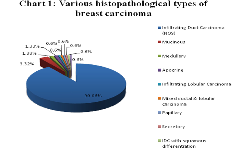
Most of the cases were reported as Infiltrating Duct Carcinoma, NOS type (90%) with 15 (10%) cases belonging to different categories which were medullary, apocrine, infilterating lobular, mixed, papillary, secretory, etc. (Figure 1).
Figure 1: Depicting Various Histopathological Types of Breast Cancer Seen in the Present Study
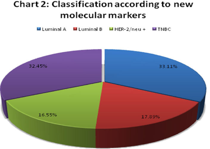
Figure 2: Classification According to New Molecular Markers
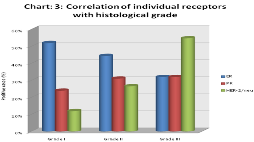
Figure 3: Correlation of Various Receptors with Tumor Grade
There was one patient with a past history of phyllodes tumour, right breast 7 year back, developing carcinoma breast left side subsequently. Another patient diagnosed as IDC (NOS) 13 years back, presented with metastatic deposits in the femur with pathological fracture of femur.
In 101 cases of IDC (NOS), modified Bloom Richardson Grading was done. 35 cases were of trucut biopsy breast and 15 cases were of histological types other than IDC (NOS). Grading was not done on them. 25 (24.75%) cases were reported as grade I, 45 (44.55%) cases as grade II and 31 (30.70%) as grade III tumours.
Majority of the cases belonged to luminal A category comprising of 50 (33.11%) cases, followed by triple negative tumours group. Triple negative group comprised of 49 (32.45%) cases. Luminal B group comprised of 27 (17.89%) cases. Least number of cases belonged to ER - ve, PR -ve and HER-2/neu +ve group comprising of 25 (16.55%) cases (Figure 2).
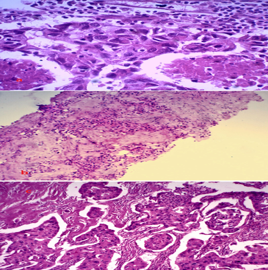
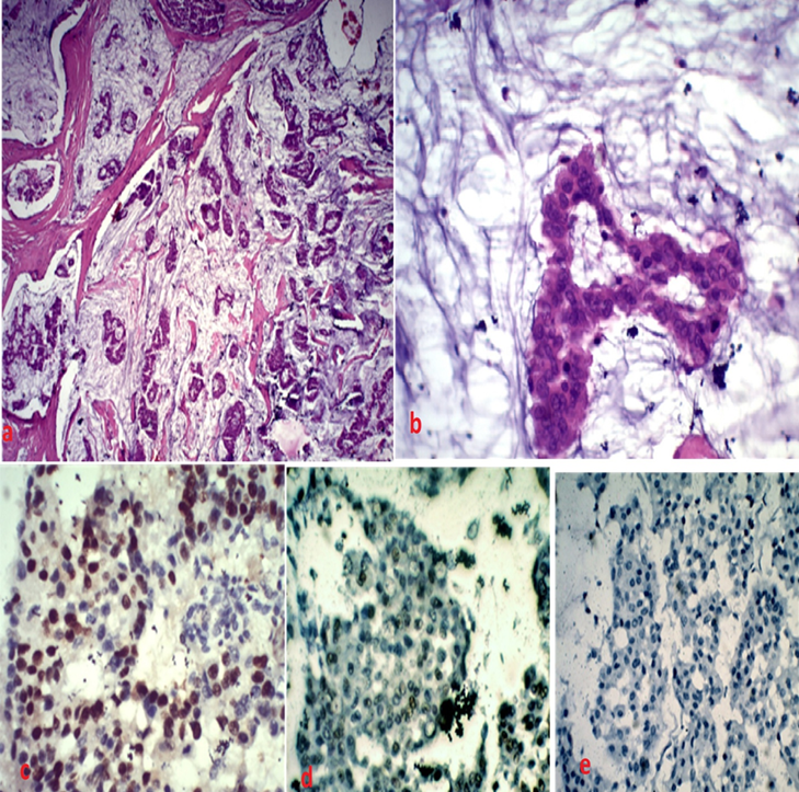
Figure 6: (a) Mucinous Carcinoma: Photomicrograph showing tubules and nests of tumour cells in pools of extracellular mucin (H&E X40). (b) Mucinous Carcinoma: High power view showing tubule lined by neoplastic cells in pool of extrcellular mucin (H&E X400). (c) Photomicrograph showing ER positivity. Allred score = 6 (3IS+3PS) (IHC X400) (d) Photomicrograph showing PR positivity. Allred score = 4 (2IS+2PS) (IHC X400). e) HER-2/neu negativity (IHC X400)
Majority of the low grade tumours expressed ER positivity. Majority of the high grade tumours showed HER-2/neu positivity (Figure 3).
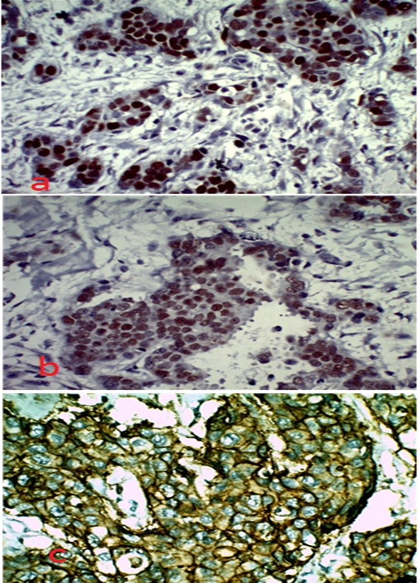
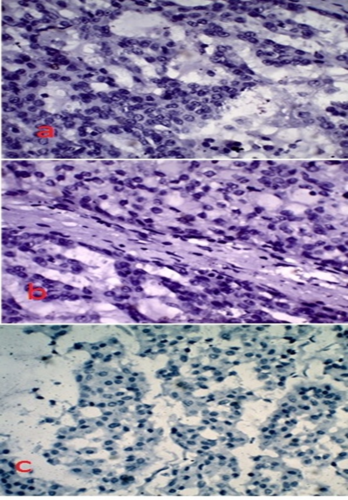
IDC (NOS) type demonstrated ER positivity in 43.38%. The next group was of mucinous carcinoma, comprising of five cases, out of which 4 cases were ER positive (Figures 4-7).
DISCUSSION
Breast cancer is considered a highly heterogeneous disease. Different types of this neoplasm exhibit variable histopathological and biological features, different clinical outcome and different response to systemic interventions.
In our study, age of the patient varied from 23-92 years with the mean age of 49 years. 37.10% were in the age group of 40-51 years, followed by, 51-60 year age group. The mean age was comparable with both national and international studies [6,7,8].
Majority of the patients were females 97.4%, males constituting only 2.6% cases. The mean age in male patients was 64.5 years. The incidence in male patient was higher as compared to various other studies, like [9,10]. This disparity could be due to large study size in both the studies. Zubedah et al. [11] and Nikhra et al. [12] in their studies showed comparable male incidence to our study.
Histopathological type is a well documented prognostic factor. In the present study the majority of the cases (90%) were of Infiltrating Duct Carcinoma (IDC) NOS, followed by mucinous/colloid carcinoma constituting 3.32% cases. This was in comparison with other Western and Indian studies wherein IDC-NOS was the most commonly seen variant of carcinoma breast [6,13,14,15].
The most commonly followed grading system is the Modified Bloom Richardson Grading system. It was largely conceived for Invasive Ductal Carcinoma, NOS type but can be used for other histopathological type also. It carries an important role in prognosis of the patients.
In our study, the grading was done only on IDC, NOS type. It was not done on trucut biopsy specimen and on histopathological types other than Infiltrating Duct Carcinoma, NOS. So a total of 101 cases were graded by Modified Bloom Richardson Grading system.In our study grade I was reported in 24.75% of the cases, grade II in 44.55% and grade III in 30.70% of the cases. The result regarding this were comparable with majority of other published studies [7,16].
In the present study majority of the cases belonged to luminal A group (33.11% cases) followed by TNBC group (32.45%). This was found to in accordance with the standard textbook of Rosai and Ackerman, according to which majority of the tumours belonged to luminal A category [17].
Figure 7:IHC on IDC (NOS) (a) ER Negative (b) PR Negative (c) HER2/neu Negative
Nikhra et al. [12], Ratnatunga et al. [18] and Onitito et al. [19] in their study also found out that majority of the tumours belonged to luminal A group.
A significant number of cases in the present study comprised of TNBC cases, 32.45% cases belonged to this category. This observation was comparable to other studies conducted in India by Sharma et al. [5], Ghosh et al. [10] and Nikhra et al. [12], where the incidence of TNBC was 29.8, 31.9 and 31.7% respectively.
In our study estrogen receptor positivity was seen in 42.40% of all the cases, which was comparable with Indian data but lower than the Western data [7,8,20,21]. PR positivity is variable in various India studies [8,12,20]. Progesterone positivity, in this study was seen in 32.5% of all the cases.
Present study, showed HER-2/neu positivity was seen in 35.10% of the total cases. In the studies conducted by Rajesh NG and Ambroise et al., HER-2/neu positivity was seen in 27.9 and 27.10% cases respectively [16,22]. These results were slightly lower than the present study, this could be due to the larger study population in the two studies. Jain et al. [8] reported HER-2/neu positivity in 35% cases. Nikhra et al. [12] observed HER-2/neu positivity in 32.5% of the cases. The results of both the studies were comparable with the present study.
In this study, HER-2/neu over expression was found to be inversely related to ER and PR expression. Majority of the tumours expressing ER/PR positivity were seen to be HER-2/neu negative and vice-versa. The inverse association between steroid hormone receptors and HER-2/neu has also been described in various clinical studies [23,24,25].
In the present study maximum number of patients in grade I showed ER positivity in 52% cases. Grade III showed maximum positivity with HER-2/neu (54.8% cases). This observation was also found to be in concordance with many studies [7,8,20].
CONCLUSION
To conclude this study showed that the maximum number of cases belonged to grade II Infiltrating Duct Carcinoma (NOS). Majority of the low grade tumours expressed the hormonal receptors. Majority of the high grade tumours showed HER-2/neu over-expression. ER/PR expression was inversely related to HER-2/neu over expression. Triple negative tumours constituted the second group after luminal A according to new molecular classification and were of high grade.
Demonstration of hormone receptors and HER-2/neu by immunohistochemistry in breast cancers is cost effective and can be done on paraffin processed tissue. Prognostic accuracy improves with incorporation of IHC into the histopathology report along with the traditional TNM staging and histological grading. The determination of receptor status is also helpful in planning the appropriate treatment of the patient.
REFERENCES
Benis, S. et al. "Prevalence of molecular subtypes and prognosis of invasive breast cancer in north-east of Morocco." BMC Research Notes, vol. 5, 2012, p. 436.
National Cancer Registry Programme. "Leading sites of cancer." Consolidated report of population based cancer registries 2001-2004 incidence and distribution of cancer, Bangalore: Coordinating Unit, National Cancer Registry Programme (ICMR), 2006, pp. 8-30.
Mudduwa, L.K. "Quick score of hormone receptor status of breast carcinoma: Correlation with the other clinicopathological prognostic parameters." Indian Journal of Pathology and Microbiology, vol. 52, no. 2, 2009, pp. 159.
Gown, A.M. "Current issues in ER and HER2 testing by IHC in breast cancer." Modern Pathology, vol. 21, no. 2, 2008, pp. S8-15.
Sharma, M. et al. "Triple negative breast cancer in people of north east India: Critical insights gained at a regional cancer centre." Asian Pacific Journal of Cancer Prevention, vol. 15, no. 11, 2014, pp. 4507-4511.
Li, J. et al. "A nation-wide multicenter 10-year (1999-2008) retrospective clinical epidemiological study of female breast cancer in China." BMC Cancer, vol. 11, 2011, p. 364.
Pathak, T.B. et al. "Estrogen and progesterone receptor expression in breast carcinoma." Journal of Pathology of Nepal, vol. 1, no. 2, 2011, pp. 100-103.
Jain, S.A. et al. "Study of ER PR and HER-2/neu reactivity pattern in the patient of breast cancer in northern part of India." IOSR Journal of Dental and Medical Sciences, vol. 13, 2014, pp. 9-19.
Al-Ahwal, M.S. "HER-2 positivity and correlations with other histopathologic features in breast cancer patients: hospital based study." Journal of Pakistan Medical Association, vol. 56, no. 2, 2006, pp. 65-68.
Ghosh, J. et al. "Estrogen progesterone and HER2 receptor expression in breast tumors of patients and their usage of HER2-targeted therapy in a tertiary care centre in India." Indian Journal of Cancer, vol. 48, no. 4, 2011, pp. 391-396.
Zubeda, S. et al. "Her-2/neu status: a neglected marker of prognostication and management of breast cancer patients in India." Asian Pacific Journal of Cancer Prevention, vol. 14, no. 4, 2013, pp. 2231-2235.
INTRODUCTION
Breast cancer causes about 5,00,000 deaths annually [1].In urban Indian population breast carcinoma is the most common cancer among women. It is second to cancer of cervix in the rural population, based on national cancer registry data (2006) [2]. The management and prognosis of breast cancer require the evaluation of hormone receptors and HER-2/neu. The hormone receptor status can predict the response to adjuvant hormone therapy [3]. HER-2/neu is an independent prognostic marker of clinical outcome [4].
Triple Negative Breast Cancer (TNBC) refers to the tumours with ER, PR and HER-2/neu negative status. They have aggressive histology, poor clinical outcome, short survival and are unresponsive to usual hormonal therapies [5].
The present study was conducted to find out the incidence of hormone receptors and HER-2/neu positivity in breast carcinoma and to assess incidence of TNBCs in the study population.
MATERIALS AND METHODS
Inclusion Criteria
Patients with carcinoma breast
Patients willing to give consent 25
Exclusion Criteria
Patients not giving consent
All benign breast diseases
About 151 cases of breast carcinoma received in the department of pathology of Indira Gandhi Medical College, Shimla over a period of 24 months (May 2014 to May 2016) were analyzed.
Method of Study
Histopathological diagnosis was established on routine haematoxylin and eosin staining of the sections
Immunohistochemistry for ER, PR and HER-2/neu was done on BioGenex Xmatrx Fully Automated Front-end Processing System
Out of the 151 cases, 147 (97.36%) were female patients and 4 (2.64%) male patients. Age of the patient varied from 23-92 years, the mean age being 49 years in females and 64.5 years in males.
Most of the cases were reported as Infiltrating Duct Carcinoma, NOS type (90%) with 15 (10%) cases belonging to different categories which were medullary, apocrine, infilterating lobular, mixed, papillary, secretory, etc. (Figure 1).
Figure 1: Depicting Various Histopathological Types of Breast Cancer Seen in the Present Study
Figure 2: Classification According to New Molecular Markers
Figure 3: Correlation of Various Receptors with Tumor Grade
There was one patient with a past history of phyllodes tumour, right breast 7 year back, developing carcinoma breast left side subsequently. Another patient diagnosed as IDC (NOS) 13 years back, presented with metastatic deposits in the femur with pathological fracture of femur.
In 101 cases of IDC (NOS), modified Bloom Richardson Grading was done. 35 cases were of trucut biopsy breast and 15 cases were of histological types other than IDC (NOS). Grading was not done on them. 25 (24.75%) cases were reported as grade I, 45 (44.55%) cases as grade II and 31 (30.70%) as grade III tumours.
Majority of the cases belonged to luminal A category comprising of 50 (33.11%) cases, followed by triple negative tumours group. Triple negative group comprised of 49 (32.45%) cases. Luminal B group comprised of 27 (17.89%) cases. Least number of cases belonged to ER - ve, PR -ve and HER-2/neu +ve group comprising of 25 (16.55%) cases (Figure 2).
Figure 6: (a) Mucinous Carcinoma: Photomicrograph showing tubules and nests of tumour cells in pools of extracellular mucin (H&E X40). (b) Mucinous Carcinoma: High power view showing tubule lined by neoplastic cells in pool of extrcellular mucin (H&E X400). (c) Photomicrograph showing ER positivity. Allred score = 6 (3IS+3PS) (IHC X400) (d) Photomicrograph showing PR positivity. Allred score = 4 (2IS+2PS) (IHC X400). e) HER-2/neu negativity (IHC X400)
Majority of the low grade tumours expressed ER positivity. Majority of the high grade tumours showed HER-2/neu positivity (Figure 3).
IDC (NOS) type demonstrated ER positivity in 43.38%. The next group was of mucinous carcinoma, comprising of five cases, out of which 4 cases were ER positive (Figures 4-7).
DISCUSSION
Breast cancer is considered a highly heterogeneous disease. Different types of this neoplasm exhibit variable histopathological and biological features, different clinical outcome and different response to systemic interventions.
In our study, age of the patient varied from 23-92 years with the mean age of 49 years. 37.10% were in the age group of 40-51 years, followed by, 51-60 year age group. The mean age was comparable with both national and international studies [6,7,8].
Majority of the patients were females 97.4%, males constituting only 2.6% cases. The mean age in male patients was 64.5 years. The incidence in male patient was higher as compared to various other studies, like [9,10]. This disparity could be due to large study size in both the studies. Zubedah et al. [11] and Nikhra et al. [12] in their studies showed comparable male incidence to our study.
Histopathological type is a well documented prognostic factor. In the present study the majority of the cases (90%) were of Infiltrating Duct Carcinoma (IDC) NOS, followed by mucinous/colloid carcinoma constituting 3.32% cases. This was in comparison with other Western and Indian studies wherein IDC-NOS was the most commonly seen variant of carcinoma breast [6,13,14,15].
The most commonly followed grading system is the Modified Bloom Richardson Grading system. It was largely conceived for Invasive Ductal Carcinoma, NOS type but can be used for other histopathological type also. It carries an important role in prognosis of the patients.
In our study, the grading was done only on IDC, NOS type. It was not done on trucut biopsy specimen and on histopathological types other than Infiltrating Duct Carcinoma, NOS. So a total of 101 cases were graded by Modified Bloom Richardson Grading system.In our study grade I was reported in 24.75% of the cases, grade II in 44.55% and grade III in 30.70% of the cases. The result regarding this were comparable with majority of other published studies [7,16].
In the present study majority of the cases belonged to luminal A group (33.11% cases) followed by TNBC group (32.45%). This was found to in accordance with the standard textbook of Rosai and Ackerman, according to which majority of the tumours belonged to luminal A category [17].
Figure 7:IHC on IDC (NOS) (a) ER Negative (b) PR Negative (c) HER2/neu Negative
Nikhra et al. [12], Ratnatunga et al. [18] and Onitito et al. [19] in their study also found out that majority of the tumours belonged to luminal A group.
A significant number of cases in the present study comprised of TNBC cases, 32.45% cases belonged to this category. This observation was comparable to other studies conducted in India by Sharma et al. [5], Ghosh et al. [10] and Nikhra et al. [12], where the incidence of TNBC was 29.8, 31.9 and 31.7% respectively.
In our study estrogen receptor positivity was seen in 42.40% of all the cases, which was comparable with Indian data but lower than the Western data [7,8,20,21]. PR positivity is variable in various India studies [8,12,20]. Progesterone positivity, in this study was seen in 32.5% of all the cases.
Present study, showed HER-2/neu positivity was seen in 35.10% of the total cases. In the studies conducted by Rajesh NG and Ambroise et al., HER-2/neu positivity was seen in 27.9 and 27.10% cases respectively [16,22]. These results were slightly lower than the present study, this could be due to the larger study population in the two studies. Jain et al. [8] reported HER-2/neu positivity in 35% cases. Nikhra et al. [12] observed HER-2/neu positivity in 32.5% of the cases. The results of both the studies were comparable with the present study.
In this study, HER-2/neu over expression was found to be inversely related to ER and PR expression. Majority of the tumours expressing ER/PR positivity were seen to be HER-2/neu negative and vice-versa. The inverse association between steroid hormone receptors and HER-2/neu has also been described in various clinical studies [23,24,25].
In the present study maximum number of patients in grade I showed ER positivity in 52% cases. Grade III showed maximum positivity with HER-2/neu (54.8% cases). This observation was also found to be in concordance with many studies [7,8,20].
CONCLUSION
To conclude this study showed that the maximum number of cases belonged to grade II Infiltrating Duct Carcinoma (NOS). Majority of the low grade tumours expressed the hormonal receptors. Majority of the high grade tumours showed HER-2/neu over-expression. ER/PR expression was inversely related to HER-2/neu over expression. Triple negative tumours constituted the second group after luminal A according to new molecular classification and were of high grade.
Demonstration of hormone receptors and HER-2/neu by immunohistochemistry in breast cancers is cost effective and can be done on paraffin processed tissue. Prognostic accuracy improves with incorporation of IHC into the histopathology report along with the traditional TNM staging and histological grading. The determination of receptor status is also helpful in planning the appropriate treatment of the patient.
REFERENCES
Benis, S. et al. "Prevalence of molecular subtypes and prognosis of invasive breast cancer in north-east of Morocco." BMC Research Notes, vol. 5, 2012, p. 436.
National Cancer Registry Programme. "Leading sites of cancer." Consolidated report of population based cancer registries 2001-2004 incidence and distribution of cancer, Bangalore: Coordinating Unit, National Cancer Registry Programme (ICMR), 2006, pp. 8-30.
Mudduwa, L.K. "Quick score of hormone receptor status of breast carcinoma: Correlation with the other clinicopathological prognostic parameters." Indian Journal of Pathology and Microbiology, vol. 52, no. 2, 2009, pp. 159.
Gown, A.M. "Current issues in ER and HER2 testing by IHC in breast cancer." Modern Pathology, vol. 21, no. 2, 2008, pp. S8-15.
Sharma, M. et al. "Triple negative breast cancer in people of north east India: Critical insights gained at a regional cancer centre." Asian Pacific Journal of Cancer Prevention, vol. 15, no. 11, 2014, pp. 4507-4511.
Li, J. et al. "A nation-wide multicenter 10-year (1999-2008) retrospective clinical epidemiological study of female breast cancer in China." BMC Cancer, vol. 11, 2011, p. 364.
Pathak, T.B. et al. "Estrogen and progesterone receptor expression in breast carcinoma." Journal of Pathology of Nepal, vol. 1, no. 2, 2011, pp. 100-103.
Jain, S.A. et al. "Study of ER PR and HER-2/neu reactivity pattern in the patient of breast cancer in northern part of India." IOSR Journal of Dental and Medical Sciences, vol. 13, 2014, pp. 9-19.
Al-Ahwal, M.S. "HER-2 positivity and correlations with other histopathologic features in breast cancer patients: hospital based study." Journal of Pakistan Medical Association, vol. 56, no. 2, 2006, pp. 65-68.
Ghosh, J. et al. "Estrogen progesterone and HER2 receptor expression in breast tumors of patients and their usage of HER2-targeted therapy in a tertiary care centre in India." Indian Journal of Cancer, vol. 48, no. 4, 2011, pp. 391-396.
Zubeda, S. et al. "Her-2/neu status: a neglected marker of prognostication and management of breast cancer patients in India." Asian Pacific Journal of Cancer Prevention, vol. 14, no. 4, 2013, pp. 2231-2235.
Nikhra, P. et al. "Study of ER (estrogen receptor) PR (progesterone receptor) and HER-2/neu (human epidermal growth factor receptor) expression by immunohistochemistry in breast carcinoma." International Journal of Biomedical and Advance Research, vol. 5, 2014, pp. 275-278.
Patil, V.W. et al. "Triple-negative (ER PR HER-2/neu) breast cancer in Indian women." Breast Cancer: Targets and Therapy, vol. 3, 2011, pp. 9-19.
Vaidyanathan, K. et al. "ErbB-2 expression and its association with other biological parameters of breast cancer among Indian women." Indian Journal of Cancer, vol. 47, no. 1, 2010, pp. 8-15.
Alfred, R. et al. "The prevalence and significance of oestrogen receptor positivity in breast cancer at the University Hospital of the West Indies Jamaica." West Indian Medical Journal, vol. 61, no. 8, 2012, pp. 795-801.
Rajesh, N.G. "Correlation of ER PR and Her2neu immunoprofile with the morphological prognostic factors in breast cancer-south Indian data." Regional Cancer Centre Bulletin, vol. 2, 2010, pp. 27-29.
Rosai, J. "Breast." Rosai and Ackerman’s Surgical Pathology, 9th ed., vol. 2, New York: Mosby (Elsevier), 2005, pp. 1763-1876.
Ratnatunga, N. and L.V. Liyanapathirana. "Hormone receptor expression and Her2/neu amplification in breast carcinoma in a cohort of Sri Lankans." The Ceylon Medical Journal, vol. 52, no. 4, 2007, pp. 133-136.
Onitilo, A.A. et al. "Breast cancer subtypes based on ER/PR and Her2 expression: Comparison of clinicopathologic features and survival." Clinical Medicine and Research, vol. 7, no. 1-2, 2009, pp. 4-13.
Bhagat, V.M. et al. "Correlation of hormonal receptor and Her-2/neu expression in breast cancer: a study at tertiary care hospital in south Gujarat." National Journal of Medical Research, vol. 2, no. 3, 2012, pp. 295-298.
Hussain, G.A. et al. "Correlations of hormone receptors (ER and PR) Her–2/neu and p53 expression in breast ductal carcinoma among Yemeni woman." The Open Cancer Immunology Journal, vol. 4, no. 1, 2011, pp. 1-9.
Ambroise, M. et al. "Immunohistochemical profile of breast cancer patients at a tertiary care hospital in South India." Asian Pacific Journal of Cancer Prevention, vol. 12, no. 3, 2011, pp. 625-629.
Zeillinger, R.A. et al. "HER-2 amplification steroid receptors and epidermal growth factor receptor in primary breast cancer." Oncogene, vol. 4, no. 1, 1989, pp. 109-114.
Marsigliante, S. et al. "Enzyme-linked immunosorbent assay of HER-2/neu gene product (p185) in breast cancer: its correlation with sex steroid receptors cathepsin D and histologic grades." Cancer Letters, vol. 75, no. 3, 1993, pp. 195-206.
Konecny, G. et al. "Quantitative association between HER-2/neu and steroid hormone receptors in hormone receptor-positive primary breast cancer." Journal of the National Cancer Institute, vol. 95, no. 2, 2003, pp. 142-153.
License
Creative Commons Attribution-NonCommercial-NoDerivatives 4.0 International License
All papers should be submitted electronically. All submitted manuscripts must be original work that is not under submission at another journal or under consideration for publication in another form, such as a monograph or chapter of a book. Authors of submitted papers are obligated not to submit their paper for publication elsewhere until an editorial decision is rendered on their submission. Further, authors of accepted papers are prohibited from publishing the results in other publications that appear before the paper is published in the Journal unless they receive approval for doing so from the Editor-In-Chief.
Himalayan Journal of Applied Medical Sciences and Research open access articles are licensed under a Creative Commons Attribution-Share A like 4.0 International License. This license lets the audience to give appropriate credit, provide a link to the license, and indicate if changes were made and if they remix, transform, or build upon the material, they must distribute contributions under the same license as the original.
Recommended Articles
Research Article
Digital Implantology: Current Concepts, Clinical Applications, Limitations and Future Directions - A Narrative Review
Anukrity Chandra
Published: 30/07/2026
Download PDF
Cite
x
APA
Chandra, A. (2026). Digital Implantology: Current Concepts, Clinical Applications, Limitations and Future Directions - A Narrative Review. Himalayan Journal of Applied Medical Sciences and Research, 7(2), 1-7.
MLA
Chandra, Anukrity. "Digital Implantology: Current Concepts, Clinical Applications, Limitations and Future Directions - A Narrative Review." Himalayan Journal of Applied Medical Sciences and Research 7.2 (2026): 1-7.
Chicago
Chandra, Anukrity. "Digital Implantology: Current Concepts, Clinical Applications, Limitations and Future Directions - A Narrative Review." Himalayan Journal of Applied Medical Sciences and Research 7, no. 2 (2026): 1-7.
Harvard
Chandra, A. (2026) 'Digital Implantology: Current Concepts, Clinical Applications, Limitations and Future Directions - A Narrative Review' Himalayan Journal of Applied Medical Sciences and Research 7(2), pp. 1-7.
Vancouver
Chandra A. Digital Implantology: Current Concepts, Clinical Applications, Limitations and Future Directions - A Narrative Review. Himalayan Journal of Applied Medical Sciences and Research. 2026 Jul;7(2):1-7.
Download PDF
Research Article
Study of the Role of Serum Interleukin-6 in Inflammation among Pregnant Women with COVID-19
Aynar Talib Samad,
Yossra Saleh Khudhur
Published: 27/07/2021
Download PDF
Cite
x
APA
Talib Samad, A. & Khudhur, Y. S. (2021). Study of the Role of Serum Interleukin-6 in Inflammation among Pregnant Women with COVID-19. Himalayan Journal of Applied Medical Sciences and Research, 2(2), 1-6.
MLA
Talib Samad, Aynar and Yossra Saleh Khudhur. "Study of the Role of Serum Interleukin-6 in Inflammation among Pregnant Women with COVID-19." Himalayan Journal of Applied Medical Sciences and Research 2.2 (2021): 1-6.
Chicago
Talib Samad, Aynar and Yossra Saleh Khudhur. "Study of the Role of Serum Interleukin-6 in Inflammation among Pregnant Women with COVID-19." Himalayan Journal of Applied Medical Sciences and Research 2, no. 2 (2021): 1-6.
Harvard
Talib Samad, A. and Khudhur, Y. S. (2021) 'Study of the Role of Serum Interleukin-6 in Inflammation among Pregnant Women with COVID-19' Himalayan Journal of Applied Medical Sciences and Research 2(2), pp. 1-6.
Vancouver
Talib Samad A, Khudhur YS. Study of the Role of Serum Interleukin-6 in Inflammation among Pregnant Women with COVID-19. Himalayan Journal of Applied Medical Sciences and Research. 2021 Jul;2(2):1-6.
Download PDF
Research Article
Evalution CD11b, CD4, IL-17A and IL-23 as Biomarkers Immunological in Patients with Behçet’s Disease
Layla Saleh Abdul-Hassan,
...
Saeed hilal khudhair
Published: 13/07/2026
Download PDF
Cite
x
APA
Abdul-Hassan, L. S., Fadel, R. H. & khudhair, S. H. (2026). Evalution CD11b, CD4, IL-17A and IL-23 as Biomarkers Immunological in Patients with Behçet’s Disease. Himalayan Journal of Applied Medical Sciences and Research, 7(2), 1-7.
MLA
Abdul-Hassan, Layla S., Radhia H. Fadel and Saeed H. khudhair. "Evalution CD11b, CD4, IL-17A and IL-23 as Biomarkers Immunological in Patients with Behçet’s Disease." Himalayan Journal of Applied Medical Sciences and Research 7.2 (2026): 1-7.
Chicago
Abdul-Hassan, Layla S., Radhia H. Fadel and Saeed H. khudhair. "Evalution CD11b, CD4, IL-17A and IL-23 as Biomarkers Immunological in Patients with Behçet’s Disease." Himalayan Journal of Applied Medical Sciences and Research 7, no. 2 (2026): 1-7.
Harvard
Abdul-Hassan, L. S., Fadel, R. H. and khudhair, S. H. (2026) 'Evalution CD11b, CD4, IL-17A and IL-23 as Biomarkers Immunological in Patients with Behçet’s Disease' Himalayan Journal of Applied Medical Sciences and Research 7(2), pp. 1-7.
Vancouver
Abdul-Hassan LS, Fadel RH, khudhair SH. Evalution CD11b, CD4, IL-17A and IL-23 as Biomarkers Immunological in Patients with Behçet’s Disease. Himalayan Journal of Applied Medical Sciences and Research. 2026 Jul;7(2):1-7.
Download PDF
Research Article
Bones in Balance: Awareness of Rickets and Nutritional Deficiencies Among the Hamirpur Community
Vishal Dhatwalia,
Swati Chandel
Published: 10/12/2024
Download PDF
Cite
x
APA
Dhatwalia, V. & Chandel, S. (2024). Bones in Balance: Awareness of Rickets and Nutritional Deficiencies Among the Hamirpur Community. Himalayan Journal of Applied Medical Sciences and Research, 5(2), 1-5.
MLA
Dhatwalia, Vishal and Swati Chandel. "Bones in Balance: Awareness of Rickets and Nutritional Deficiencies Among the Hamirpur Community." Himalayan Journal of Applied Medical Sciences and Research 5.2 (2024): 1-5.
Chicago
Dhatwalia, Vishal and Swati Chandel. "Bones in Balance: Awareness of Rickets and Nutritional Deficiencies Among the Hamirpur Community." Himalayan Journal of Applied Medical Sciences and Research 5, no. 2 (2024): 1-5.
Harvard
Dhatwalia, V. and Chandel, S. (2024) 'Bones in Balance: Awareness of Rickets and Nutritional Deficiencies Among the Hamirpur Community' Himalayan Journal of Applied Medical Sciences and Research 5(2), pp. 1-5.
Vancouver
Dhatwalia V, Chandel S. Bones in Balance: Awareness of Rickets and Nutritional Deficiencies Among the Hamirpur Community. Himalayan Journal of Applied Medical Sciences and Research. 2024 Jul;5(2):1-5.
Raina, N., Negi, L., Kaushal, V., Rana, A. & Sharma, R. (2021). Immuno-Histochemical Profile of Breast Cancer Patients at I.G.M.C. Shimla: A Tertiary Care Hospital in North India. Himalayan Journal of Applied Medical Sciences and Research, 2(2), 1-7.
MLA
Raina, Nidhi, et al. "Immuno-Histochemical Profile of Breast Cancer Patients at I.G.M.C. Shimla: A Tertiary Care Hospital in North India." Himalayan Journal of Applied Medical Sciences and Research 2.2 (2021): 1-7.
Chicago
Raina, Nidhi, Lalita Negi, Vijay Kaushal, Akshay Rana and Ritu Sharma. "Immuno-Histochemical Profile of Breast Cancer Patients at I.G.M.C. Shimla: A Tertiary Care Hospital in North India." Himalayan Journal of Applied Medical Sciences and Research 2, no. 2 (2021): 1-7.
Harvard
Raina, N., Negi, L., Kaushal, V., Rana, A. and Sharma, R. (2021) 'Immuno-Histochemical Profile of Breast Cancer Patients at I.G.M.C. Shimla: A Tertiary Care Hospital in North India' Himalayan Journal of Applied Medical Sciences and Research 2(2), pp. 1-7.
Vancouver
Raina N, Negi L, Kaushal V, Rana A, Sharma R. Immuno-Histochemical Profile of Breast Cancer Patients at I.G.M.C. Shimla: A Tertiary Care Hospital in North India. Himalayan Journal of Applied Medical Sciences and Research. 2021 Jul;2(2):1-7.


 Download PDF
 Download XML