Central retinal vein occlusion (CRVO) is one of the major causes of severe vision impairment and blindness. The prevalence of retinal artery or vein occlusion in the general population increases with age.ADPKD patients are at risk for developing central retinal vascular occlusions at a younger age. Here we report a case of ADPKD with central retinal vein occlusion.
Keywords
Central Retinal Vein
ADPKD
Vascular Occlusions
INTRODUCTION
Central retinal vein occlusion (CRVO) is one of the major causes of severe vision impairment and blindness [1]. The prevalence of retinal artery or vein occlusion in the general population increases with age [2]. The prevalence of central retinal vein occlusion (CRVO) is 0.1 to 0.7 percent in population-based studies [3]. CRVO occurs at 0.7% in persons between 49 and 60 years of age. The prevalence progressively increases to 4.6% in persons older than 80 years [4]. ADPKD patients are at risk for developing central retinal vascular occlusions at a younger age [5]. Autosomal dominant polycystic kidney disease (ADPKD) is a systemic disease with renal and extra renal, cystic and non-cystic manifestations. The cystic disease involves kidney, liver, pancreas, seminal vesicles and meninges [6]. The risk factors for developing CRVO include hypertension, diabetes, hyperlipidaemia, cigarette smoking, hyperviscosity or hypercoagulable states and hyperhomocysteinaemia [7].
Case Report
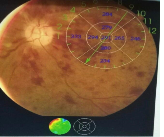
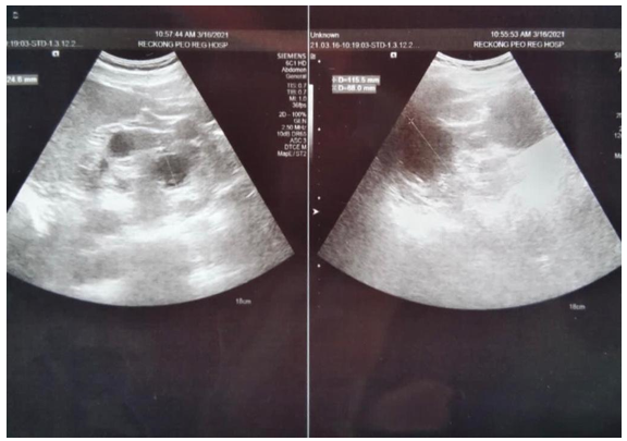
A 53-year-old male presented with a sudden loss of vision in his left eye for last 3 days. The vision loss occurred without any associated symptoms or notable preceding event.He had no prior vision problem. On physical examination, blood pressure was 158/88 mmHg. Visual acuity in left eye was counting finger at 1 feet; the right eye was unaffected (OD: 20/20). Pupil examination showed relative afferent pupillary defect in left eye. Intraocular pressures were 12 mm of Hg in right and 18 mm of Hg in left eye. Extraocular movements were normal. Slit examination showed normal anterior segments. Ophthalmoscopic examination of the left eye revealed pale disc, multiple areas of retinal haemorrhages with venous dilatations adjacent to the disc and cotton wool patches. ( Figure 1). These findings were consistent with combined central retinal vein occlusion (CRVO). Basic investigations were done which revealed azotemia (S.creatinine 2.2 mg/dl). Rest of the baseline investigations were normal. USG Kidneys were done which showed multiple renal cysts with mild raised cortical echogenicity consistent with signs of polycystic kidney disease. Case was referred to medicine department for same. Diagnosis of adult polycystic kidney disease was made. He was prescribed amlodipine and chlorothiazide for better control of hypertension. He was advised intravitreal injections of bevacizumab and was put on topical antiglaucoma medications.
Figure 1: Fundus Photograph Showing Pale Disc, Multiple Retinal Hemorrhages and Cotton Wool Spots
Apart from a diagnosis of ADPKD, the only predisposing condition was hypertension. We propose that retinal vascular occlusion may be a manifestation of the vasculopathy associated with ADPKD ( Autosomal dominant polycystic kidney disease).
DISCUSSION
CRVO has two types: a) Nonischemic (70%): which is characterized by vision that is better than 20/200, 16% progress to nonperfused; 50% resolve completely without treatment; defined as <10 disk diameter (DD) of capillary nonperfusion. b)Ischemic (30%): which is defined as more than 10 DD of nonperfusion; patients are usually older and have worse vision; 60% develop iris NV; up to 33% develop neovascular glaucoma; 10% are combined with branch retinal arterial occlusion (usually cilioretinal artery due to low perfusion pressure of choroidal system) [8]. Ophthalmoscopic examination typically shows retinal haemorrhages with venous dilatation and cotton wool spots for CRVO; and retinal/macular oedema. Long-term complications include retinal neovascularization, macular detachment with vitreous haemorrhage and neovascular glaucoma. The final visual acuity depends on the visual acuity at presentation, the duration of visual impairment and the severity of the subsequent complications [9-10].Qi Qian et al reported occurrence of central retinal vascular occlusions in eight patients with ADPKD at a median age of 51.2 years, contrasted with an approximately 63–75 years in the general population [2,11]. Retinal vascular occlusion is another manifestation of the systemic vasculopathy in ADPKD. Increased awareness of this manifestation could lead to an earlier diagnosis and treatment before irreversible vision loss develops [11].
CONCLUSION
When a patient less than 50 years of age presents with central retinal vein occlusion, other causes such as ADPKD should be specifically considered. Detailed history taking, supportive examinations and imaging should be performed. Thus thorough examination helps in early diagnosis and prompt treatment may prevent progress of disease and loss of vision.
Financial support and sponsorship: NIL
Conflict of interest: There is no conflict of interest
REFERENCE
The Central Vein Occlusion Study Group. "Natural history and clinical management of central retinal vein occlusion." Arch Ophthalmol, vol. 115, no. 4, 1997, pp. 486–491.
Lorentzen S.E. "Occlusion of the central retinal artery: a follow-up." Acta Ophthalmol (Copenh), vol. 47, no. 6, 1969, pp. 690–703.
Mohamed Q. et al. "Interventions for central retinal vein occlusion: an evidence-based systematic review." Ophthalmology, vol. 114, no. 3, 2007, pp. 507–519. doi:10.1016/j.ophtha.2006.11.011.
Mitchell P. et al. "Prevalence and associations of retinal vein occlusion in Australia: the Blue Mountains Eye Study." Arch Ophthalmol, vol. 114, no. 10, 1996, pp. 1243–1247.
Gabow P.A. et al. "Polycystic kidney disease: prospective analysis of nonazotemic patients and family members." Ann Intern Med, vol. 101, no. 2, 1984, pp. 238–247.
Torres V. et al. "Epidemiology, problems in diagnosis and management of polycystic kidney disease." In: Proceedings of the PKR Foundation, Kansas City, 1985, pp. 49–69.
Colucciello M. "Retinal vascular disease in hypertension: risk factor modification optimizes vision outcomes." Postgrad Med, vol. 117, no. 1, 2005, pp. 33–38, 41–42.
Friedman N. et al. "Central retinal vein occlusion." In: Review of Ophthalmology, 2005, ch. 11, p. 329.
Augsburger J.J., Magargal L.E. "Visual prognosis following treatment of acute central retinal artery obstruction." Br J Ophthalmol, vol. 64, no. 12, 1980, pp. 913–917.
The Central Vein Occlusion Study Group. "Natural history and clinical management of central retinal vein occlusion." Arch Ophthalmol, vol. 115, no. 4, 1997, pp. 486–491.
Qian Q., Younge B.R., Torres V.E. "Retinal arterial and venous occlusions in patients with ADPKD." Nephrol Dial Transplant, vol. 22, no. 6, 2007, pp. 1769–1771.
License
Creative Commons Attribution-NonCommercial-NoDerivatives 4.0 International License
All papers should be submitted electronically. All submitted manuscripts must be original work that is not under submission at another journal or under consideration for publication in another form, such as a monograph or chapter of a book. Authors of submitted papers are obligated not to submit their paper for publication elsewhere until an editorial decision is rendered on their submission. Further, authors of accepted papers are prohibited from publishing the results in other publications that appear before the paper is published in the Journal unless they receive approval for doing so from the Editor-In-Chief.
Himalayan Journal of Applied Medical Sciences and Research open access articles are licensed under a Creative Commons Attribution-Share A like 4.0 International License. This license lets the audience to give appropriate credit, provide a link to the license, and indicate if changes were made and if they remix, transform, or build upon the material, they must distribute contributions under the same license as the original.
Recommended Articles
Research Article
Ethnomedicinal Plants for Obesity Management - Phytochemicals, Molecular Targets and Prospects of Green Nanotechnology
Arpana Ashokrao Durge,
Utpal Jagdish Dongre
Published: 25/03/2026
Download PDF
Cite
x
APA
Durge, A. A. & Dongre, U. J. (2026). Ethnomedicinal Plants for Obesity Management - Phytochemicals, Molecular Targets and Prospects of Green Nanotechnology. Himalayan Journal of Applied Medical Sciences and Research, 7(1), 1-6.
MLA
Durge, Arpana Ashokrao and Utpal Jagdish Dongre. "Ethnomedicinal Plants for Obesity Management - Phytochemicals, Molecular Targets and Prospects of Green Nanotechnology." Himalayan Journal of Applied Medical Sciences and Research 7.1 (2026): 1-6.
Chicago
Durge, Arpana Ashokrao and Utpal Jagdish Dongre. "Ethnomedicinal Plants for Obesity Management - Phytochemicals, Molecular Targets and Prospects of Green Nanotechnology." Himalayan Journal of Applied Medical Sciences and Research 7, no. 1 (2026): 1-6.
Harvard
Durge, A. A. and Dongre, U. J. (2026) 'Ethnomedicinal Plants for Obesity Management - Phytochemicals, Molecular Targets and Prospects of Green Nanotechnology' Himalayan Journal of Applied Medical Sciences and Research 7(1), pp. 1-6.
Vancouver
Durge AA, Dongre UJ. Ethnomedicinal Plants for Obesity Management - Phytochemicals, Molecular Targets and Prospects of Green Nanotechnology. Himalayan Journal of Applied Medical Sciences and Research. 2026 Jan;7(1):1-6.
Download PDF
Research Article
Digital Implantology: Current Concepts, Clinical Applications, Limitations and Future Directions - A Narrative Review
Anukrity Chandra
Published: 30/07/2026
Download PDF
Cite
x
APA
Chandra, A. (2026). Digital Implantology: Current Concepts, Clinical Applications, Limitations and Future Directions - A Narrative Review. Himalayan Journal of Applied Medical Sciences and Research, 7(2), 1-7.
MLA
Chandra, Anukrity. "Digital Implantology: Current Concepts, Clinical Applications, Limitations and Future Directions - A Narrative Review." Himalayan Journal of Applied Medical Sciences and Research 7.2 (2026): 1-7.
Chicago
Chandra, Anukrity. "Digital Implantology: Current Concepts, Clinical Applications, Limitations and Future Directions - A Narrative Review." Himalayan Journal of Applied Medical Sciences and Research 7, no. 2 (2026): 1-7.
Harvard
Chandra, A. (2026) 'Digital Implantology: Current Concepts, Clinical Applications, Limitations and Future Directions - A Narrative Review' Himalayan Journal of Applied Medical Sciences and Research 7(2), pp. 1-7.
Vancouver
Chandra A. Digital Implantology: Current Concepts, Clinical Applications, Limitations and Future Directions - A Narrative Review. Himalayan Journal of Applied Medical Sciences and Research. 2026 Jul;7(2):1-7.
Download PDF
Research Article
Study of the Role of Serum Interleukin-6 in Inflammation among Pregnant Women with COVID-19
Aynar Talib Samad,
Yossra Saleh Khudhur
Published: 27/07/2021
Download PDF
Cite
x
APA
Talib Samad, A. & Khudhur, Y. S. (2021). Study of the Role of Serum Interleukin-6 in Inflammation among Pregnant Women with COVID-19. Himalayan Journal of Applied Medical Sciences and Research, 2(2), 1-6.
MLA
Talib Samad, Aynar and Yossra Saleh Khudhur. "Study of the Role of Serum Interleukin-6 in Inflammation among Pregnant Women with COVID-19." Himalayan Journal of Applied Medical Sciences and Research 2.2 (2021): 1-6.
Chicago
Talib Samad, Aynar and Yossra Saleh Khudhur. "Study of the Role of Serum Interleukin-6 in Inflammation among Pregnant Women with COVID-19." Himalayan Journal of Applied Medical Sciences and Research 2, no. 2 (2021): 1-6.
Harvard
Talib Samad, A. and Khudhur, Y. S. (2021) 'Study of the Role of Serum Interleukin-6 in Inflammation among Pregnant Women with COVID-19' Himalayan Journal of Applied Medical Sciences and Research 2(2), pp. 1-6.
Vancouver
Talib Samad A, Khudhur YS. Study of the Role of Serum Interleukin-6 in Inflammation among Pregnant Women with COVID-19. Himalayan Journal of Applied Medical Sciences and Research. 2021 Jul;2(2):1-6.
Download PDF
Research Article
Evalution CD11b, CD4, IL-17A and IL-23 as Biomarkers Immunological in Patients with Behçet’s Disease
Layla Saleh Abdul-Hassan,
...
Saeed hilal khudhair
Published: 13/07/2026
Download PDF
Cite
x
APA
Abdul-Hassan, L. S., Fadel, R. H. & khudhair, S. H. (2026). Evalution CD11b, CD4, IL-17A and IL-23 as Biomarkers Immunological in Patients with Behçet’s Disease. Himalayan Journal of Applied Medical Sciences and Research, 7(2), 1-7.
MLA
Abdul-Hassan, Layla S., Radhia H. Fadel and Saeed H. khudhair. "Evalution CD11b, CD4, IL-17A and IL-23 as Biomarkers Immunological in Patients with Behçet’s Disease." Himalayan Journal of Applied Medical Sciences and Research 7.2 (2026): 1-7.
Chicago
Abdul-Hassan, Layla S., Radhia H. Fadel and Saeed H. khudhair. "Evalution CD11b, CD4, IL-17A and IL-23 as Biomarkers Immunological in Patients with Behçet’s Disease." Himalayan Journal of Applied Medical Sciences and Research 7, no. 2 (2026): 1-7.
Harvard
Abdul-Hassan, L. S., Fadel, R. H. and khudhair, S. H. (2026) 'Evalution CD11b, CD4, IL-17A and IL-23 as Biomarkers Immunological in Patients with Behçet’s Disease' Himalayan Journal of Applied Medical Sciences and Research 7(2), pp. 1-7.
Vancouver
Abdul-Hassan LS, Fadel RH, khudhair SH. Evalution CD11b, CD4, IL-17A and IL-23 as Biomarkers Immunological in Patients with Behçet’s Disease. Himalayan Journal of Applied Medical Sciences and Research. 2026 Jul;7(2):1-7.
Negi, B. D. & Negi, S. (2021). Case Report: Central Retinal Vein Occlusion in Adpkd. Himalayan Journal of Applied Medical Sciences and Research, 2(1), 1-2.
MLA
Negi, Bhagwan D. and Shaloo Negi. "Case Report: Central Retinal Vein Occlusion in Adpkd." Himalayan Journal of Applied Medical Sciences and Research 2.1 (2021): 1-2.
Chicago
Negi, Bhagwan D. and Shaloo Negi. "Case Report: Central Retinal Vein Occlusion in Adpkd." Himalayan Journal of Applied Medical Sciences and Research 2, no. 1 (2021): 1-2.
Harvard
Negi, B. D. and Negi, S. (2021) 'Case Report: Central Retinal Vein Occlusion in Adpkd' Himalayan Journal of Applied Medical Sciences and Research 2(1), pp. 1-2.
Vancouver
Negi BD, Negi S. Case Report: Central Retinal Vein Occlusion in Adpkd. Himalayan Journal of Applied Medical Sciences and Research. 2021 Jan;2(1):1-2.


 Download PDF
 Download XML