Contents
Download PDF
pdf Download XML
979 Views
451 Downloads
Share this article
Research Article | Volume 4 Issue 2 (July-Dec, 2023) | Pages 1 - 4
Use of Ablative CO2 Laser For Treatment of Active Cutaneous Leishmaniasis
 ,
 ,
1
Department of Dermatology and Venereology, Baquba Teaching Hospital, Diyala, Iraq
Under a Creative Commons license
Open Access
Received
April 3, 2023
Revised
May 9, 2023
Accepted
June 19, 2023
Published
July 19, 2023
Abstract

Background: Cutaneous leishmaniasis is a major health problem in Iraq especially in the last decade because of forced internal displacement of population and bad infrastructure. Pentavalent antimonial drugs used as 1st line of management had many drawbacks, multiple courses, painfulness, unavailability and emergence of resistance. Carbon dioxide laser had been used for treatment of cutaneous leishmaniasis in many studies with shorter course and better cosmetic result. Objective: To evaluate the efficacy of ablative CO2 laser for treatment of active lesions of cutaneous lieshmaniasis in Iraqi patients. Patients and Methods: In this prospective therapeutic study conducted at department of dermatology in Baquba teaching hospital beginning from March 2019 to November 2020, 28 patients with total of 70 lesions of cutaneous leishmaniasis diagnosed clinically and by positive smear treated with continues ablative co2 laser and assessed at 2, 4, 8 and 12 weeks. Compared to group of 30 patients with 76 lesions treated with weekly intralesional injection of pentavalent antimonial injection. Healing defined as complete flattening of skin for both groups. Results: For group A mean age was 22.25±16.8 years, in group B mean of age is 16.10±16.4 years. There is no significant difference in number of lesions and duration of disease prior to treatment between both groups (p-value = 0.915) and (p-value = 0.057) respectively. Average duration of healing for group A (2.75±0.6 weeks), while in group B (5.70±1.5 weeks) with significant p-value of ˂0.001. Conclusion: Ablative co2 laser is an effective treatment for active cutaneous leishmaniasis with mostly single session treatment and shorter duration of healing.

Keywords
INTRODUCTION

Cutaneous Leishmaniasis (CL) is a major health problem in many developed countries including Iraq, Syria, Saudi Arabia and Iran it is mainly zoonotic disease (although anthroponotic transmission reported in some species) transmitted to human through bite of female phelobotomus sandflies [1]. CL divided in to two types: old world and new world CL [1].


Old World CL Endemic in Middle East (Include Iraq) and Africa 

During disasters like wars, forced internal displacement of population lead to collapse of health care system, inability of patients to reach health care centers and unsanitary life conditions [2-4].


According to WHO recommendations 1st line of treatment of CL is pentavalent antimonilial drugs meglumine antimoniate and sodium stibogluconate (intralesional or systemic) according to number and distribution of lesions. Drawback of this treatment include cardiotoxicity, painful, multiple injections and emergence of resistance [2]. 


Carbon dioxide laser had been evaluated in many studies as a type of physical treatment either alone in Continuous Wave (CW) mode [4,5-8] or in combination with other modalities of treatment in fractional mode [9,10], all revealed that CO2 laser was safe and effective compared to medical treatment regarding both duration of healing and better cosmetic result, because the main sequel of CL is disfiguring scars [5,6-11].

MATERIALS AND METHODS

This prospective therapeutic study was conducted in the department of dermatology and venereology in Baquba teaching hospital, beginning from March 2019 to November 2020, include 58 patients (32 males and 26 females) presented with 164 lesions of active cutaneous leishmaniasis, proved clinically and by positive smear.


Purpose of This Study and Procedure had been Explained for Each Patient

Patients divided in to two groups: Group A treated by continuous ablative CO2 laser (COPRO 3 machine) each lesion examined and pictured by I phone camera, then lesion and surrounded skin had been cleansed with normal saline, anesthesia done by injection of lidocaine 2%. Patient and doctor used protective eyeglasses then whole lesion and 3mm of apparently normal surrounding skin were ablated in layers till no more necrotic tissue and color of tissue turn to brown, wound covered by Fusidic sodium 2% ointment applied twice daily.


Lesions assessed after 2 weeks if there is an induration or new lesions appear at the periphery then after 4, 8 and 12 weeks.


After reepithelization and flattening of lesion patient allowed to send pictures for follow up.


Group B treated by local infiltration of pentavalent antimonial (sodium stibogluconate) injection weekly until blanching of lesion. For both groups complete flattening of the lesion was indicator of healing. Patients followed two years after treatment to exclude reoccurrence.


Patients with duration of lesions less than 4 weeks and patients received systemic treatment (systemic sodium stibogluconate, ketoconazole, fluconazole) excluded from study, while who received intralesional treatment with no response should stop treatment 4 weeks before ablation by CO2 laser.


Uncooperative patients mentally retarded, hyperactive easily irritated children (cannot wear protective goggles), patients with debilitating chronic illness and those with large lesions (more than 3 cm) in lower extremities and near the eyes were excluded from the study.


Statistical Analysis 

Data were entered into an excel sheet, then coded and analyzed using Statistical Package for the Social Sciences (SPSS) version 22, numerical data were presented in means and Standard Deviations (M±SD) while categorical data were presented in frequency and percentages, independent t-test and chi-square test were used accordingly to show the difference between the two groups of the study. In all statistical analysis p-value equal to or less than 0.05 is considered statistically significant.

RESULTS

The current study included 28 in group 1 (underwent CO2 laser therapy) compared to 30 in group 2 (underwent regular pentostam therapy). 32 (55.2%) of the sample were males and the remaining 26(44.8%) were females.


There was no significant difference between the two studied groups regarding age, number, position and duration of lesions prior to therapy as shown in Tables 1-3 respectively.


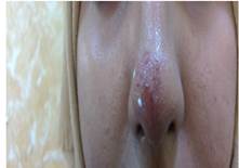
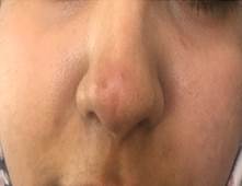
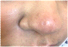
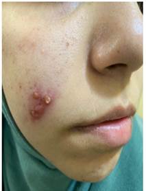
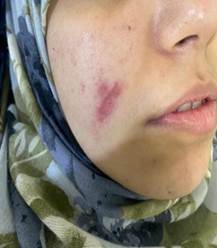
The average duration needed for healing in group 1: CO2 laser therapy was 2.75±0.6 weeks ranging from 2 to 4 weeks. Whereas in group 2: Pentostam therapy, it was 5.70±1.5 weeks, ranging between 3 to 10 weeks. There was a highly significant difference in healing duration between the two groups (p-value<0.001). Table 4 demonstrates the distribution of the two studied groups by the duration of healing (Figures 1-5).


Table 1: The Characteristics of the Studied Sample

VariablesGroup 1Group 2p-value

CO2 laser therapy

Pentostam therapy

N = 28

N = 30

Age in years

Mean ± SD (range)

22.25±16.8 (5-61)

16.10±16.4 (5-65)

0.165ǂ

Gender

N(%)

N(%)


Male

15(46.9%)

17(53.1%)

0.813§

Female

13(50.0%)

13(50.0%)

ǂIndependent t-test, §Chi-square test


Table 2: The Number and Position of Lesions in both Groups Pre-Treatment

PlaceGroup 1Group 2
CO2 laser therapypentostam therapy

Face

15 (21.4%)

17 (22.4%)

Neck

3 (4.3%)

2 (2.6%)

Trunk

5 (7.1%)

3 (3.9%)

Upper Arm

42 (60%)

44 (57.9%)

Lower Arm

5 (7.1%)

10 (13.2%)

Total number of lesions

70 (100%)

76(100%)


Table 3: Distribution of the Studied Sample by Lesions Characteristics   

VariablesGroup 1Group 2p valueǂ

CO2 laser therapy

Pentostam therapy

N = 28

N = 30

Total number of lesions

Mean ± SD (range)

2.50±1.4 (1-7)

2.5±0.8 (1-5)

0.915

Disease Duration in weeks

Mean ± SD (range)

7.85±3.5 (4-16)

6.23±2.8 (2-12)

0.057

ǂ independent t-test


Table 4; Distribution of the Studied Sample by the Duration of Healing

VariablesGroup 1Group 2p value

CO2 laser therapy

Pentostam therapy

N = 28

N = 30

Duration of healing in weeks

Mean ± SD (range)

2.75±0.6 (2-4)

5.70±1.5 (3-10)

0.000ǂ

ǂ Independent t-test


Figure 1: Lieshmania on dorsum of the nose before treatment


Figure 2:  Same Patient 4 Weeks after Contineous Ablative Laser


A close up of a person's ear

Description automatically generated with low confidence


Figure 2: Same Patient Small Atrophic Scar Compared to Lesion Size


Figure 4: Lieshmania on Check Before Treatment


Figure 5: Same Patient 4 Weeks after Contineous Ablative CO2 Laser

DISCUSSION

Aim of treatment in CL especially old-world disease is to shorten disease duration and prevent disfiguring scars because disease in general is self-limited and will be overcome by immunity in about 6-18 months [4] ablation of lesions by CW COlaser found to be effective and safe, selective destruction of diseased tissue, minimal effect on healthy surrounding skin, thermal effect extends beyond ablated tissue result in; best cosmetic result, control of bleeding and ensure sterilization which decrease possibility of infection; which result in shorter duration of healing and better cosmetic result [11].


In this research the main advantages of continuous ablative CO2 laser over the 1st line medical treatment (sodium stibogluconate) are single session treatment and shorter healing time, Babajev et al. [5], Asilian et al. [6] and Meymandi et al. [7] agree with these results; number of patients was larger in these studies. 


Shortage supply of sodium stibogluconate forced us to try other experimental modalities of treatment like intralesional metronidazole, same drawbacks of other medical treatment multiple treatment sessions, pain during injections and variable unpredictable response most important factors convincing patients (or their parents) to enrolled in this study was decreased number of visits or failure of intralesional treatment with both metronidazole and sodium stibogluconate.


Most patients enrolled to laser group were forcibly displaced from their homes and living in camps, it was hard for them to come weekly to receive treatment pain and edema were not a concern for patients, they advised to take simple anti-inflammatory drugs if it was annoying, infection not reported in any patient in laser treated group.


Unfortunately, scar could not be evaluated clinically in laser treated group, because most patients preferred to send pictures, but in general scar was small in comparison to the lesion by visual assessment.


Good and rapid response in laser treated group in this study could be attributed to small sample size, long duration of disease before treatment (at least 4 weeks), parasite species.

CONCLUSION

Continuous ablative CO2 laser is effective in treatment of active cutaneous lieshmaniasis with single session and short healing time.


Recommendation

further studies with larger sample size and correlation to lieshmania species are recommended.

REFERENCES
  1. Ali, Magda A. et al. "Epidemiological study of cutaneous leishmaniasis in some Iraqi provinces." Journal of Men’s Health, vol. 14, no. 4, 2018, pp. e18–e24.

  2. Alsamarai, Abdulghani Mohamed, et al. "Cutaneous leishmaniasis in Iraq: A continuing endemic disease." Journal of Drug Design and Research, vol. 3, no. 1, 2016, p. 1024.

  3. Hussein, Nawfal R. et al. "A clinical study of cutaneous leishmaniasis in a new focus in the Kurdistan region, Iraq." PLOS ONE, 31 May 2019.

  4. Salam, Nasir, Waleed Mohammed Al-Shaqha and Arezki Azzi. "Leishmaniasis in the Middle East: Incidence and epidemiology." PLOS Neglected Tropical Diseases, vol. 8, no. 10, 2014, e3208.

  5. Babajev, K.B. et al. "Treatment of cutaneous leishmaniasis using a carbon dioxide laser." Bulletin of the World Health Organization, vol. 69, no. 1, 1991, pp. 103–106.

  6. Asilian, A. et al. "Evaluation of CO2 laser efficacy in the treatment of cutaneous leishmaniasis." International Journal of Dermatology, vol. 43, 2004, pp. 736–738.

  7. Shamsi Meymandi, S. et al. "Efficacy of CO2 laser for treatment of anthroponotic cutaneous leishmaniasis, compared with combination of cryotherapy and intralesional meglumine antimoniate." Journal of the European Academy of Dermatology and Venereology, vol. 25, 2011, pp. 587–591.

  8. Zaib, Alam et al. "Comparison between efficacy of CO2 laser alone versus combination of cryotherapy and intralesional meglumine antimoniate in the treatment of cutaneous leishmaniasis." Journal of Pakistan Association of Dermatologists, vol. 29, no. 1, 2019, pp. 8–12.

  9. Jaffary, Fariba et al. "A comparison between the effects of Glucantime, topical trichloroacetic acid 50% plus Glucantime, and fractional carbon dioxide laser plus Glucantime on cutaneous leishmaniasis lesions." Dermatology Research and Practice, vol. 2016, Article ID 6462804.

  10. Artzi, Ofir, et al. "Fractional ablative CO2 laser followed by topical application of sodium stibogluconate for treatment of active cutaneous leishmaniasis: A randomized controlled trial." Acta Dermato-Venereologica, vol. 99, 2019, pp. 53–57.

  11. Sardana, Kabir, and Vijay K. Garg. Lasers in Dermatological Practice. Jaypee Brothers Medical Publishers, 2014.

License
CC BY-NC-ND
Creative Commons Attribution-NonCommercial-NoDerivatives 4.0 International License
Use of Ablative CO2 Laser For Treatment of Active Cutaneous Leishmaniasis © 2026 by Attaa A. Alhajji, Saad D. Hameed, Khaleel A. Al-Tameemi licensed under CC BY-NC-ND 4.0
All papers should be submitted electronically. All submitted manuscripts must be original work that is not under submission at another journal or under consideration for publication in another form, such as a monograph or chapter of a book. Authors of submitted papers are obligated not to submit their paper for publication elsewhere until an editorial decision is rendered on their submission. Further, authors of accepted papers are prohibited from publishing the results in other publications that appear before the paper is published in the Journal unless they receive approval for doing so from the Editor-In-Chief.
Himalayan Journal of Applied Medical Sciences and Research open access articles are licensed under a Creative Commons Attribution-Share A like 4.0 International License. This license lets the audience to give appropriate credit, provide a link to the license, and indicate if changes were made and if they remix, transform, or build upon the material, they must distribute contributions under the same license as the original.
Recommended Articles
Research Article
Digital Implantology: Current Concepts, Clinical Applications, Limitations and Future Directions - A Narrative Review
Published: 30/07/2026
Download PDF
Research Article
Study of the Role of Serum Interleukin-6 in Inflammation among Pregnant Women with COVID-19
Published: 27/07/2021
Download PDF
Research Article
Evalution CD11b, CD4, IL-17A and IL-23 as Biomarkers Immunological in Patients with Behçet’s Disease
...
Published: 13/07/2026
Download PDF
Research Article
Bones in Balance: Awareness of Rickets and Nutritional Deficiencies Among the Hamirpur Community
Published: 10/12/2024
Download PDF
Flowbite Logo
Najmal Complex,
Opposite Farwaniya,
Kuwait.
Email: support@himjournals.com

Useful Links
Order Hard Copy
Privacy policy
Terms and Conditions
Refund Policy
Others
About Us
Team Members
Contact Us
Online Payments
Join as Editor
Join as Reviewer
Subscribe to our Newsletter
Follow us
MOST SEARCHED KEYWORDS
scientific journal
 | 
business journal
 | 
medical journals
 | 
Scientific Journals
 | 
Academic Publisher
 | 
Peer-reviewed Journals
 | 
Open Access Journals
 | 
Impact Factor
 | 
Indexing Services
 | 
Journal Citation Reports
 | 
Publication Process
 | 
Impact factor of journals
 | 
Finding reputable journals for publication
 | 
Submitting a manuscript for publication
 | 
Copyright and licensing of published papers
 | 
Writing an abstract for a research paper
 | 
Manuscript formatting guidelines
 | 
Promoting published research
 | 
Publication in high-impact journals
Copyright © Himalayan Journals . All Rights Reserved.