Clinicopathological Profile and Outcomes of Unilateral Nasal Disease: A Prospective Study
Abdulkareem Hussien Dabi
1
,
Mohammed Naji Yaseen
2
,
Watheq A. Mohammed Ali
1
1
M.B.Ch.B., F.I.B.M.S., Ministry of Health, Al-Rusafa Health Directorate, Baghdad, Iraq
2
M.B.Ch.B., F.I.B.M.S. (ORL–HN), Ministry of Higher Education and Scientific Research. Ibn Sina University for Medical and Pharmaceutical Sciences, Ibn Sina Medical College, Iraq
Background: Unilateral nasal disease presents a diagnostic challenge in otolaryngology due to the potential risk of neoplastic pathology despite the predominance of inflammatory conditions. Early differentiation between benign and malignant causes is essential to prevent delayed diagnosis and optimize outcomes. Objective: To evaluate the clinicopathological profile, radiological characteristics, management strategies, and outcomes of unilateral nasal disease among adult patients attending Al-Kindy Teaching Hospital, Baghdad. Methods: This prospective observational study was conducted from February 1 to December 30, 2025. Seventy adult patients presenting with unilateral nasal symptoms and radiologically confirmed unilateral sinonasal pathology were included. All patients underwent detailed clinical evaluation, nasal endoscopy, and computed tomography (CT) scanning. Surgical management using Functional Endoscopic Sinus Surgery (FESS) was performed when indicated. Histopathological examination established definitive diagnoses. Data were analyzed using SPSS version 26, and statistical significance was set at p<0.05. Results: The mean age was 45.8±9.6 years, with a predominance of middle-aged patients (p = 0.032). Females constituted 38 (54.29%) of cases. Nasal obstruction was the most common presenting symptom 63 (90.00%) (p<0.001). Inflammatory lesions predominated, with nasal polyposis 18 (25.71%) and chronic rhinosinusitis 13 (18.57%) being the most frequent diagnoses. Benign lesions accounted for 65 (92.86%), while malignant tumors represented 5 (7.14%) (p<0.001). Squamous cell carcinoma was the most common malignancy. CT findings revealed soft tissue mass in 52 (74.29%) and bone erosion in 8 (11.43%) (p = 0.018). FESS was performed in 46 (65.71%) patients (p = 0.004). At 6-month follow-up, recurrence occurred in 6 (8.57%) cases (p = 0.041). Complete clinical resolution was achieved in 58 (82.86%) patients (p<0.001). Conclusion: Although inflammatory conditions represent the majority of unilateral nasal diseases, a clinically significant proportion of neoplastic lesions exists. Comprehensive evaluation combining endoscopy, CT imaging, and histopathological confirmation is essential. Early surgical intervention with FESS demonstrates favorable outcomes and low complication rates.
Keywords
Unilateral Nasal Disease
Nasal Polyposis
Chronic Rhinosinusitis
Functional Endoscopic Sinus Surgery
Computed Tomography
INTRODUCTION
Unilateral nasal disease represents a diagnostic challenge in routine otolaryngology practice. Patients commonly present with unilateral nasal obstruction, discharge, epistaxis, facial pain, or anosmia. While bilateral sinonasal symptoms are typically associated with inflammatory conditions such as chronic rhinosinusitis or allergic rhinitis, unilateral manifestations warrant a higher index of suspicion, particularly for neoplastic lesions [1]. Inflammatory etiologies remain the most common causes of unilateral nasal disease. Nasal polyposis, chronic rhinosinusitis, and antrochoanal polyps frequently manifest unilaterally, especially in early stages [2]. These conditions often respond well to medical therapy, including corticosteroids and antibiotics. However, failure to respond to conservative management necessitates further investigation [3]. Inverted papilloma is a benign but locally aggressive tumor that characteristically presents unilaterally and carries a risk of recurrence and malignant transformation [4]. Early identification is crucial due to its association with squamous cell carcinoma. Similarly, benign lesions such as mucoceles, concha bullosa, rhinoliths, and vascular tumors may produce unilateral symptoms and require surgical intervention [5]. Malignant tumors of the sinonasal tract, though relatively rare, account for a significant proportion of unilateral nasal masses. Squamous cell carcinoma is the most common malignancy of the sinonasal region, followed by adenocarcinoma, lymphoma, melanoma, and other rare tumors [6]. Early-stage malignant lesions may clinically resemble inflammatory polyps, leading to delayed diagnosis if biopsy is not performed [7]. The advent of nasal endoscopy and high-resolution computed tomography (CT) has significantly improved diagnostic accuracy. Endoscopic visualization allows direct assessment of lesion characteristics, site of origin, and extent, while CT imaging evaluates bony erosion and sinus involvement [8]. Histopathological confirmation remains the gold standard for definitive diagnosis [9]. Functional endoscopic sinus surgery (FESS) has revolutionized the management of sinonasal disease. It allows precise excision of lesions with minimal morbidity and facilitates both diagnostic biopsy and therapeutic intervention [10]. Careful postoperative follow-up is essential, especially in cases of inverted papilloma and malignancy. In Iraq, limited recent data exist regarding the etiological distribution of unilateral nasal disease. Given regional variations in environmental exposure, infection rates, and healthcare accessibility, local data are important for guiding clinical practice. Therefore, this study aimed to evaluate the causes, presentation patterns, management strategies, and outcomes of unilateral nasal disease among patients attending Al-Kindy Teaching Hospital in Baghdad during 2025.
MATERIALS AND METHODS
Patients and Methods
This prospective observational study was conducted at Al-Kindy Teaching Hospital, Baghdad, Iraq, over a one-year period from 1st February 2025 to 30th December 2025. The study aimed to evaluate the etiological spectrum, clinical characteristics, and management outcomes of unilateral nasal disease.
A total of 70 consecutive adult patients presenting with unilateral nasal symptoms were enrolled during the study period.
Inclusion Criteria
Patients were eligible for inclusion if they met the following criteria:
Age ≥ 18 years
Presence of unilateral nasal symptoms including obstruction, discharge, epistaxis, or visible nasal mass
Radiological evidence of unilateral sinonasal pathology confirmed by computed tomography (CT)
Exclusion Criteria
Patients were excluded if they had:
Bilateral sinonasal disease
History of previously diagnosed sinonasal malignancy
Incomplete clinical, radiological, or histopathological data
Clinical Assessment
All enrolled patients underwent a standardized evaluation protocol that included:
Detailed medical history focusing on symptom duration, associated complaints, smoking history, and prior medical or surgical treatment
Comprehensive otorhinolaryngological examination
Diagnostic nasal endoscopy using rigid endoscopes to assess lesion characteristics, site of origin, and extent
Computed tomography (CT) scan of the paranasal sinuses to evaluate sinus involvement, bony changes, and possible extension to adjacent structures
Surgical Management
Patients requiring surgical intervention underwent Functional Endoscopic Sinus Surgery (FESS) under general anesthesia. The procedure was performed either for complete excision of the lesion or for obtaining an incisional biopsy when malignancy was suspected. Surgical planning was guided by endoscopic findings and CT imaging.
Histopathological Examination
All excised or biopsied specimens were immediately preserved in formalin and sent to the pathology department for histopathological evaluation. Final diagnosis was established based on microscopic examination and, when indicated, immunohistochemically analysis.
Statistical Analysis
Data were entered and analyzed using the Statistical Package for the Social Sciences (SPSS), version 26. Continuous variables were expressed as mean±standard deviation (SD), while categorical variables were presented as frequency and percentage n (%). A p-value of <0.05 was considered statistically significant where applicable.
RESULTS
The study showed that the highest proportion of patients belonged to the 41–50-year age group 20 (28.57%), followed by 31–40 years 15 (21.43%) and 51–60 years 14 (20.00%). Patients older than 60 years represented 9 (12.86%) of the cohort. The mean age was 45.8±9.6 years. There was a statistically significant predominance of middle-aged patients compared to younger age groups (p = 0.032) (Table 1).
Table 1: Age Distribution of the Studied Patients (n = 70)
Age Group (years)
n (%)
18–30
12 (17.14)
31–40
15 (21.43)
41–50
20 (28.57)
51–60
14 (20.00)
>60
9 (12.86)
Total
70 (100.00)
Females constituted a slightly higher proportion of cases 38 (54.29%) compared to males 32 (45.71%), with no statistically significant gender difference (p = 0.421) (Table 2).
Table 2: Gender Distribution
Gender
n ()
Male
32 (45.71)
Female
38 (54.29)
Total
70 (100.00)
Unilateral nasal obstruction was the most common presenting symptom 63 (90.00%), followed by nasal discharge 48 (68.57%). Facial pain was reported in 25 (35.71%), while epistaxis occurred in 14 (20.00%). Anosmia was present in 18 (25.71%). The predominance of nasal obstruction was statistically significant compared with other symptoms (p<0.001) (Table 3).
Table 3: Presenting Symptoms
Symptom
n (%)
Nasal obstruction
63 (90.00)
Nasal discharge
48 (68.57)
Epistaxis
14 (20.00)
Facial pain
25 (35.71)
Anosmia
18 (25.71)
Nasal polyposis was the most frequent diagnosis 18 (25.71%), followed by chronic rhinosinusitis 13 (18.57%) and antrochoanal polyp 11 (15.71%). Inverted papilloma accounted for 9 (12.86%). Malignant lesions included squamous cell carcinoma 3 (4.29%), adenocarcinoma 1 (1.43%), and non-Hodgkin lymphoma 1 (1.43%). The distribution of benign inflammatory causes was significantly higher than neoplastic causes (p<0.001) (Table 4).
Table 4: Final Diagnoses
Diagnosis
n (%)
Nasal polyposis
18 (25.71)
Chronic rhinosinusitis
13 (18.57)
Antrochoanal polyp
11 (15.71)
Inverted papilloma
9 (12.86)
Mucocele
5 (7.14)
Concha bullosa
4 (5.71)
Squamous cell carcinoma
3 (4.29)
Adenocarcinoma
1 (1.43)
Non-Hodgkin lymphoma
1 (1.43)
Others
5 (7.14)
Total
70 (100.00)
Benign lesions were predominant 65 (92.86%), while malignant lesions accounted for 5 (7.14%). This difference was highly statistically significant (p<0.001) (Table 5).
Table 5: Nature of the Lesions
Nature
n ()
Benign
65 (92.86)
Malignant
5 (7.14)
Total
70 (100.00)
Most patients underwent FESS excision or biopsy 46 (65.71%), whereas 18 (25.71%) were managed medically. Six patients 6 (8.57%) required oncologic referral. Surgical management was significantly more frequent than conservative treatment (p = 0.004) (Table 6).
Table 6: Management Strategy
Treatment
n (%)
Medical treatment only
18 (25.71)
FESS excision/biopsy
46 (65.71)
Oncologic referral
6 (8.57)
Total
70 (100.00)
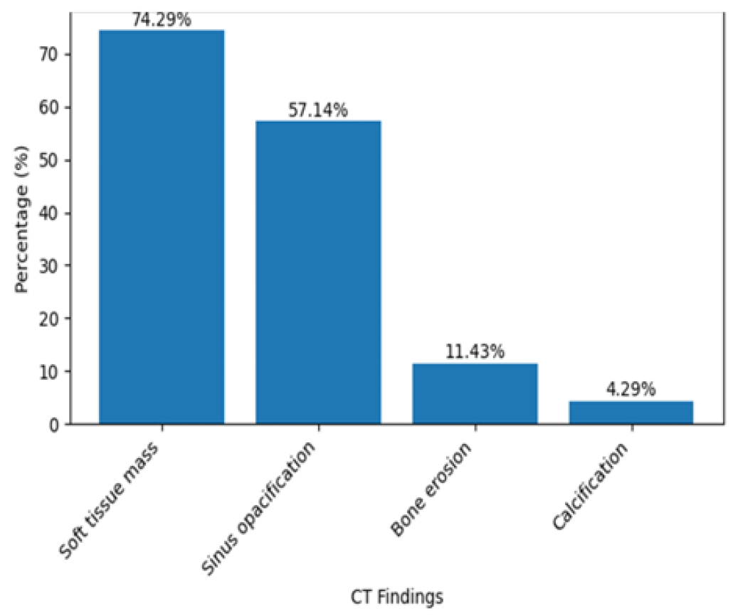
CT imaging revealed soft tissue mass in 52 (74.29%) patients and sinus opacification in 40 (57.14%). Bone erosion was observed in 8 (11.43%), mainly associated with neoplastic lesions (p = 0.018) (Table 7).
Table 7: CT Scan Findings
Finding
n (%)
Soft tissue mass
52 (74.29)
Sinus opacification
40 (57.14)
Bone erosion
8 (11.43)
Calcification
3 (4.29)
The majority of patients had no postoperative complications 60 (85.71%). Minor bleeding occurred in 6 (8.57%) and infection in 4 (5.71%). Overall complication rate was low and not statistically significant (p = 0.312) (Table 8, Figure 1).
Figure 1: CT Scan Findings
Table 8: Postoperative Complications
Complication
n (%)
None
60 (85.71)
Minor bleeding
6 (8.57)
Infection
4 (5.71)
Total
70 (100.00)
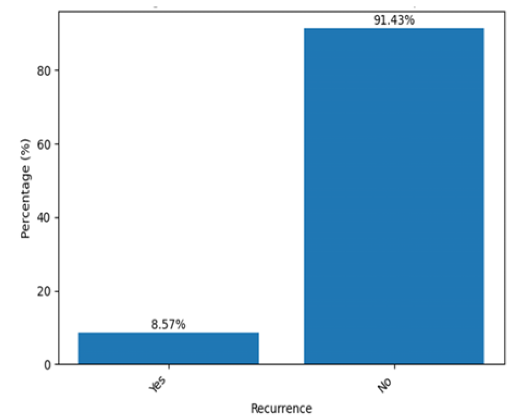
At 6-month follow-up, recurrence was observed in 6 (8.57%) patients, mainly among inverted papilloma and malignant cases. Most patients 64 (91.43%) remained recurrence-free (p = 0.041) (Table 9, Figure 2).
Figure 2: Recurrence at 6-Month Follow-Up
Table 9: Recurrence at 6-Month Follow-Up
Recurrence
n (%)
Yes
6 (8.57)
No
64 (91.43)
Total
70 (100.00)
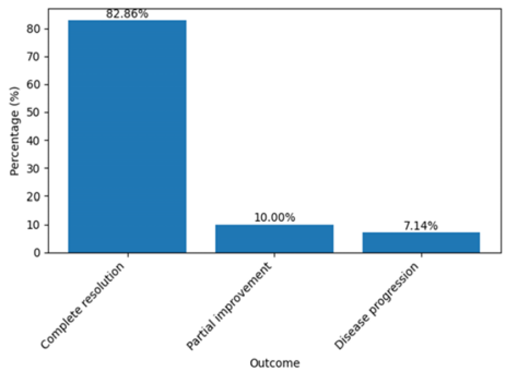
Complete resolution was achieved in 58 (82.86%) patients, while 7 (10.00%) showed partial improvement. Disease progression occurred in 5 (7.14%), corresponding to malignant cases. The difference in favorable versus unfavorable outcomes was statistically significant (p<0.001) (Table 10, Figure 3).
Table 10: Clinical Outcome
Outcome
n (%)
Complete resolution
58 (82.86)
Partial improvement
7 (10.00)
Disease progression
5 (7.14)
Total
70 (100.00)
Figure 3: Clinical Outcome
DISCUSSION
Unilateral nasal disease remains a frequent yet diagnostically challenging presentation in daily otolaryngology practice. The concern surrounding unilateral symptoms lies in the relatively higher likelihood of neoplastic pathology compared with bilateral sinonasal disease, although inflammatory conditions still predominate overall [1,2]. The present prospective study conducted at Al-Kindy Teaching Hospital in Baghdad confirmed that the majority of unilateral nasal lesions were benign and inflammatory in nature, while malignant tumors represented a small but clinically significant proportion. In our cohort, inflammatory pathologies such as nasal polyposis and chronic rhinosinusitis constituted the largest diagnostic categories. This finding is consistent with previous regional and international reports indicating that reactive inflammatory disorders account for most unilateral sinonasal lesions [1,3,4]. Nair et al. [1] and Chung et al. [5] similarly reported that nasal polyposis and chronic rhinosinusitis were the most common etiologies in unilateral nasal masses. However, some retrospective series have shown a relatively higher proportion of neoplastic lesions, particularly in tertiary oncologic referral centers [6,7], which may explain minor discrepancies between studies. Age distribution in our study demonstrated predominance among middle-aged adults, particularly those between 41–50 years. This aligns with previous findings that inflammatory sinonasal diseases are most common in middle adulthood [8,9]. Conversely, malignant neoplasms are typically reported in the fifth to seventh decades of life with a male predominance [10,11]. Although females slightly predominated in our overall sample, malignant cases in our cohort were more frequently observed in males, supporting earlier observations [12,13]. This gender tendency has been attributed to environmental exposure, occupational hazards, and smoking prevalence [14]. Nasal obstruction was the most common presenting symptom (90%), followed by rhinorrhea. These findings agree with multiple previous reports [1,4,15], which identify obstruction as the hallmark symptom of unilateral sinonasal pathology. Lee [16] and Paz Silva et al. [17] similarly reported nasal obstruction in more than 80% of cases. Epistaxis, although less frequent, was more commonly associated with neoplastic lesions in our study, which corresponds to the findings of Belli et al. [7]. Some studies, however, have reported higher rates of facial pain and headache, particularly in cases of fungal sinusitis or mucoceles [18], which were less common in our sample. One of the most important clinical messages emphasized in previous literature is the potential for malignant sinonasal tumors to mimic inflammatory polyps in early stages [2,10,19]. Delayed diagnosis significantly worsens prognosis due to late-stage presentation and local invasion [11,20]. In our study, CT findings such as bone erosion were strongly associated with malignant pathology, consistent with observations by Dammann et al. [21] and Eckhoff et al. [22], who highlighted the importance of radiologic red flags in unilateral disease. Inverted papilloma was the most common benign neoplasm in our cohort, in agreement with prior reports [1,23]. This tumor is known for its local aggressiveness and risk of malignant transformation ranging from 5–15% [24]. Our recurrence rate was low but primarily observed in inverted papilloma cases, supporting literature emphasizing the need for long-term follow-up [25]. Some studies report higher recurrence rates exceeding 15% depending on surgical technique and tumor staging [23], which may differ from our findings due to careful endoscopic excision and short follow-up duration. Regarding malignant tumors, squamous cell carcinoma was the most frequent malignancy detected, which is consistent with most sinonasal cancer epidemiology studies [10,11,26]. Adenocarcinoma and non-Hodgkin lymphoma were rare in our series, similar to previous documentation of their low incidence in unilateral nasal presentations [27,28]. However, certain European studies report relatively higher rates of adenocarcinoma related to occupational wood dust exposure [29], a factor less prominent in our population. The widespread use of nasal endoscopy has significantly improved diagnostic accuracy. Endoscopic visualization allows precise localization, characterization, and guided biopsy of suspicious lesions [17,22,30]. In our study, endoscopy combined with CT scanning provided both diagnostic clarity and therapeutic guidance, reinforcing current recommendations for systematic evaluation of unilateral sinonasal disease [22,30]. Overall, our findings confirm that although inflammatory etiologies predominate in unilateral nasal disease, the risk of malignancy remains clinically relevant. Comprehensive clinical assessment, radiological imaging, endoscopic examination, and histopathological confirmation are mandatory to avoid delayed diagnosis.
CONCLUSION
Although inflammatory conditions represent the majority of unilateral nasal diseases, a clinically significant proportion of neoplastic lesions exists. Comprehensive evaluation combining endoscopy, CT imaging, and histopathological confirmation is essential. Early surgical intervention with FESS demonstrates favorable outcomes and low complication rates.
REFERENCE
Nair, S. et al. “A review of the clinicopathological and radiological features of unilateral nasal mass.” Indian Journal of Otolaryngology and Head and Neck Surgery vol. 65 no. Suppl 2 2013, pp. 199–204.
Kaplan, B.A. and Kountakis, S.E. “Diagnosis and pathology of unilateral maxillary sinus opacification with or without contralateral disease.” The Laryngoscope vol. 114 no. 6 2004, pp. 981–985.
Tritt, S. et al. “Unilateral nasal polyposis: clinical presentation and pathology.” American Journal of Otolaryngology vol. 29 no. 4 2008, pp. 230–232.
Iqbal, S.M. and Hussain, S.I. “Unilateral nasal obstruction caused by sinonasal neoplastic lesions.” Journal of Liaquat University of Medical and Health Sciences vol. 5 no. 1 2006, pp. 18–23.
Chung, H.K. et al. “Analysis of disease patterns in patients with unilateral sinonasal diseases.” Mid Taiwan Journal of Medicine vol. 13 2008, pp. 82–88.
Lee, J.Y. “Unilateral paranasal sinus diseases: analysis of clinical characteristics and pathology.” Acta Oto-Laryngologica vol. 128 no. 6 2008, pp. 621–626.
Belli, S. et al. “Single-sided sinonasal mass: a retrospective study.” Northern Clinics of Istanbul vol. 5 no. 2 2018, pp. 139–143.
Larsen, K. and Tos, M. “The estimated incidence of symptomatic nasal polyps.” Acta Oto-Laryngologica vol. 122 no. 2 2002, pp. 179–182.
Fokkens, W.J. et al. “European position paper on rhinosinusitis and nasal polyps 2020.” Rhinology vol. 58 no. Suppl S29 2020, pp. 1–464.
Euteneuer, S. et al. “Malignomas of the nasal cavity and paranasal sinuses: clinical characteristics and prognosis.” Laryngo-Rhino-Otologie vol. 83 no. 1 2004, pp. 33–39.
Zbaren, P. et al. “Malignant neoplasms of the nasal cavity and paranasal sinuses.” HNO vol. 35 no. 6 1987, pp. 246–249.
Gullane, P.J. and Conley, J. “Carcinoma of the maxillary sinus.” Journal of Otolaryngology vol. 12 no. 3 1983, pp. 141–145.
Dulguerov, P. et al. “Nasal and paranasal sinus carcinoma: prognosis and treatment.” Cancer vol. 92 no. 12 2001, pp. 3012–3029.
Blot, W.J. and Fraumeni, J.F. “Cancers of the nasal cavity and paranasal sinuses.” Cancer Surveys vol. 8 no. 3 1989, pp. 439–455.
Klossek, J.M. et al. “Unilateral sinus disease: diagnostic algorithm.” Annals of Otology, Rhinology & Laryngology vol. 106 no. 9 1997, pp. 760–766.
Eckhoff, A. et al. “Unilateral versus bilateral sinonasal disease: differential diagnosis considerations.” The Laryngoscope vol. 130 no. 4 2020, pp. E116–E121.
Paz Silva, M. et al. “Diagnostic algorithm for unilateral sinus disease: 15-year review.” International Forum of Allergy & Rhinology vol. 5 no. 7 2015, pp. 590–596.
Chakrabarti, A. et al. “Fungal rhinosinusitis: classification and management.” Clinical Microbiology Reviews vol. 22 no. 2 2009, pp. 286–319.
Lund, V.J. et al. “Sinonasal malignancy: disease overview and management.” The Journal of Laryngology & Otology vol. 124 no. 5 2010, pp. 475–480.
Turner, J.H. and Reh, D.D. “Incidence and survival in patients with sinonasal cancer.” Head & Neck vol. 34 no. 6 2012, pp. 877–885.
Dammann, F. et al. “Inverted papilloma of the nasal cavity and paranasal sinuses: CT for diagnosis and follow-up.” American Journal of Roentgenology vol. 172 no. 2 1999, pp. 543–548.
DelGaudio, J.M. and Wise, S.K. “Endoscopic management of sinonasal tumors.” Otolaryngologic Clinics of North America vol. 48 no. 5 2015, pp. 821–836.
Lawson, W. and Patel, Z.M. “The evolution of management for inverted papilloma.” The Laryngoscope vol. 119 no. 5 2009, pp. 989–997.
Mirza, S. et al. “Sinonasal inverted papillomas: recurrence and malignant transformation rates.” The Laryngoscope vol. 117 no. 7 2007, pp. 1288–1291.
Kamel, R. “Endoscopic transnasal surgery in inverted papilloma.” Archives of Otolaryngology – Head & Neck Surgery vol. 121 no. 5 1995, pp. 512–517.
Dulguerov, P. et al. “Esthesioneuroblastoma: review of 70 cases.” The Laryngoscope vol. 111 no. 3 2001, pp. 502–508.
Nowara, E. et al. “Primary non-Hodgkin’s lymphoma of the maxillary sinus.” Otolaryngologia Polska vol. 51 no. 4 1997, pp. 418–423.
Dubal, P.M. et al. “Sinonasal malignancies: epidemiology and survival trends.” The Laryngoscope vol. 126 no. 6 2016, pp. 1304–1313.
d’Errico, A. et al. “Occupational risk factors for sinonasal adenocarcinoma.” Occupational and Environmental Medicine vol. 66 no. 7 2009, pp. 448–455.
Wise, S.K. and Harvey, R.J. “Endoscopic sinus surgery for management of sinonasal disease.” Otolaryngologic Clinics of North America vol. 43 no. 3 2010, pp. 605–612.
License
Creative Commons Attribution-NonCommercial-NoDerivatives 4.0 International License
All papers should be submitted electronically. All submitted manuscripts must be original work that is not under submission at another journal or under consideration for publication in another form, such as a monograph or chapter of a book. Authors of submitted papers are obligated not to submit their paper for publication elsewhere until an editorial decision is rendered on their submission. Further, authors of accepted papers are prohibited from publishing the results in other publications that appear before the paper is published in the Journal unless they receive approval for doing so from the Editor-In-Chief.
Himalayan Journal of Community Medicine and Public Health open access articles are licensed under a Creative Commons Attribution-Share A like 4.0 International License. This license lets the audience to give appropriate credit, provide a link to the license, and indicate if changes were made and if they remix, transform, or build upon the material, they must distribute contributions under the same license as the original.
Recommended Articles
Research Article
Curves of Concern: Public Awareness of Scoliosis and Its Impact on Childhood Development in Hamirpur
Vishal Dhatwalia,
Swati Chandel
Published: 10/12/2024
Download PDF
Cite
x
APA
Dhatwalia, V. & Chandel, S. (2024). Curves of Concern: Public Awareness of Scoliosis and Its Impact on Childhood Development in Hamirpur. Himalayan Journal of Community Medicine and Public Health, 5(2), 1-6.
MLA
Dhatwalia, Vishal and Swati Chandel. "Curves of Concern: Public Awareness of Scoliosis and Its Impact on Childhood Development in Hamirpur." Himalayan Journal of Community Medicine and Public Health 5.2 (2024): 1-6.
Chicago
Dhatwalia, Vishal and Swati Chandel. "Curves of Concern: Public Awareness of Scoliosis and Its Impact on Childhood Development in Hamirpur." Himalayan Journal of Community Medicine and Public Health 5, no. 2 (2024): 1-6.
Harvard
Dhatwalia, V. and Chandel, S. (2024) 'Curves of Concern: Public Awareness of Scoliosis and Its Impact on Childhood Development in Hamirpur' Himalayan Journal of Community Medicine and Public Health 5(2), pp. 1-6.
Vancouver
Dhatwalia V, Chandel S. Curves of Concern: Public Awareness of Scoliosis and Its Impact on Childhood Development in Hamirpur. Himalayan Journal of Community Medicine and Public Health. 2024 Jul;5(2):1-6.
Download PDF
Research Article
Etiological Analysis of the Incident of Computer Vision Syndrome (Cvs) in Students of the Faculty of Medical Muhammadiyah University Semarang
Anang Kriswantoro,
...
Swasty
Published: 20/01/2026
Download PDF
Cite
x
APA
Kriswantoro, A., Martiningsih, W. R., Novitasari, A. & None, S. (2026). Etiological Analysis of the Incident of Computer Vision Syndrome (Cvs) in Students of the Faculty of Medical Muhammadiyah University Semarang. Himalayan Journal of Community Medicine and Public Health, 7(1), 1-5.
MLA
Kriswantoro, Anang, et al. "Etiological Analysis of the Incident of Computer Vision Syndrome (Cvs) in Students of the Faculty of Medical Muhammadiyah University Semarang." Himalayan Journal of Community Medicine and Public Health 7.1 (2026): 1-5.
Chicago
Kriswantoro, Anang, Wahju R. Martiningsih, Andra Novitasari and Swasty . "Etiological Analysis of the Incident of Computer Vision Syndrome (Cvs) in Students of the Faculty of Medical Muhammadiyah University Semarang." Himalayan Journal of Community Medicine and Public Health 7, no. 1 (2026): 1-5.
Harvard
Kriswantoro, A., Martiningsih, W. R., Novitasari, A. and None, S. (2026) 'Etiological Analysis of the Incident of Computer Vision Syndrome (Cvs) in Students of the Faculty of Medical Muhammadiyah University Semarang' Himalayan Journal of Community Medicine and Public Health 7(1), pp. 1-5.
Vancouver
Kriswantoro A, Martiningsih WR, Novitasari A, Swasty S. Etiological Analysis of the Incident of Computer Vision Syndrome (Cvs) in Students of the Faculty of Medical Muhammadiyah University Semarang. Himalayan Journal of Community Medicine and Public Health. 2026 Jan;7(1):1-5.
Download PDF
Research Article
Silent Threats Unveiled: Gauging Public Awareness and Understanding of Blood Clotting Disorders in Ambala, Haryana
Ashmita Joshi,
...
Sanjeev Kumar
Published: 30/07/2024
Download PDF
Cite
x
APA
Joshi, A., kaur, J. & Kumar, S. (2024). Silent Threats Unveiled: Gauging Public Awareness and Understanding of Blood Clotting Disorders in Ambala, Haryana. Himalayan Journal of Community Medicine and Public Health, 5(2), 1-6.
MLA
Joshi, Ashmita, Jasleen kaur and Sanjeev Kumar. "Silent Threats Unveiled: Gauging Public Awareness and Understanding of Blood Clotting Disorders in Ambala, Haryana." Himalayan Journal of Community Medicine and Public Health 5.2 (2024): 1-6.
Chicago
Joshi, Ashmita, Jasleen kaur and Sanjeev Kumar. "Silent Threats Unveiled: Gauging Public Awareness and Understanding of Blood Clotting Disorders in Ambala, Haryana." Himalayan Journal of Community Medicine and Public Health 5, no. 2 (2024): 1-6.
Harvard
Joshi, A., kaur, J. and Kumar, S. (2024) 'Silent Threats Unveiled: Gauging Public Awareness and Understanding of Blood Clotting Disorders in Ambala, Haryana' Himalayan Journal of Community Medicine and Public Health 5(2), pp. 1-6.
Vancouver
Joshi A, kaur J, Kumar S. Silent Threats Unveiled: Gauging Public Awareness and Understanding of Blood Clotting Disorders in Ambala, Haryana. Himalayan Journal of Community Medicine and Public Health. 2024 Jul;5(2):1-6.
Download PDF
Research Article
Knowledge About HBV Infection and Its Association with HBSAG Positivity among Residents of Kaza Sub- Division Of District Lahaul & Spiti In Himachal Pradesh
Brij Sharma,
...
Amit Sachdeva
Published: 30/07/2024
Download PDF
Cite
x
APA
Sharma, B., Gupta, A., Sharma, R., Bodh, V., Chauhan, A., None, D., Sharma, N., Sharma, S. & Sachdeva, A. (2024). Knowledge About HBV Infection and Its Association with HBSAG Positivity among Residents of Kaza Sub- Division Of District Lahaul & Spiti In Himachal Pradesh. Himalayan Journal of Community Medicine and Public Health, 5(2), 1-8.
MLA
Sharma, Brij, et al. "Knowledge About HBV Infection and Its Association with HBSAG Positivity among Residents of Kaza Sub- Division Of District Lahaul & Spiti In Himachal Pradesh." Himalayan Journal of Community Medicine and Public Health 5.2 (2024): 1-8.
Chicago
Sharma, Brij, Anmol Gupta, Rajesh Sharma, Vishal Bodh, Aashish Chauhan, Dipesh , Neetu Sharma, Sidhant Sharma and Amit Sachdeva. "Knowledge About HBV Infection and Its Association with HBSAG Positivity among Residents of Kaza Sub- Division Of District Lahaul & Spiti In Himachal Pradesh." Himalayan Journal of Community Medicine and Public Health 5, no. 2 (2024): 1-8.
Harvard
Sharma, B., Gupta, A., Sharma, R., Bodh, V., Chauhan, A., None, D., Sharma, N., Sharma, S. and Sachdeva, A. (2024) 'Knowledge About HBV Infection and Its Association with HBSAG Positivity among Residents of Kaza Sub- Division Of District Lahaul & Spiti In Himachal Pradesh' Himalayan Journal of Community Medicine and Public Health 5(2), pp. 1-8.
Vancouver
Sharma B, Gupta A, Sharma R, Bodh V, Chauhan A, Dipesh D, Sharma N, Sharma S, Sachdeva A. Knowledge About HBV Infection and Its Association with HBSAG Positivity among Residents of Kaza Sub- Division Of District Lahaul & Spiti In Himachal Pradesh. Himalayan Journal of Community Medicine and Public Health. 2024 Jul;5(2):1-8.
Hussien Dabi, A., Naji Yaseen, M. & A. Mohammed Ali, W. (2026). Clinicopathological Profile and Outcomes of Unilateral Nasal Disease: A Prospective Study. Himalayan Journal of Community Medicine and Public Health, 7(1), 1-5.
MLA
Hussien Dabi, Abdulkareem, Mohammed Naji Yaseen and Watheq A. Mohammed Ali. "Clinicopathological Profile and Outcomes of Unilateral Nasal Disease: A Prospective Study." Himalayan Journal of Community Medicine and Public Health 7.1 (2026): 1-5.
Chicago
Hussien Dabi, Abdulkareem, Mohammed Naji Yaseen and Watheq A. Mohammed Ali. "Clinicopathological Profile and Outcomes of Unilateral Nasal Disease: A Prospective Study." Himalayan Journal of Community Medicine and Public Health 7, no. 1 (2026): 1-5.
Harvard
Hussien Dabi, A., Naji Yaseen, M. and A. Mohammed Ali, W. (2026) 'Clinicopathological Profile and Outcomes of Unilateral Nasal Disease: A Prospective Study' Himalayan Journal of Community Medicine and Public Health 7(1), pp. 1-5.
Vancouver
Hussien Dabi A, Naji Yaseen M, A. Mohammed Ali W. Clinicopathological Profile and Outcomes of Unilateral Nasal Disease: A Prospective Study. Himalayan Journal of Community Medicine and Public Health. 2026 Jan;7(1):1-5.


 Download PDF
 Download XML