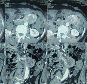
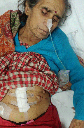
NF1 is one of the most common autosomal dominant conditions in which affected individuals have an increased risk of malignancy. It is mostly characterized as a neurocutaneous disorder, with both café au lait patches and cutaneous neurofibromas occurring at frequencies greater than 99% [1,2]. However, NF1 is a multi-system and variable disease with clinicopathological features that encompass a much broader spectrum.
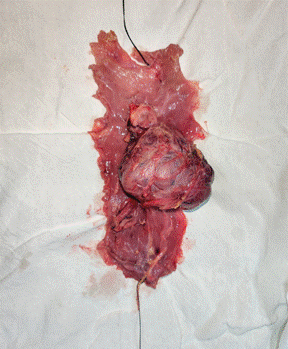
Figure 3: Showing resected growth with diverticula
The genetic basis of this disease is a mutation in the NF1 gene located on chromosome 17q11.2. NF1 encodes neurofibromin, a tumor suppressor protein. Neurofibromin regulates cellular proliferation via inactivation of RAS, a protein that stimulates signal transduction through the MAP-kinase pathway when activated. The resultant loss of function of neurofibromin predisposes to the development of benign and malignant tumors [1,2,7].
The major neurological disease manifestations can be divided between the Central Nervous System (CNS) and the Peripheral Nervous System (PNS). CNS involvement includes cognitive impairment, learning disability, Attention Deficit Hyperactivity Disorder (ADHD), epilepsy, structural malformations and cerebrovascular abnormalities. The most frequent CNS malignancy observed is glioma, which has a predisposition for the optic pathway, the brainstem and the cerebellum. Neurofibromas are the chief feature of PNS. They are benign tumors that develop along the peripheral nerve sheath and occur in multiple types: Focal cutaneous or subcutaneous, and diffuse or nodular plexiform. Malignant Peripheral Nerve Sheath Tumors (MPNST) are the predominant PNS cancers, with an 8-13% lifetime risk in NF1. MPNST usually arise from preexisting plexiform or subcutaneous neurofibromas [1,2].
Those affected with NF1 also carry the potential for disease manifestations beyond the neurological system. Lisch nodules, which are benign hamartomas of the iris, constitute the major pathology of the eye. The musculoskeletal system can also be involved with scoliosis, tibial bowing and pseudoarthrosis most frequently observed. The predominant dermatologic features are café au lait spots, followed by skin-fold freckling [1,2]. Cardiovascular disease, including congenital heart disease, vasculopathy and hypertension, as well as other malignancies, such as leukemia, rhabdomyosarcoma and pheochromocytoma, are reported to be associated with NF1 at greater frequencies than the general population [7,11].
In addition to the aforementioned disease hallmarks, GI involvement in NF1 has been reported in as many as 25% of affected individuals [17]. NF1-related GI disease has been described to occur in three principle forms: neurogenic tumors, stromal tumors and neuroendocrine tumors [10-18]. The most common of these GI manifestations in NF1 is GIST. Further, NF1 has been associated with an increased risk of developing GIST, with one study indicating an incidence of 7% in the NF1 population and another study reporting a 150-fold increased risk as compared to the general population [7,19].
GIST is a soft-tissue sarcoma that likely arises from the Interstitial Cells of Cajal (ICC), GI pacemaker cells with a role in motility. Its origin was elucidated by the discovery that both the ICC and the majority of GISTs express the receptor tyrosine kinase KIT (CD 117) [5,6]. Approximately 85% of GISTs are characterized by a sporadic activating mutation in the gene KIT, leading to the constitutive activation of KIT. KIT mutations most commonly occur in exon 11 (60%), but can also occur in exons 9 (15%), 13 or 17 (5%). Less commonly, GISTs harbor a mutation in the platelet derived growth factor receptor alpha (PDGFRA) gene, which also encodes a receptor tyrosine kinase. These mutations can occur in exons 12 or 18 [5,6]. Both tyrosine kinases play key roles in the tumorigenesis of GIST via signal transduction through the PI3K-AKT, MAP-kinase, and JAK-STAT3 pathways [12].
In addition to mutation analysis, GIST is further characterized by its pathology. The majority of cases occur as a solitary tumor, with the stomach as the most common site (60%), followed by the small intestine (25%). Tumor morphology can be classified as one of three types: spindle cell-type (70%), epithelioid-type (20%) and mixed-type (10%). Approximately 95% of tumors have CD 117 (KIT) expression. GIST can also demonstrate expression of BCL-2 [human proto-oncogene located on chromosome 18] (80%), CD34 [hematopoietic progenitor cell antigen CD34] (70%), muscle-specific actin (50%), smooth muscle actin (35%), S-100 (10%) and desmin (5%).
The cases of GIST observed in the NF1 population also demonstrated CD 117 expression; however, in contrast, the NF1-related cases appear to constitute a subset of wild-type cases, in which neither KIT or PDGFRA are mutated, implying a different molecular pathogenesis [5,7,8]. NF-1 related GIST occurs as a result of somatic inactivation of the wild-type NF1 allele in the tumor, leading to increased signal transduction via the MAP-kinase pathway [7,8]. There has been evidence to suggest that the NF1 mutation can result in the overexpression of KIT and/or PDGFRA; thus, explaining positive CD 117 (KIT) expression on immunohistochemistry, despite the absence of a mutation in either KIT or PDGFRA [20]. NF1-related GIST differs from its sporadic counterpart in several other ways, including its propensity to be multifocal and to occur in the small bowel, as well as its higher frequency of spindle cell morphology and CD34 expression [6,7].
The management of GIST depends on the extent of disease. Surgery constitutes the mainstay treatment for primary resectable disease. For unresectable and metastatic disease, targeted-therapy with imatinib mesylate, a tyrosine kinase inhibitor with activity at both KIT and PDGFRA, has shown promising effects. In 2002, the US Food and Drug Administration (FDA) approved targeted-therapy with imatinib, following the landmark CSTIB2222 trial, which demonstrated significantly improved median Time-To-Progression (TTP), median Overall Survival (OS) and 5-year survival rates. Imatinib was also approved as adjunctive therapy for primary resected GISTS >3 cm in size, after a randomized phase III trial conducted by the American College of Surgeons Oncology Group, demonstrated an improved 1-year recurrence-free survival, compared to placebo. With regards to predictive markers, the CSTIB2222 trial demonstrated that imatinib has highest efficacy in GISTs with exon 11 KIT mutations, when compared to GISTs with exon 9 mutations or wild-type KIT. In addition, imatinib was shown to have no effect in patients with PDGFRA D842V point mutations [5,6]. Specifically for wild-type cases, such as NF1, adjuvant therapy with imatinib should be considered on an individual case-by-case basis, as the benefit may be variable. The dosage of imatinib is approved at 400 mg per day; however, studies have demonstrated increased benefit for exon 9 mutations at a dose of 800 mg per day. The optimal duration of treatment is undetermined. Initial trials demonstrated benefit with one year of imatinib treatment, whereas the Scandinavian Sarcoma Group recently demonstrated an increased benefit with 3 years of treatment, specifically in the setting of advanced disease [21].
With regard to surveillance for disease progression and recurrence, it has been recommended to perform CT imaging every 6 months during imatinib therapy. The highest risk of GIST recurrence is in the 2 years following treatment, so patients should be followed with CT imaging every 3 to 4 months. After the initial 2-year period, CT imaging should be performed every 6 months for 3 years and then annually. The risk of recurrence after the 10 years substantially decreases, such that the risk of radiation associated with CT imaging may out-weigh its benefit after that period [21].


 Download PDF
 Download XML