Contents
Download PDF
pdf Download XML
1998 Views
268 Downloads
Share this article
Research Article | Volume 3 Issue 2 (July-Dec, 2022) | Pages 1 - 4
Gastrointestinal Stromal Tumor (GIST) in a Patient with Neurofibromatosis Type 1: Case Report
 ,
1
M.B.B.S, M.S General Surgery Civil Hospital Theog, Shimla, Himachal Pradesh, India
2
M.B.B.S, M.S General Surgery Civil Hospital Arki, Solan, Himachal Pradesh, India
Under a Creative Commons license
Open Access
Received
May 1, 2022
Revised
May 25, 2022
Accepted
June 20, 2022
Published
July 10, 2022
Abstract

Neurofibromatosis type 1 (NF1) is a hereditary cancer predisposition syndrome which is characterized by neurologic, dermatologic and orthopaedic manifestations. There is a spectrum of tumors that affects individuals with NF1 at an increased incidence compared to the general population, such as neurofibromas, Malignant Peripheral Nerve Sheath Tumors (MPNST) and gliomas. There has been a growing number of literature reporting an association between NF1 and Gastrointestinal Stromal Tumors (GIST). It is the most common genetic syndrome with an incidence of 1 in 2,500 to 3,000 and a prevalence of 1 in 4,000 to 5,000 [1]. It has an autosomal dominant pattern of inheritance with complete penetrance; however, as many as 50% of affected individuals have been reported to be sporadic cases [2,3]. NF1 is further defined by its variable expression, resulting in a wide array of clinicopathological features and a varying, unpredictable disease course [1,2]. Occurring at an incidence of 6 to 15 per one million GIST is the most common mesenchymal tumor of the gastrointestinal (GI) tract [5]. It is only in recent years that the molecular pathogenesis of GIST has been demonstrated, with aberrant tyrosine kinase activity identified as the key modulator in the majority of cases. This advancement has allowed for molecularly targeted therapy via imatinib, a tyrosine kinase inhibitor [5,6,9]. NF1-associated GIST, however, has been described to comprise a minority of cases, in which there is an alternative molecular pathogenesis. This difference between NF1-related GISTs and that of the general population has important therapeutic implications [9]. We herein report a case of symptomatic GIST in a 62-year-old woman with a neurofibromatosis type 1.

Keywords
INTRODUCTION

Case Report

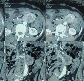
A 62 year old female with NF1 presented with history of pain epigastric region for past 2 years. Computed Tomographic (CT) imaging revealed Heterogenously enhancing exophytic mass of 45×53×59 mm arising from DJ flexure proximal adjacent jejunal loop with no intraluminal extension with areas of necrosis within it. Posteriorly, abutting left renal vein and IVC and anteriorly abutting descending colon? Jejunal GIST:


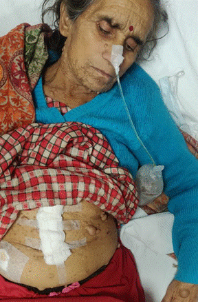
  • GPE: Multiple neurofibromas all over body

  • Lisch nodules present in iris

  • Café-au lait spots present on abdomen

  • Axillary freckling present


Exploratory Laparotomy with segmental resection of growth bearing segment with duodenojejunostomy done.

Findings: 


  • A growth of size 5X4 cm present at DJ with two diverticula present distal and proximal to it, displacing IMV antero-laterally and posteriorly abutting aorta

  • Multiple small nodules (~0.5 cm) present all over the gut

  • Segmental resection of growth bearing segment with duodenojejunostomy (end to end, double-layered) done


HPE: fusiform tumor of mesenchymal nature. The tumor grows into the mucous membrane of the jejunum. Moderate cellularity is noted. No necrosis. Low mitotic index (3 mitoses x 50 HPF).

In order to verify the tumor, immunohistochemistry was performed using antibodies CD117, CD34, SMA, which demonstrated overexpression CD117 (c-kit) and SMA in the cytoplasm of tumor cells, the absence of expression in tumor cells CD34.


Figure 1: CECT Abdomen showing exophytic mass arising from DJ flexure


Figure 2: Showing Multiple neurofibromas and Café-au lait spots present on abdomen


DISCUSSION

NF1 is one of the most common autosomal dominant conditions in which affected individuals have an increased risk of malignancy. It is mostly characterized as a neurocutaneous disorder, with both café au lait patches and cutaneous neurofibromas occurring at frequencies greater than 99% [1,2]. However, NF1 is a multi-system and variable disease with clinicopathological features that encompass a much broader spectrum.
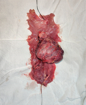


Figure 3: Showing resected growth with diverticula


The genetic basis of this disease is a mutation in the NF1 gene located on chromosome 17q11.2. NF1 encodes neurofibromin, a tumor suppressor protein. Neurofibromin regulates cellular proliferation via inactivation of RAS, a protein that stimulates signal transduction through the MAP-kinase pathway when activated. The resultant loss of function of neurofibromin predisposes to the development of benign and malignant tumors [1,2,7].


The major neurological disease manifestations can be divided between the Central Nervous System (CNS) and the Peripheral Nervous System (PNS). CNS involvement includes cognitive impairment, learning disability, Attention Deficit Hyperactivity Disorder (ADHD), epilepsy, structural malformations and cerebrovascular abnormalities. The most frequent CNS malignancy observed is glioma, which has a predisposition for the optic pathway, the brainstem and the cerebellum. Neurofibromas are the chief feature of PNS. They are benign tumors that develop along the peripheral nerve sheath and occur in multiple types: Focal cutaneous or subcutaneous, and diffuse or nodular plexiform. Malignant Peripheral Nerve Sheath Tumors (MPNST) are the predominant PNS cancers, with an 8-13% lifetime risk in NF1. MPNST usually arise from preexisting plexiform or subcutaneous neurofibromas [1,2].


Those affected with NF1 also carry the potential for disease manifestations beyond the neurological system. Lisch nodules, which are benign hamartomas of the iris, constitute the major pathology of the eye. The musculoskeletal system can also be involved with scoliosis, tibial bowing and pseudoarthrosis most frequently observed. The predominant dermatologic features are café au lait spots, followed by skin-fold freckling [1,2]. Cardiovascular disease, including congenital heart disease, vasculopathy and hypertension, as well as other malignancies, such as leukemia, rhabdomyosarcoma and pheochromocytoma, are reported to be associated with NF1 at greater frequencies than the general population [7,11].


In addition to the aforementioned disease hallmarks, GI involvement in NF1 has been reported in as many as 25% of affected individuals [17]. NF1-related GI disease has been described to occur in three principle forms: neurogenic tumors, stromal tumors and neuroendocrine tumors [10-18]. The most common of these GI manifestations in NF1 is GIST. Further, NF1 has been associated with an increased risk of developing GIST, with one study indicating an incidence of 7% in the NF1 population and another study reporting a 150-fold increased risk as compared to the general population [7,19].


GIST is a soft-tissue sarcoma that likely arises from the Interstitial Cells of Cajal (ICC), GI pacemaker cells with a role in motility. Its origin was elucidated by the discovery that both the ICC and the majority of GISTs express the receptor tyrosine kinase KIT (CD 117) [5,6]. Approximately 85% of GISTs are characterized by a sporadic activating mutation in the gene KIT, leading to the constitutive activation of KIT. KIT mutations most commonly occur in exon 11 (60%), but can also occur in exons 9 (15%), 13 or 17 (5%). Less commonly, GISTs harbor a mutation in the platelet derived growth factor receptor alpha (PDGFRA) gene, which also encodes a receptor tyrosine kinase. These mutations can occur in exons 12 or 18 [5,6]. Both tyrosine kinases play key roles in the tumorigenesis of GIST via signal transduction through the PI3K-AKT, MAP-kinase, and JAK-STAT3 pathways [12].


In addition to mutation analysis, GIST is further characterized by its pathology. The majority of cases occur as a solitary tumor, with the stomach as the most common site (60%), followed by the small intestine (25%). Tumor morphology can be classified as one of three types: spindle cell-type (70%), epithelioid-type (20%) and mixed-type (10%). Approximately 95% of tumors have CD 117 (KIT) expression. GIST can also demonstrate expression of BCL-2 [human proto-oncogene located on chromosome 18] (80%), CD34 [hematopoietic progenitor cell antigen CD34] (70%), muscle-specific actin (50%), smooth muscle actin (35%), S-100 (10%) and desmin (5%).


The cases of GIST observed in the NF1 population also demonstrated CD 117 expression; however, in contrast, the NF1-related cases appear to constitute a subset of wild-type cases, in which neither KIT or PDGFRA are mutated, implying a different molecular pathogenesis [5,7,8]. NF-1 related GIST occurs as a result of somatic inactivation of the wild-type NF1 allele in the tumor, leading to increased signal transduction via the MAP-kinase pathway [7,8]. There has been evidence to suggest that the NF1 mutation can result in the overexpression of KIT and/or PDGFRA; thus, explaining positive CD 117 (KIT) expression on immunohistochemistry, despite the absence of a mutation in either KIT or PDGFRA [20]. NF1-related GIST differs from its sporadic counterpart in several other ways, including its propensity to be multifocal and to occur in the small bowel, as well as its higher frequency of spindle cell morphology and CD34 expression [6,7].


The management of GIST depends on the extent of disease. Surgery constitutes the mainstay treatment for primary resectable disease. For unresectable and metastatic disease, targeted-therapy with imatinib mesylate, a tyrosine kinase inhibitor with activity at both KIT and PDGFRA, has shown promising effects. In 2002, the US Food and Drug Administration (FDA) approved targeted-therapy with imatinib, following the landmark CSTIB2222 trial, which demonstrated significantly improved median Time-To-Progression (TTP), median Overall Survival (OS) and 5-year survival rates. Imatinib was also approved as adjunctive therapy for primary resected GISTS >3 cm in size, after a randomized phase III trial conducted by the American College of Surgeons Oncology Group, demonstrated an improved 1-year recurrence-free survival, compared to placebo. With regards to predictive markers, the CSTIB2222 trial demonstrated that imatinib has highest efficacy in GISTs with exon 11 KIT mutations, when compared to GISTs with exon 9 mutations or wild-type KIT. In addition, imatinib was shown to have no effect in patients with PDGFRA D842V point mutations [5,6]. Specifically for wild-type cases, such as NF1, adjuvant therapy with imatinib should be considered on an individual case-by-case basis, as the benefit may be variable. The dosage of imatinib is approved at 400 mg per day; however, studies have demonstrated increased benefit for exon 9 mutations at a dose of 800 mg per day. The optimal duration of treatment is undetermined. Initial trials demonstrated benefit with one year of imatinib treatment, whereas the Scandinavian Sarcoma Group recently demonstrated an increased benefit with 3 years of treatment, specifically in the setting of advanced disease [21].


With regard to surveillance for disease progression and recurrence, it has been recommended to perform CT imaging every 6 months during imatinib therapy. The highest risk of GIST recurrence is in the 2 years following treatment, so patients should be followed with CT imaging every 3 to 4 months. After the initial 2-year period, CT imaging should be performed every 6 months for 3 years and then annually. The risk of recurrence after the 10 years substantially decreases, such that the risk of radiation associated with CT imaging may out-weigh its benefit after that period [21].

REFERENCES
  1. Ferner, R.E. "Neurofibromatosis 1 and neurofibromatosis 2: A twenty first century perspective." Lancet Neurol, vol. 6, no. 340-351, 2007.

  2. Ferner, R.E., et al. "Guidelines for the diagnosis and management of individuals with neurofibromatosis." J Med Genet, vol. 44, no. 81-88, 2007.

  3. Huson, S.M., et al. "Von Rechlinghausen neurofibromatosis: Clinical and population study in southeast Wales." Brain, vol. 111, no. 55-81, 1988.

  4. Neurofibromatosis Conference Statement. "National Institutes of Health Consensus Development Conference." Arch Neurol, vol. 45, no. 575-578, 1988.

  5. Corless, C., et al. "Biology of gastrointestinal stromal tumors." Journal of Clinical Oncology, vol. 22, no. 3813-3825, 2004.

  6. Ho, M.Y. and Blanke, C.D. "Gastrointestinal stromal tumors: Disease and treatment update." Gastroenterology, vol. 140, no. 1372-1376, 2011.

  7. Anderson, J., et al. "NF1-associated gastrointestinal stromal tumors have unique clinical, phenotypic and genotypic characteristics." Am J Surg Pathol, vol. 29, no. 1170-1176, 2005.

  8. Maertans, O., et al. "Molecular pathogenesis of multiple gastrointestinal stromal tumors in NF1 patients." Human Molecular Genetics, vol. 15, no. 1015-1023, 2006.

  9. Heinrich, M.C., et al. "Kinase mutations and imatinib response in patients with metastatic gastrointestinal stromal tumors." Journal of Clinical Oncology, vol. 21, no. 4342-4349, 2003.

  10. Cheng, S., et al. "Neurofibromatosis with gastrointestinal stromal tumors: Insights into the association." Digestive Diseases and Sciences, vol. 49, no. 1165-1169, 2004.

  11. Miettinen, M., et al. "Gastrointestinal stromal tumors in patients with neurofibromatosis 1." Am J Surg Pathol, vol. 30, no. 90-96, 2006.

  12. Maertans, O., et al. "Molecular pathogenesis of multiple gastrointestinal stromal tumors in NF1 patients." Human Molecular Genetics, vol. 15, no. 1015-1023, 2006.

  13. Theos, A. and Korf, B.R. "Pathophysiology of neurofibromatosis type 1." Am Intern Med, vol. 144, no. 842-849, 2006.

  14. Friedman, J.M., et al. "Cardiovascular disease in neurofibromatosis type 1: Report of the NF1 cardiovascular task force." Genetics in Medicine, vol. 4, no. 105-111, 2002.

  15. Fletcher, C.D., et al. "Diagnosis of gastrointestinal stromal tumors: A consensus approach." Int J Surg Pathol, vol. 10, no. 81-89, 2002.

  16. Hochberg, F.H., et al. "Gastrointestinal involvement in von Recklinghausen's neurofibromatosis." Neurology, vol. 24, no. 1144-1149, 1974.

  17. Fuller, C.E. and Williams, G. T. "Gastrointestinal manifestations of type 1 neurofibromatosis (von Recklinghausen's disease)." Histopathology, vol. 19, no. 1-12, 1991.

  18. Agaimy, A., et al. "Gastrointestinal manifestations of neurofibromatosis type 1 (Recklinghausen's disease): Clinicopathologic spectrum with pathogenetic considerations." Int J Clin Exp Pathol, vol. 5, no. 852-862, 2012.

  19. Zoller, M., et al. "Malignant and benign tumors in patients with neurofibromatosis type 1 in a defined Swedish population." Cancer, vol. 79, no. 2125-2131, 1997.

  20. Badache, A., et al. "Expression of kit in neurofibromin-deficient human Schwann cells: Role in Schwann cell hyperplasia associated with type 1 neurofibromatosis." Oncogene, vol. 17, no. 795-800, 1998.

  21. Reichardt, P., et al. "Adjuvant therapy in primary GIST: State of the art." Annals of Oncology, vol. 23, no. 2776-2781, 2012.

License
CC BY-NC-ND
Creative Commons Attribution-NonCommercial-NoDerivatives 4.0 International License
Gastrointestinal Stromal Tumor (GIST) in a Patient with Neurofibromatosis Type 1: Case Report © 2026 by Rohit Sharma, Abhey Minhas licensed under CC BY-NC-ND 4.0
All papers should be submitted electronically. All submitted manuscripts must be original work that is not under submission at another journal or under consideration for publication in another form, such as a monograph or chapter of a book. Authors of submitted papers are obligated not to submit their paper for publication elsewhere until an editorial decision is rendered on their submission. Further, authors of accepted papers are prohibited from publishing the results in other publications that appear before the paper is published in the Journal unless they receive approval for doing so from the Editor-In-Chief.
Himalayan Journal of Community Medicine and Public Health open access articles are licensed under a Creative Commons Attribution-Share A like 4.0 International License. This license lets the audience to give appropriate credit, provide a link to the license, and indicate if changes were made and if they remix, transform, or build upon the material, they must distribute contributions under the same license as the original.
Recommended Articles
Research Article
Curves of Concern: Public Awareness of Scoliosis and Its Impact on Childhood Development in Hamirpur
Published: 10/12/2024
Download PDF
Research Article
Etiological Analysis of the Incident of Computer Vision Syndrome (Cvs) in Students of the Faculty of Medical Muhammadiyah University Semarang
...
Published: 20/01/2026
Download PDF
Research Article
Silent Threats Unveiled: Gauging Public Awareness and Understanding of Blood Clotting Disorders in Ambala, Haryana
...
Published: 30/07/2024
Download PDF
Research Article
Knowledge About HBV Infection and Its Association with HBSAG Positivity among Residents of Kaza Sub- Division Of District Lahaul & Spiti In Himachal Pradesh
...
Published: 30/07/2024
Download PDF
Flowbite Logo
Najmal Complex,
Opposite Farwaniya,
Kuwait.
Email: support@himjournals.com

Useful Links
Order Hard Copy
Privacy policy
Terms and Conditions
Refund Policy
Others
About Us
Team Members
Contact Us
Online Payments
Join as Editor
Join as Reviewer
Subscribe to our Newsletter
Follow us
MOST SEARCHED KEYWORDS
scientific journal
 | 
business journal
 | 
medical journals
 | 
Scientific Journals
 | 
Academic Publisher
 | 
Peer-reviewed Journals
 | 
Open Access Journals
 | 
Impact Factor
 | 
Indexing Services
 | 
Journal Citation Reports
 | 
Publication Process
 | 
Impact factor of journals
 | 
Finding reputable journals for publication
 | 
Submitting a manuscript for publication
 | 
Copyright and licensing of published papers
 | 
Writing an abstract for a research paper
 | 
Manuscript formatting guidelines
 | 
Promoting published research
 | 
Publication in high-impact journals
Copyright © Himalayan Journals . All Rights Reserved.