Contents
Download PDF
pdf Download XML
663 Views
76 Downloads
Share this article
Case Report | Volume 2 Issue 2 (July-Dec, 2021) | Pages 1 - 3
A Vertebral Cysthidatic Case with Pleural and Spinal Involvement
 ,
1
Ersin Arslan Education and Research Hospital Thoracic Surgery Department, Gaziantep, Turkey
2
Ersin Arslan Education and Research Hospital Neurosurgery Department, Gaziantep, Turkey
Under a Creative Commons license
Open Access
Received
April 16, 2021
Revised
May 24, 2021
Accepted
June 12, 2021
Published
July 10, 2021
Abstract

Vertebral cysthidatic phenomenon is a very rare disease. Spinal involvement is usually seen with pulmonary, abdominal and pelvic involvement spreading in the thoracic region. A 35-year-old female patient presented with right-sided back pain and increased muscle weakness and loss of sensation for the last 6 months. By performing thoracotomy after MRI and CT evaluations; The cystic lesion involving vertebra, spinal canal and pleura was excised. There were no complications in the early postoperative period. The patient was administered albendazole 2x400 mg for three months and physical therapy. At the end of the first postoperative year, it was observed that there was almost complete recovery.

Keywords
INTRODUCTION

Cyst hydatic which caused by echinococcal parasites in humans most commonly seen in liver and lungs and least commonly in bone tissue. The hydatid cyst; with portovertebral venous shunt within the bone tissue is most commonly located in the vertebrae and the spinal canal is affected in half of the cases [1]. Pleural or chest wall involvement was reported in 0.9-7.4% of all cysthydatic cases [2]. Due to its rarity We report a case of complicated primary vertebral cysthidatic with spinal canal and pleural involvement.


Case Report

A 35-year-old female patient presented with complaints of back pain and increasing muscle weakness and loss of sensation on the right side for the last 6 months. On physical examination, vital signs and respiratory system examinations were normal.In the neurological examination, muscle strength was normal in the upper extremity, sensation of touch on the right was significantly reduced, pain sensation was present on both sides, muscle strength on the lower extremity was 3 on the left, 4 on the right, touch sensation was absent on the right side and decreased on the left side. No pathological findings were found in biochemical tests. on the X-ray Paravertebral opacity was detected, T4-5 vertebra corpus T2W hyperintense, 3 cm mass lesion extending to the right corpus and right loculated pleural effusion (Figure 1) were detected on Magnetic Resonance Imaging (MRI). Computed Tomography (CT) revealed a 5´4 cm solid mass lesion with lobulated contours causing a lytic destructive change in the 4th and 5th ribs with T5 vertebra corpus right half and T6 right transverse proces (Figure 2 and 3) The patient required immediate decompression due to hemiparesis and increasing muscle strength loss. It was decided that the approach with right thoracotomy would provide much better exploration and total excision would be possible. The patient was deeply sedated and neuromonitored in the left decubitus position. signal recordings were taken and right posterolateral thoracotomy through the 5th intercostal space was performed under general anesthesia. Complete exploration was achieved after partial decortication. An intrathoracic extraparenchymal 4´5 cm sized soft tissue lesion that destroyed the 5th vertebra and caused lysis of the right 4th and 5th Costovertebral junction and involving the right posterior and mediastinal pleura was reached from the antevertebral area (Figure 4). Pleural part of the lesion   was   punctured   and   clear   liquid   was   aspirated. 
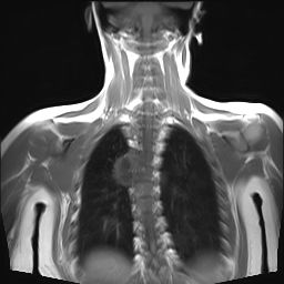


Figure 1: Preoperative MRI view
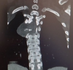


Figure 2: Preoperative CT view
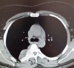


Figure 3: Preoperative CT view


About 3% NaCl-impregnated sponges were placed around the intrathoracic area and operation site considering it may be cysthydatic. The cavity was opened with sharp dissections and the fluid inside it was aspirated. Numerous cystic lesions with an extradural localization extending from the  paravertebral   area  to   the  pleura, the  smallest 0.5 cm  and   the  largest  3 cm  in  diameter,  were   removed.
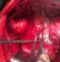


Figure 4: Right thoracotomy exploration view


No other pathological findings were found in the intrathoracic area. the patient who did not require vertebral cage; Significant increase in neuromonitorization in the right lower extremity after stopping muscle relaxant was reported. Albendazole treatment was started postoperatively 2´400 mg/3 months with no early complication was seen. physical therapy was started on the second postoperative day. In the second postoperative month, the right lower extremity muscle strength was 4 and the decrease in tactile sensation continued and other neurological findings were normalized.


DISCUSSION

Cysthydatic is most commonly located in the liver (60-70%), lung (20-30%) and bone (0.5-4%), most common bone tissue in the vertebra (30-50%) and pelvis (10-15%) [3]. As a result of portovertebral venous shunt, the corpus body of the vertebra is primarily affected and other cancellous bones such as lamina and pedicle are affected. Spinal involvement is present in approximately 50% of patients with bone involvement [1]. Spinal involvement is most commonly seen in the thoracic region. In vertebral cysthidatics, a real cyst cannot be formed because the fibrous adventitial layer does not form and its borders cannot be clearly selected. Polycystic and diffuse cysthydatism are seen in the bone. Our case is a polycystic cysthydatic case with spinal involvement and pleural involvement located in the thoracic vertebrae [4]. Although the symptoms vary according to the location of the cysthydatic involvement, it is often asymptomatic. Vertebral involvement is associated with pain, secondary infection, collapse fracture and spinal canal compression and pleural involvement involves trachea, esophagus, heart and large vessel compression. Our patient presented with complaints of back pain, numbness in the right arm, weakness in the right lower extremity and numbness. In addition to serological tests, radiological radiographs can be used to detect rare cases. Peripheral tissue involvement can be detected by Ultrasonography (USG) and bone and lung involvement can be detected by CT. T1a hypointense and T2a hyperintense are seen on MRI and extradural intradural distinction is made with this method [5]. In the pre-diagnosis of our patient, serological test was not performed because it was evaluated as a mass. Direct X-ray, CT and MRI were taken and evaluated as a mass. Mass, tuberculosis, abscess, spondylodiscitis are considered in the differential diagnosis, but cysthydatic should be considered. The incidence of recurrence in patients who underwent surgical treatment alone was reported to be more frequent compared to the combination of surgery and medical treatment. Medical treatment alone is not recommended as antiparasitic drugs are switched to bone tissue and have little efficacy [6]. In our patient, the lesion was totally removed by right thoracotomy and postop albendazole treatment was applied.

CONCLUSION

Surgical approach with thoracotomy should be considered in lesions affecting the thoracic vertebra. It makes exploration of the thorax and vertebra very easy for the surgeon. In case of intrathoracic complications, it provides a chance for simultaneous intervention and reduces the risk of complications.


Conflict of Interest

The authors declared that there is no conflict of interest.


Funding

No financial support has been received for the study.


Ethical Approval

Informed consent form in accordance with international hospital standards and patient approval has been obtained for scientific publication.


Authors Contribution

Bayrakçi Onur: Designed the study and wrote the first draft of the manuscript. Gökten Murat: Managed the literature searches. Both authors readand approved the final manuscript. The study has not been sent to another journal.

REFERENCES
  1. Sapkas, G.S., et al. "Spinal Hydatid Disease, a Rare but Existent Pathological Entity: Case Report and Review of the Literature." South Med J, vol. 99, 2006, pp. 178–83.

  2. Şahin, F., et al. "Plevral Efüzyonun Ayırıcı Tanısında Kist Hidatik." TTD Plevra Bülteni, vol. 2, no. 2, 2008, pp. 53–58.

  3. Moro, P. and P.M. Schantz. "Echinococcosis: A Review." Int. J. Infect. Dis., vol. 13, no. 2, 2009, pp. 125–33.

  4. Kafaji, A., et al. "Spinal Hydatid Disease: Case Report and Review." World Neurosurg, vol. 80, no. 5, 2013.

  5. Normelli, H., et al. "Spinal Hydatid Cysts." European Spine Journal, vol. 7, no. 2, 1998.

  6. Neumayr, A., et al. "Spinal Cystic Echinococcosis–A Systematic Review." PLoS Neglected Tropical Diseases, vol. 7, no. 9, 2013.
License
CC BY-NC-ND
Creative Commons Attribution-NonCommercial-NoDerivatives 4.0 International License
A Vertebral Cysthidatic Case with Pleural and Spinal Involvement © 2026 by BAYRAKÇI Onur, GÖKTEN Murat licensed under CC BY-NC-ND 4.0
All papers should be submitted electronically. All submitted manuscripts must be original work that is not under submission at another journal or under consideration for publication in another form, such as a monograph or chapter of a book. Authors of submitted papers are obligated not to submit their paper for publication elsewhere until an editorial decision is rendered on their submission. Further, authors of accepted papers are prohibited from publishing the results in other publications that appear before the paper is published in the Journal unless they receive approval for doing so from the Editor-In-Chief.
Himalayan Journal of Medicine and Surgery open access articles are licensed under a Creative Commons Attribution-Share A like 4.0 International License. This license lets the audience to give appropriate credit, provide a link to the license, and indicate if changes were made and if they remix, transform, or build upon the material, they must distribute contributions under the same license as the original.
Recommended Articles
Research Article
Phenotypic Characterization and Antimicrobial Resistance Patterns of Clinical Klebsiella pneumoniae Isolates from Al-Najaf Teaching Hospital
Published: 30/06/2026
Download PDF
Research Article
Impact of Gut-Liver Axis: Hepatic Biochemical and Metabolic Changes Associated with Chronic Gastritis in Iraqi Patients with Helicobacter Pylori
Published: 20/02/2026
Download PDF
Research Article
Effect of Land Degradation on Livelihood
Published: 04/01/2024
Download PDF
Research Article
Awareness and Practices among Professional Healthcare Workers towards COVID-19 in Iraq
...
Published: 30/03/2024
Download PDF
Flowbite Logo
Najmal Complex,
Opposite Farwaniya,
Kuwait.
Email: support@himjournals.com

Useful Links
Order Hard Copy
Privacy policy
Terms and Conditions
Refund Policy
Others
About Us
Team Members
Contact Us
Online Payments
Join as Editor
Join as Reviewer
Subscribe to our Newsletter
Follow us
MOST SEARCHED KEYWORDS
scientific journal
 | 
business journal
 | 
medical journals
 | 
Scientific Journals
 | 
Academic Publisher
 | 
Peer-reviewed Journals
 | 
Open Access Journals
 | 
Impact Factor
 | 
Indexing Services
 | 
Journal Citation Reports
 | 
Publication Process
 | 
Impact factor of journals
 | 
Finding reputable journals for publication
 | 
Submitting a manuscript for publication
 | 
Copyright and licensing of published papers
 | 
Writing an abstract for a research paper
 | 
Manuscript formatting guidelines
 | 
Promoting published research
 | 
Publication in high-impact journals
Copyright © Himalayan Journals . All Rights Reserved.








