Introduction: Gall bladder disease is a common health problem throughout the world. The earliest gallstone case was found in Gotland, in the remnants of a tomb dating back to the Stone Age, approximately 4000 years ago. The incidence of cholelithiasis is reported to vary between 5.9% and 21.9% of the general population. The clinical presentation of choledocholithiasis may vary widely, as CBD stones may be asymptomatic in 5-10% of cases. Material and Methods: This cross-sectional descriptive study was carried out in the department of General surgery in concordance with Department of Gastroenterology Indira Gandhi Medical College Shimla (H.P.) on patients admitted with diagnosis of Cholelithiasis with choledocholithiasis from 1-August-2018 to 31-July-2019. Results: A Total of 108 patients were included in the study. The age of patients in the present study ranged from 20 years to 70 years. The mean age of the patients was 47.60 years. Out of 108 cases, 21 (19.44%) cases were male and 87 (80.56%) were females with M: F of 1:4.14. Conclusions: Majority of the patients presented with cholelithiasis and choledocholithiasis are from 51-60 years age group females. All patients included in this study demonstrated cholelithiasis with dilated common bile duct.
Gall bladder disease is a common health problem throughout the world. The earliest gallstone case was found in Gotland, in the remnants of a tomb dating back to the Stone Age, approximately 4000 years ago. The incidence of cholelithiasis is reported to vary between 5.9% and 21.9% of the general population [1]. The clinical presentation of choledocholithiasis may vary widely, as CBD stones may be asymptomatic in 5-10% of cases. Because of the smaller diameter of the distal CBD at the Vater papilla, they may remain in the choledochus. Common bile duct produces colicky pain, jaundice or potentially life-threatening complications, such as ascending cholangitis or acute pancreatitis [2].
The management of concomitant gall bladder and CBD stones has evolved significantly over the past 20-30 years. In the era of open surgery, open CBD exploration (choledocholithotomy) used to be performed if any common bile duct stones were identified at cholangiography. Following the introduction of ERCP, open CBD exploration was reserved for patients with failed ERCP. With the advent of various modern technologies, open surgical procedures, such as CBD exploration or biliary-enteric bypasses, are now usually considered where ERCP fails to retrieve stones or where facilities of advanced laparoscopic surgery are not available. A relative indication for open exploration is large or multiple stones or the need to perform a trans duodenal sphincteroplasty. Generally, in well-equipped centers of the world, ERCP followed by laparoscopic cholecystectomy is recommended as a safe and cost-effective procedure [3]. However, various centers advocate laparoscopic cholecystectomy with common bile duct (CBD) exploration or a Rendezvous technique where endoscopy and laparoscopy are performed simultaneously. They both have a similar efficacy profile with the added benefit of saving time.
The objective of this study was to describe the socio epidemiological profile of the patients presented with cholelithiasis along with choledocholithiasis at department of surgery, IGMC Shimla.
This cross-sectional descriptive study was carried out in the department of General surgery in concordance with Department of Gastroenterology Indira Gandhi Medical College Shimla (H.P.) on patients admitted with diagnosis of Cholelithiaisis with choledocholithiasis from 1-August-2018 to 31-July-2019.This study comprised of 108 patients of cholelithiasis and choledocholithiasis diagnosed by ultrasound or MRCP. Consent of all patients was taken. All diagnosed cases of cholelithiasis with choledocholithiasis were included in the study whereas patients below 20 and above 70 years and patients with empyema gall bladder and cholangitis were excluded from the study. Socio epidemiological characteristics of the patients were collected using a structured closed ended questionnaire [4].
Statistical Analysis
Data was entered in Microsoft excel spreadsheet, cleaned for errors and was analyzed using Stata Software version 15. Descriptive statistics were used to summarize the demographic data. Frequencies, percentages and their 95% confidence intervals were used to describe categorical variables [5].
A Total of 108 patients were included in the study. The age of patients in the present study ranged from 20 years to 70 years. The mean age of the patients was 47.60 years. Out of 108 cases, 21 (19.44%) cases were male and 87 (80.56%) were females with M: F of 1:4.14 (Table 1).
In study of 108 patients, four patients had hypertension, five patients were diabetic and two patients had hypothyroidism. Pain was the chief complaint for which patients visited hospital and was presented in 98(90.74%) patients. Jaundice was present in 65(60.19%) patients. Forty-two (38.89%) patients had nausea and vomiting. None of the patients in our present study had cholangitis, pancreatitis, acute cholecystitis and shock.
Table 1: Age and gender distribution among study participants
Sr. No. | Category | Number Of Patients (%) |
Age Groups (years) | ||
| 21-30 31-40 41-50 51-60 61-70 | 14 (12.97) 21 (19.44) 21 (19.44) 37 (34.26) 15 (13.89) |
Gender | ||
Male Female | 21 (19.44) 87 (80.56) | |
Table 2: USG findings in different patients
Usg Findings | Number | (%) |
Cholelithiasis with choledocholithiasis | 24 | 22.22 |
Cholelithiasis with dilated CBD | 84 | 77.78 |
Total | 108 | 100 |

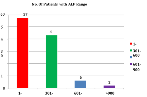
Figure 1: Value of ALP in different patient
All 108 patients included in this study demonstrated cholelithiasis with dilated common bile duct. CBD stones were observed in 24 patients on ultrasound. In rest of the 84 patients’ common bile duct stones were detected on MRI abdomen and MRCP (Table 2).
ALP was raised in 76 (70.37%) patients in our study and in other 32 patients it was less than 3 times the upper limit of normal value (Figure 1).
Cholelithiasis with choledocholithiasis is a disease of young and middle age. The age of patients in our study ranged from 20 years to 70 years with mean age of 47.60 years. M: F ratio in our study was 1: 4.14. The age and sex ratio in our study was similar to a study done by Mohamed Mohsen Salem et al., in 2019 on 50 patients in which ages ranged between 21 and 70 years with a mean age of 47.24 years and the ratio of M: F was 1: 4.56 [5].
In the present study 64 patients (59.26%) complained of upper abdominal pain depicting pain as the predominant clinical presentation. Shelat et al. reported abdominal pain as a major presenting complaint (55.9%) which is almost equal to that in our study [6]. Stanley et al., reported upper abdominal pain in 80% of the patients. Pain was present in 98 (90.74%) patients [7]. This in comparison to a study done by Ehab El Hanafy et al. in which pain was the presenting feature in 90.5% of patients. Pain is a feature of cholelithiasis and choledocholithiasis and not in malignant causes of jaundice [8]. In our study jaundice was present in 65 (60.19%) of patients i.e. almost equal (64%) to a study done by Rajkumar Sharma et al. When stones in CBD obstruct the lumen then obstructive jaundice occurs. This jaundice regresses when obstruction is relieved [9].
Ultrasound was able to diagnose cholelithiasis in all (100%) patients and choledocholithiasis in 24 (22.22%) patients out of 108 patients. Detection of CBD stones by USG is low because distal CBD is not properly visualized on USG due to overlying gas filled loops of small bowel. Sensitivity of detection of gallstones by USG was 100%. And sensitivity of detection of CBD stones was 23% whereas the specificity was 92%. Gall bladder was distended in 84 patients and contracted gall bladder was found in 4 patients. Intra hepatic biliary radical dilation was present in 58 patients.
Mark A Stott et al., did a study in which the sensitivity and specificity of USG for detection was 36% and 98% respectively [10]. Gurusamy et al. concluded that many people may have CBD stones in spite of having a negative ultrasound or liver function test [11]. Einstein DM et al. concluded that definite diagnosis of choledocholithiasis could be made on the basis of the sonograms in 22% of cases [12].
All patients underwent MRI abdomen with MRCP following initial ultrasound study. It demonstrated choledocholithiasis in all 108 patients who were missed on ultrasound. Sensitivity and specificity of MRCP in detection of CBD stone was 100%. Hjartarson et al., supported that the use of MRCP as a tool for exclusion of choledocholithiasis as MRI has high sensitivity and specificity to diagnose choledocholithiasis [13]. Grubnik et al., reported that a total of 256 patients with CBD stones were operated laparoscopically, bile duct stones were visualized pre-operatively by means of MRCP in all (100%) patients [14]. Results are comparable to present study showing MRI has high sensitivity and specificity to diagnose choledocholithiasis. Our study has similar findings as in other studies.

Majority of the patients presented with cholelithiasis and choledocholithiasis are from 51-60 years age group females. All patients included in this study demonstrated cholelithiasis with dilated common bile duct.
Ahlberg, J. and S. Sahlin. "The still frustrating mysterious gallstone: a century of gallstone disease in lakartidningen." Lakartidningen, vol. 101, no. 51-52, 1 Dec. 2004, pp. 4238–41.
Aerts, R. and F. Penninckx. "The Burden of Gallstone Disease in Europe." Alimentary Pharmacology and Therapeutics, vol. 18, Nov. 2003, pp. 49–53.
Sarli, L. et al. "Asymptomatic bile duct stones: selection criteria for intravenous cholangiography and/or endoscopic retrograde cholangiography prior to laparoscopic cholecystectomy." European Journal of Gastroenterology and Hepatology, vol. 12, no. 11, 1 Nov. 2000, pp. 1175–80.
Ghazanfor, R. et al. "Choledocholithiasis: Treatment options in a tertiary care setup in Pakistan." Cureus, vol. 9, no. 8, Aug. 2017.
Salem, M.M. et al. "Comparative study between laparoscopic common bile duct exploration and endoscopic retrograde cholangiopancreatography plus laparoscopic cholecystectomy for choledocholithiasis." International Surgery Journal, vol. 6, no. 7, 29 June 2019, pp. 2250–57.
Shelat, V.G. et al. "Laparoscopic exploration can salvage failed endoscopic bile duct stone extraction." Singapore Medical Journal, vol. 53, no. 5, 1 May 2012, pp. 313.
Rogers, S.J. et al. "Prospective randomized trial of lc + lcbde vs ercp/s + lc for common bile duct stone disease." Archives of Surgery, vol. 145, no. 1, 1 Jan. 2010, pp. 28–33.
El Hanafy, E. et al. "Is primary closure a feasible and acceptable option in the era of t-tube-free common bile duct exploration for choledocholithiasis?" The Egyptian Journal of Surgery, vol. 35, no. 3, 1 July 2016, pp. 254.
Sharma, R. et al. "Is It time to write obituary for open choledocholithotomy in the present era of endoscopic management? outcome of surgery for failed endoscopic extraction, retrospective study." International Surgery Journal, vol. 4, no. 4, 25 Mar. 2017, pp. 1350–54.
Stott, M.A. et al. "Ultrasound of the common bile duct in patients undergoing cholecystectomy." Journal of Clinical Ultrasound, vol. 19, no. 2, Feb. 1991, pp. 73–76.
Gurusamy, K.S. et al. "T‐Tube drainage versus primary closure after laparoscopic common bile duct exploration." Cochrane Database of Systematic Reviews, no. 6, 2013.
Einstein, D.M. et al. "The insensitivity of sonography in the detection of choledocholithiasis." American Journal of Roentgenology, vol. 142, no. 4, 1 Apr. 1984, pp. 725–28.
Hjartarson, J.H. et al. "The value of magnetic resonance cholangiopancreatography for the exclusion of choledocholithiasis." Scandinavian Journal of Gastroenterology, vol. 51, no. 10, 2 Oct. 2016, pp. 1249–56.