Patients with laryngeal cancer have a high risk of developing lung cancer in the future. A patient treated for supraglottic cancer in 2011 presented with complaint of shortness of breath and right sided chest pain for the last 4-5 months. Chest X-ray posterior-anterior view showed a homogeneous opacity in the right hilar region. CECT thorax revealed a heterogeneously enhancing soft tissue density mass lesion in hilar region of the right lung. On histopathology, it was adenocarcinoma of the lung. Patients with laryngeal cancer should be routinely screened during follow-up with X-ray chest or CECT thorax for the early detection of lung cancer.
Because of the common etiological agent of smoking, patients with head and neck cancers including laryngeal cancer have a high risk of developing second primary lung cancers. Supraglottic laryngeal cancer is more commonly associated with second primary lung cancer as compared to other head and neck cancers. So, patients with head and neck cancer should be routinely screened with chest X-ray or CT scan during follow up visits for the early detection of lung cancer.
A 67-year-old male patient presented with a complaint of right-sided chest pain for the last 4-5 months with shortness of breath and haemoptysis. On examination, vitals were normal with no lymphadenopathy. Systemic examination was also normal. Routine blood investigations were within normal range. Chest X-ray posterior-anterior view showed homogeneous opacity in the right hilar region. CECT thorax was suggestive of a heterogeneously enhancing soft tissue density mass lesion in the hilar region of right lung (Figure 1). On histopathological examination of the mass lesion, it was diagnosed as a case of adenocarcinoma ca lung.
In 2011, patient was treated for ca supraglottis with chemoradiation. The patient was diagnosed as a case of second primary ca lung with supraglottic squamous cell carcinoma as index tumour. He was referred to the radiotherapy department for further management.
Head and neck cancers constitute about 10.3% of all cancers [1]. Tobacco and alcohol are the two major etiological agents associated with the head and neck cancers. Because of common etiological factors, patients with head and neck cancer are at risk for developing second primary malignancies at a rate of 4-7% per year [2]. According to the theory of ‘field cancerization’ continuous exposure to common etiological agents leads to the development of second primary cancer. According to Wu et al. [4], carcinogenic agents such as tobacco and alcohol cause irreversible genetic and histologic changes leading to the development of second primary cancer. The lung is the most common site for the development of a second primary tumour after head and neck cancer [5]. Oro-hypopharynx or larynx are the most common sites of index head and neck tumour. Squamous cell carcinoma is the most common histological type of index head and neck tumour associated with second primary lung cancer [6].
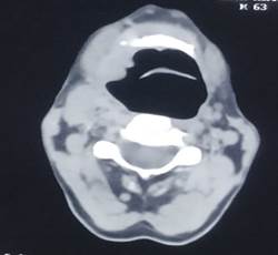
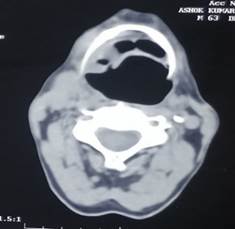
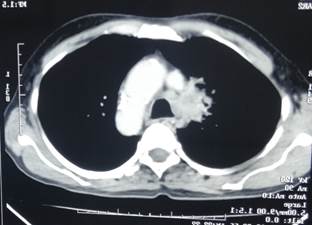
Figure 1: Axial CT images of the neck and thorax demonstrating cross-sectional anatomy of the oropharynx, cervical airway and mediastinal structures
Supraglottic laryngeal cancer is more commonly associated with second primary lung cancer as compared to glottic cancer [7,8]. The prognosis for second primary lung cancer in association with a head and neck carcinoma is poor because it is diagnosed in advanced stage [9].
All head and neck cancer patients should be routinely screened using chest X-rays for early detection of a second primary lung cancer [10]. But screening by chest X-rays has failed to improve survival in patients with secondary lung cancer [11]. Screening for lung cancer by CT scan has a higher detection rate as compared to chest X-rays [12,13]. CECT thorax or PET-CT scan should be used for early detection of second primary lung cancer, especially if the site of head and neck carcinoma are the Oro-hypopharynx or larynx, if the cell type is squamous cell carcinoma and if initial chest X-ray findings are abnormal [14].

Lefebvre, J.L. "Current clinical outcomes demand new treatment options for SCCHN." Annals of Oncology, vol. 16, suppl. 6, 2005, pp. vi7–vi12.
Leon, X. et al. "Second primary tumors in head and neck cancer patients." Acta Otolaryngologica, vol. 122, 2002, pp. 765–778.
Slaughter, D.P. et al. "Field cancerization in oral stratified squamous epithelium: Clinical implications of multicentric origin." Cancer, vol. 6, 1953, pp. 963–968.
Wu, X, et al. "Upper aerodigestive tract cancers." Multiple Primary Cancers, edited by A.I. Neugut, A.T. Meadows and E. Robinson, Lippincott Williams and Wilkins, 1999, pp. 319–346.
Vikram, B. "Changing patterns of failure in advanced head and neck cancer." Archives of Otolaryngology, vol. 110, 1984, pp. 564–565.
Jones, A.S. et al."Second primary tumors in patients with head and neck squamous cell carcinoma." Cancer, vol. 75, 1995, pp. 1343–1353.
Hofmann, H.S. et al."Primary lung cancer and extrapulmonary malignancy." European Journal of Cardio-Thoracic Surgery, vol. 32, 2007, pp. 653–658.
Kim, E.B. et al. "Clinical factors related to suspected second primary lung cancer development in patients with head and neck cancer." Cancer Research and Treatment, vol. 40, 2008, pp. 178–183.
Wagenfeld, D.J.H. et al. "second primary respiratory tract malignancies in glottic carcinoma." Cancer, vol. 46, 1980, pp. 1883–1886.
Wagenfeld, D.J.H. et al. "Second primary respiratory tract malignant neoplasms in supraglottic carcinoma." Archives of Otolaryngology, vol. 107, 1981, pp. 135–137.
Atabek, U. et al. "Lung cancer in patients with head and neck cancer: Incidence and long-term survival." American Journal of Surgery, vol. 154, 1987, pp. 434–438.
Ritoe, S.C. et al."Screening for second primary lung cancer after treatment of laryngeal cancer." Laryngoscope, vol. 112, 2002, pp. 2002–2008.
Loh, K.S. et al. "A rational approach to pulmonary screening in newly diagnosed head and neck cancer." Head and Neck, vol. 27, 2005, pp. 990–994.
Henschke, C.I. and Yankelevitz, D.F. "CT screening for lung cancer." Radiologic Clinics of North America, vol. 38, 2000, pp. 487–495.









