Introduction: Surgical vascular emergencies are infrequent, compared to bone and digestive emergencies [1]. A distinction should be made between minor vascular emergencies and major ones. The latter being the subject of our study, the objective of which is to describe the epidemiology and surgical aspects. Material and Method: We carried out a retrospective and descriptive study over a period of 4 years in the general surgery, otolaryngology and emergency departments of the Omar Bongo Ondimba Army Instruction Hospital. Results: 30 patients were collected, i.e. a prevalence of 1.6%. The male sex predominated at 67.8% and a sex ratio of 2.11. The young adult was the most affected age group with an average age of 35, 4 years. The stabbing agent was the main wounding agent followed by the arteriovenous fistula aneurysm. Vascular lesions mainly affected the limbs. Morbidity was marked by sensitive motor paralysis, joint tiffs and 10% mortality. Conclusion: Vascular wounds are major surgical emergencies, of which the stabbing is the main agent of vulnerability.
Surgical vascular emergencies are common clinical situations. They are underestimated in tropical environments because few articles are devoted to them. [1,2].
Etiopathogenically, in civil practice, sharp objects are the main cause of lesions. These latter consist mainly of penetrating wounds [2,3]. But currently, the frequency of iatrogenic trauma is constantly increasing due to the increasingly common practice of catheterizations and other invasive cardiac and vascular explorations [1-4]. Vascular emergencies represent a heterogeneous set of clinical situations. From which it is necessary to exclude medical emergencies (thromboembolic disease, vascular accidents, etc.
Vascular emergencies according to the literature are part of a traumatic context.
We will focus on two main aspects:
The acute context of the trauma is the case of acute post-traumatic ischemia. He This is an emergency that involves the functional and vital prognosis. The priority is to stop the bleeding and restore normal circulation, regardless of their location
The subacute context: this is the case of obstruction by cruoric embolus or aneurysm pathology
We report here our experience over 4 years in practice at GABON in order to identify, on a general level, the epidemiological and surgical aspects and in a specific way, to describe the prevalence of vascular emergencies, the injury mechanisms, to assess the morbidity and mortality. describe the different surgical procedures performed.
Patients and Methods
The visceral surgery department, the ENT department and the emergency department of the OMAR BONGO ONDIMBA Army Instruction Hospital (H. I. A. O. B. O) served as the location for this study.
This was a retrospective and descriptive study, spanning a period of 4 years, from January 2012 to December 2016 (60 months).
All patients admitted in emergency for a traumatic or non-life-threatening vascular wound and/or functional were included in our study.
Minor vascular injuries and medical vascular emergencies were not included.
The data was collected from hospitalization registers from surgery and ENT departments, patient medical files and other data was found in the operating report register.
The data collected concerned the patient (age, sex, profession, etc.), the vascular lesion (the mechanism, the type of lesion, etc.) and the treatment (pre-hospital, origin of the patient, the time taken to take in charge, the surgery performed and its evolution).
These data were recorded on an individual file, which was previously designed.
All the statistical analyzes were carried out by the statistical software Epi info. The data was collected and saved in an Excel table from the Excel 2013 software.
Numeric variables were described by the mean and the standard deviation if they were normally distributed, otherwise by the median and the inter-type. Categorical variables were described by their size and percentage. Bar and pie charts were shown to visualize the distribution of patients according to the different categories of study variables.
Epidemiological Aspects
From January 2012 to December 2016, 30 patients were collected out of 1,800 hospitalized patients, i.e. a prevalence of 1.6%.
We saw 19 men 11 women, the sex ratio was 1.7. In our series the mean age was 35.4 years with extremes between 19 years and 65 years. 73% of the patients were aged between 20 and 40 years and 12.8% below 20 years. Thus 86% of patients were young (<40 years) (Table 1).
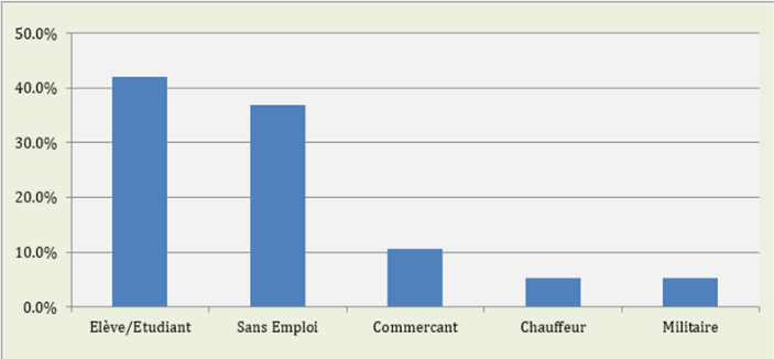
Among the socio-professional categories present in our study, 42.1% were pupils/students, 36.8% were unemployed, 10.5% were traders, 5.3% were drivers and 5.3% were soldiers (Figure 1).
The stabbing was the main wounding agent in 50% of cases, followed by arteriovenous fistula aneurysm rupture and gunshot wound at 25% and 7.14% respectively (Table 2).
Medical transport (10%) was used very little for the transport of patients (Table 3).
About 75% of our patients were treated beyond the 6 hours required (Table 4).
Table 1: Distribution of cases by sex and age
| Sex | Workforce (%) | Age moyen (± ET) | Age extrêms | P |
| Women | 11 (36,7) | 35,1 ans (± 18,5 ans) | [19 ans-65 ans] | 0,43 |
| Men | 19 (63,3) | 30,7 ans (± 8,4 ans) | [19 ans– 49 ans] | |
| Total | 30(100,0) | 32,1 ans (± 12,1 ans) | [19 ans-65 ans] |
Table 2: Circumstances of occurrence and vulnerable agents
Circumstances of occurrence and vulnerable agents | Effectives | % |
| Wound by weapon white | 14 | 46,7 |
| Gunshot wound | 2 | 6,7 |
| Anévrism rupture | 9 | 30 |
| Acute ischemia by embolus | 1 | 3,3 |
| sports / work accident | 1 | 3,3 |
| Public road accident | 2 | 6,7 |
| Luxation | 1 | 3,3 |
| Total | 30 | 100,00 |
Vascular limb injury was predominant in our study at 50%, followed by cervical vascular injury at 35.7% (Table 5).
The duration of hospitalization was less than 6 days for peripheral vascular lesion.
She excelled the 10 days for serious vascular lesions involving the large arterial trunks with several associated lesions.
The postoperative follow-up was straightforward in the majority of cases. The main complications were neurological and hematological.
Table 3: Mode of transport of patients (n = 28 Modes of transport
Modes of transport | N = 28 | % |
Samu | 3 | 10,7 |
Personnel cars | 3 | 10,7 |
Others | 22 | 78,6 |
Table 4: Breakdown of the period of care
Breakdown of period of care | N = 28 | % |
More 6 hours | 21 | 75 |
Less 6 hours | 7 | 25 |
Table 5: The topography of the lesions (n = 28)
Topography of lesions | N=28 |
Cervical | 10 |
Thoracic Inferior member | 8 |
Superior member | 8 |
Table 6: The different lesions observed
Topography of lesion | N = 30 | % |
Ruptured abdominal infra-renal aneurysm | 2 | 7,1 |
Fissured right internal carotid aneurysm | 1 | 3,6 |
Aneurysm of humero-basilaire fistula | 3 | 3,6 |
Left popliteal aneurysm | 1 | 3,6 |
Right radio-radial AVF aneurysm | 1 | 3,6 |
1 | 3,6 | |
axillary artery and et vein left | 2 | 7,1 |
External carotid collateral | 1 | 3,6 |
Left Ulnar artery | 1 | 3,6 |
Right humeral artery | 1 | 3,6 |
Right humeral Artery ans vein | 1 | 3,6 |
arteries and veins Subclavian | 2 | 7,1 |
Left subclavian arteries | 1 | 3,6 |
Right popliteal and sub-articular arteries | 4 | 14,3 |
Jugulo-carotid | 1 | 3,6 |
Lower vena cava | 1 | 3,6 |
Lower vena cava / sawmill work accident | 1 | 3,6 |
Superior vena cava | 1 | 3,6 |
External jugular vein | 1 | 3,6 |
Internal jugular vein | 1 | 3,6 |
Internal jugular vein and collateral external carotid arteries | 2 | 7,1 |
Table 7: associated lesions
Lesions associates | Effectives | % |
Hemorragy | 4 | 23,5 |
Bleeding / disturbance of consciousness | 2 | 11,7 |
Hemothorax | 1 | 5,8 |
Hemodynamic instability associated with mesentery wound and eventration | 1 | 5,8 |
Ischémia | 2 | 5,8 |
Pharyngeal lesions transfixing wound of the thyroid cartilage | 1 | 5,8 |
Tendon lesions | 1 | 5,8 |
Wound of the right colon | 1 | 5,8 |
Right atrium sores | 1 | 5,8 |
Section of the branches of the brachial plexus | 1 | 5,8 |
Tendon section | 1 | 5,8 |
Thrombosis of the artery | 1 | 5,8 |
Total | 17 | 100,0 |
Table 8: the surgical procedures applied
The surgical procedures applied | N |
Conservative procedures: revascularization | 18 |
Lateral arterial et venous suture under left Clavier | 1 |
aorto-bifemoral bypass | 1 |
End to end artériel suture | 1 |
Trimming and end-to-end anastomosis | 1 |
Lateral arterial et venous suture under left Clavier | 1 |
Saphenous vein bypass and humeral vein ligation | 1 |
Femora-popliteal bypass with PTFE prosthesis and left popliteal vein ligation | 2 |
Right femora-popliteal PTFE prosthetic bypass | 1 |
Left ulnar artery end-to-end anastomosis | 1 |
Flattening and prosthetic bypass of the right internal carotid artery | 1 |
Flattening and interposition by saphenous vein | 1 |
Subclavian arterial and venous suture + thoracic drainage | 1 |
Lateral vena cava suture | 1 |
Lateral suture of the inferior vena cava and end-to-end anastomosis of the right primary iliac artery | 1 |
Double arterial and venous shunt | 1 |
External fixator and saphenous bypass | 1 |
Non-conservative gestures | 10 |
Ligation of the internal jugular vein and ligation of the middle thyroid artery | 1 |
Flattening and suturing end-to-end radio-radial | 2 |
Ligatures of the internal jugular vein and suture of the left carotid arteries collaterals | 1 |
Vascular suture; resection + prosthetic bypass by saphenous vein | 1 |
tracheostomy and external carotid collateral ligation | 1 |
Internal jugular vein ligatures | 4 |
Amputation et désobstruction | 1 |
Table 9: Breakdowns by length of hospitalization
Duration of hospitalization | N = 30 | % |
0-6 days | 14 | 50 |
6-10 days | 5 | 17,8 |
more 10 days | 9 | 31,1 |
Table 10: Post operative consequences
Post operative consequences | N = 30 | % |
Simple | 23 | 75 |
Complicated | 7 | 25 |
Table 11: Distribution according to the characteristics of the complications
Characteristics of the complications | N = 7 | 100% |
Joint stiffness | 2 | 28,5 |
Sensory-motor palsy left upper limb | 2 | 28,5 |
Deaths from acute anemia | 3 | 42,8 |

Figure 1: Distribution of cases by professional status
We will consider a discussion successively on the epidemiology and the surgical strategy.
Vascular emergencies represent a heterogeneous set of clinical situations. From which medical emergencies (thromboembolic disease, vascular accidents, etc.) should be excluded. In our analysis, we will consider all emergency situations, traumatic or not, which will lead to a solution of continuity at the level of a vascular axis whatever its situation. The physiological consequences of these emergencies will engage in the short or medium term the vital prognosis and the functional prognosis.
Epidemiology
Surgical vascular trauma is a frequent situation in our region. With a hospital frequency of 1.55% or approximately 2 out of 100 patients admitted to the emergency departments. They have underestimated because few reports are devoted.
In sub-Saharan Africa, few data exist only a few posts in war zones have been made. This is how Kedir et al. [5], in Ethiopia, reported in 2004 their experience in the management of vascular lesions seen late in the Eritrean War (1997-2000).
Fokou et al. [6] report 38 cases of vascular lesions in 5 years in Cameroon. We have collected 30 cases in 4 years.
These clinical entities are poorly evaluated for several reasons:
The seriousness of these lesions which are quickly fatal
The lack of pre-hospital medical care, allowing rapid transfer of the injured vascular patient. 10.7% against 78.6 benefited from medical transport
The lack of service practicing vascular surgery
All these are therefore at the origin of the wandering of patients, from late treatment to the required 6 hours. Patients then enter the lethal triangle of MOORE, responsible for postoperative complications in the short, medium and long term.
Age and Gender
In our series, we note a male predominance at 67.8%, the female sex being both very little represented at 32.1%.
The average age was 35.4 years with extremes between 19 and 65 years. 86% of patients were under 40 years old.
Our results are similar to those of Fokoul and Al [6], who report male predominance with a mean age of 28.3 years and extremes of 14 and 38 years.
Vascular emergencies remain the responsibility of adolescents and young adults. These data could be explained by the fact that young people represent the most active segment of the population.
The Injury Mechanisms
We can distinguish two nomological types. Vascular emergencies of traumatic and non-traumatic origin.
Traumatic emergencies predominate. They are the most encountered they are dominated by direct trauma by stabbing 50% of cases. They are characteristic of petty banditry, or related to crime.
Fokou et al. [6] observed them 52.43%, the lesions were caused by contending objects: knife, broken bottles.
7.14% of firearm injuries were recorded. They are part of a particular context of post-election crisis.
Road accidents were found in 7.14% of cases. This frequency is well below Soma Sekar et al. [7] who observe a higher frequency with 39% in a western series.
An anecdotal mechanism is reported, the lesion of the popliteal artery, following a dislocation of the knee in an obese patient following a fall.
Non-traumatic emergencies, this second entomopathogenic category is related to aneurysm ruptures on arteriovenous, aortic and popliteal fistulas, etc. at 21.5%. In our series, these vascular ruptures are very serious with a mortality rate of nearly 50%. They are often unrecognized or poorly evaluated [8].
Surgical Aspects
Support Time
75% of patients were treated more than 6 hours after the accident and only 10.7% received medical transport.
These results corroborate those found in the literature in particular Fokou et al. [6] in Cameroon found an average delay of 8.7 h, likewise Soma Sekar et al. [7] 9.3 h. These delays contrast with those of certain series of developing countries which are more common, notably India or Ricco and Fébrer [9] find a delay of 3.8 hours.
Surgical vascular pathology still suffers from a delay in management. Patients still arrive at an advanced ischemic stage, requiring us to perform a conservative or non-conservative procedure.
Surgical Procedures
Usually, the surgical procedure depends on the affected blood vessels. Almost all the variants of reconstruction procedures were performed according to the anatomical location, the mechanism and the type of lesion, or in relation to the lesions found. Most of these are complete vascular lesions associating a more or less significant solution of parietal continuity. We find it in 87%. Fokou et al. [6] more than 70%.
We have made either conservative or radical gestures. The conservative gestures consisted of interpositions of the great saphenous vein reversed or more rarely prosthesis. We performed a venous graft in 17% of cases. Fokou et al. [6] performed prosthetic bypass in 9.75%.
This graft remains the best solution for revascularization in an emergency situation. It can be removed quickly and limits the risk of secondary infection and thrombosis. Ricco and Fébrer [9] have also noted. We took it preferentially from the leg. Except for a lesion of the axillary cavity where we used the thigh portion.
Prosthetic substitutes are rarely used in emergency. Due to the potential infectious risk. But also, by its availability. In our series, the use of prosthesis was justified by the long admission period and therefore a sample would increase the ischemia time. But also, by the caliber of the traumatized vessel. This was the case with an axillary artery and popliteal artery lesion. The caliber of the large saphenous veins was small. This strategy is observed in the literature.
Another revascularization strategy, implemented: the temporary vascular shunt. The vascular shunt provides vascular continuity through a hollow tubular device. Several types exist: JAVID, ARGYLE, or PRUITT.
This strategy is inspired by war surgery, in the larger concept of vascular damage control. It allows for rapid hemostasis and revascularization, before evacuation to the infrastructure hospital.
We performed this tactic in a young patient admitted in emergency, with ischemia of the forearm following a ruptured arteriovenous humeral-basilar fistula aneurysm.
The placement of a temporary vascular shunt enabled us to remove ischemia and remove the venous graft. This tactic is not widely used in civilian settings. But could find a place in our therapeutic arsenal.
We report a peculiarity; the rupture of aneurysms developed on arteriovenous fistula paths for dialysis. This extremely serious complication of the vascular accesses for hemodialysis is known but often little reported because a prophylactic surgical treatment is often carried out. This entity is characterized by its unpredictability and the difficulty of a surgical strategy. In this situation we achieved on the one hand hemostasis and on the other hand the closure of the arteriovenous fistula.
These procedures relate in our study to two ruptured abdominal sub-renal aneurysms, a right internal carotid aneurysm fissured, a left popliteal aneurysm, aneurysm on right arteriovenous fistula radio-radial.
The etiological mechanisms are certainly different, but vascular lesions can be grouped together according to the parietal involvement. Vascular lesions can be partial, affecting only part Complete parietal lesions, ranging from the transfixing wound to the solution of continuity with more or less significant loss of substance. Let us note first, that with regard to the type of vessels: the traumatic vascular lesions most found in this study are arterial lesions with 61%, our results agree with those of the literature as demonstrated respectively by Ricco and Fébrer [9] 90% of arterial vascular damage and Soma Sekar et al. [7] 94% of arterial vascular involvement.
The type of lesion is dominated by arterial lesions with loss of substance. We report this type of lesion in 74% of patients. The partial lesions are marginal 26%. This is observed by Ricco and Fébrer [9], who found nearly 90% of arterial damage. This type of lesion defines clinical manifestations on admission of the vascular wall: intimal lesions, or sub-adventitious.
The radical gestures, ligature, amputations, were carried out in our series,
26% (ligatures). They are 12.19% ligatures in the series by Fokou et al. [6]. These actions are often gestures of necessity, that is to say justified by the local condition of the limb and the risk of infection.
A trans femoral amputation was performed in a late seen patient with acute lower limb ischemia.
In our series we did not note any endovascular surgery. This technique is generally used for the treatment of unruptured arterial aneurysms due to the low mortality including in fragile patients. What is involved in the decision is the complexity of the endovascular act which essentially arises from the anatomy of the aneurysm.
Stents require a prior choice of design, diameter and length. These parameters determine the success of the intervention.
Morbi-Mortality
For the majority of patients seen in our study, the prognosis was earlier favorable but we encountered complications and those most encountered were deaths from acute anemia 33.3% which are due to long delays in treatment, non-transport. medicalization of patients to the appropriate health structures and difficulty of emergency access to labile blood products. Joint stiffness 33.3%, sensory-motor paralysis of the upper limb 33.3%. Other authors in the literature Menakuru et al. [2] and Soma Sekar [7] find infectious complications 7% and vascular thrombosis 4%. Ricco and Fébrer [9] found in their series 9 amputee patients. The remaining patients were successfully revascularized.

Vascular wounds, due to the spread of urban violence and growing socio-economic difficulties, will constitute more of a challenge for our healthcare system because of the vital and functional emergency they constitute.
By far the most frequent aetiology is stabbing attacks, young male adults being the most affected population because they are more active.
The early medicalization of care from the pre-hospital phase is an essential asset for the management of vascular wounds, making it possible to improve the quality of care and to provide for the subsequent therapeutic stage, which is the surgical act. This attitude has been adopted very little in our practice, the majority of our wounded being transported in non-medical vehicles.
Oxygen therapy, vascular filling, serovaccination, antibiotic therapy and monitoring surveillance are fundamental gestures, essential for subsequent surgical decision-making, for improving the death rate of the injured.
Doody, O. et al. (2008). “Extremities Indications and techniques of treatment of extremity vascular injuries”. Injury, 39, 1295–1303.
Menakuru, S.R. et al. (2005). “Extremity vascular trauma in civilian population: A seven-year review from North India”. Injury, 36, 400–406.
Diamond, S. et al. (2003). “Vascular injuries in the extremities in a suburban trauma center”. American Surgeon, 69, 848–851.
Wood, D.B. et al. (2001). “Non-arteriographic evaluation of penetrating vascular injuries. In C. B. Ernst and J. C. Stanley (Eds.)”, Current therapy in vascular surgery (pp. 580–583). Mosby.
Kedir, M. and Bekele, A. (2004). “Surgery of traumatic peripheral arterial injury with delayed transfer during the Ethio-Eritrean War (1997–2000)”. East and Central African Journal of Surgery, 9, 20–23.
Fokou, M. et al. (2011). “Les traumatisms vascularizes périphériques en pratique civile: À propose de 41 lésions opérées au Cameroun”. Chirurgie Thoracique Cardio-Vasculaire, 15(3), 145–149.
Soma Sekar, R. et al. (2005). “Extremity vascular trauma in civilian population: A seven-year review from North India”. Injury, 36, 400–406.
Wani, M.L. et al. (2012). “Peripheral vascular injuries due to blunt trauma (road traffic accident): Management and outcome”. International Journal of Surgery, 10, 560–562.