Contents
Download PDF
pdf Download XML
2815 Views
419 Downloads
Share this article
Research Article | Volume 2 Issue 2 (July-Dec, 2021) | Pages 1 - 4
Surgical Treatment of Patients with Pelvic Organ Prolapse
 ,
 ,
1
Ministry of Health, Thi Qar Health Office, Shatra Hospital, Thi-Qar, Iraq
2
Ministry of Health, Thi Qar Health Office, Al-Rifai Hospital, Thi-Qar, Iraq
3
Ministry of Health, Department of Health Al-Anbar, Fallujah Maternity and Children's Hospital, Al-Anbar, Iraq
Under a Creative Commons license
Open Access
Received
April 28, 2021
Revised
May 26, 2021
Accepted
June 22, 2021
Published
July 31, 2021
Abstract

The study and complete data collection were conducted in Shatra Hospital, Thi-Qar, Iraq. The purpose of this study was to evaluate the effectiveness of surgical procedures using nets in pelvic prolapse. Where it was 150 and the average woman's age was 50 ± 5.4 from Shatra Hospital, Thi-Qar, where a questionnaire was collected from patients in the hospital and it was identified that there are some complications from the surgery that weaken the muscles and ligaments that support the pelvic organs in r body, the pelvic organs can slip out of place and hang down insider body Vagina (pelvic organ prolapse). To treat pelvic organ, prolapse, a surgical mesh may be implanted to support the weakened vaginal wall. This surgery can be performed through the abdomen, using a net, or through the vagina without using a net. And surgery through the vagina using mesh is no longer an option. Each use of surgical mesh has its risks and benefits. The use of transvaginal surgical mesh to treat pelvic organ prolapse is associated with high rates of mesh-related complications, including mesh protrusion through the vaginal tissues and pelvic pain.

Keywords
INTRODUCTION

Pelvic organ prolapse in women (POP) is a common condition that can reach up to 40% in multiparous women. Their associated conditions, such as urinary and fecal incontinence are important problems to address when studying these patients.


Adequate knowledge of the anatomy and physiology of the pelvic floor, as well as an understanding of the pathophysiology of POP, are essential when planning treatment for these pathologies.


There are multiple treatment alternatives for POP, not all of them are surgical.


The diagnosis of each of the defects will allow planning the most appropriate surgical technique. The repair of each of the levels involved is essential to achieve a better result over time.


Pelvic organ prolapse (POP) is one of the most common indications in gynecological surgery. In the United States, it is estimated that 200,000 surgeries are performed annually for this reason. Between 7.5-14% of hysterectomies are caused by POP [1]. A Women's Health Initiative (WHI) study describes that 41% of non-hysterectomies women and 38% of hysterectomies women had some type of prolapse [2,3]. In this same study, anterior chamber prolapse was always the most common, both in women who did not give birth, multiparous women and in women who had a hysterectomy versus women without a uterus [4,5].      


The incidence of POP surgery increases with age. It is estimated that the risk of having an intervention for this reason at age 80 is 11.1% and that the risk of re-surgery is 30%. Given that the life expectancy of the US population has been increasing, it is estimated that by 2030 20% of the population will be over 65 years of age, the volume of POP surgeries will therefore increase dramatically (45%) (1). The same situation is expected to occur in Chile, were, according to data published by the National Institute of Statistics (INE), by 2025, 20.1% of the population will be over 60 years [6,7].


It is difficult to estimate the incidence and prevalence of POPs, as they only become symptomatic once they are removed from the entrance.


It is important to keep in mind that the prevalence of urinary incontinence in women in the major reproductive period is still 40% and rises to 50% in the postpartum period [8]. Anal enuresis  is  found  among 6-28% of the female population in the United States [10]. Finally, the relationship between urinary incontinence and anal incontinence is as high as 9% [9].


It is important to take these latter elements into account when confronting a patient with POP, as urinary and rectal diseases should not be overlooked due to the high percentage of association between them [11]. 


This prolapse may cause the vagina or uterus to protrude, or both. The development of pelvic organ prolapse can also be attributed to other factors such as age, obesity, hysterectomy, chronic stress and connective tissue abnormalities that lead to rupture or stretching or dysfunction of vaginal connective tissue appendices, which leads to pelvic organ prolapse [12].


There are different types of pelvic organ prolapse depending on which organ is affected. The most common types are:


  • Cystocele, the most common type of pelvic prolapse that occurs when the bladder falls into or out of the vagina.

  • A rectocele occurs when the rectum bulges into or out of the vagina for a variety of reasons.


Uterine prolapse occurs when the uterus swells inside or outside the vagina and this may sometimes be associated with a collapse of the small intestine called an enterocele, in which part of the small intestine swells and protrudes into the vagina. Pelvic prolapse can also occur after a hysterectomy, although infrequently, any part of the vaginal wall is lowered, resulting in swelling inside or outside the vagina [13].


Low to medium quality evidence suggests the benefit of using a permanent (prosthetic) transvaginal mesh over restoration using original tissue. These benefits include decreased prolapse sensation, re-progression and recurrence [of occurrence] of prolapse found on examination. Evidence suggests that while 19% of women will experience prolapse after recovering with the use of their tissue, 10% to 15% of women will experience prolapse after recovering with permanent mesh. If the frequency of recurrent prolapse detected during the examination, after reconstructive surgery using tissues, is 38%, then, after re-installation with a permanent transvaginal mesh, this risk will be between 11% and 20%. However, there are problems associated with the use of permanent vaginal nets. If the frequency of surgical operations due to prolapse, incontinence, or traces of mesh use - after restoration with special tissues is 5%, then this risk after restoration with permanent mesh will be from 7% to 18%. Eight percent of the women in the networked groups required a restart due to network effects. Then after recovering with a static network, this risk will range from 7% to 18%. Eight percent of the women in the networked groups required a restart due to network effects. Then after recovering with a static network, this risk will range from 7% to 18%. Eight percent of the women in the networked groups required a restart due to network effects [14-18].


The pelvic floor (SP) is a complex anatomical and functional unit located in the lower part of the pelvic cavity. In addition to maintaining a balance between standing, intra-abdominal pressure and support of the abdominal organ's pelvic organs, it has important functions, among which its contribution to sexual response and its great distension at the time of delivery [19]. Far from being a static phenomenon, pelvic floor dysfunction is an alteration of the adequate interaction of all the elements that form it.3 Therefore, the weakness of any of them especially predisposes women due to their characteristics anatomical to present affections at this level, which cause symptoms in multiple occasions by combining different problems such as incontinence urinary and fecal, genital prolapse, chronic pelvic pain and sexual dysfunction, among other [20-24].


MATERIALS AND METHODS

Suggested Surgical Methods

Several methods of surgical treatment of prolapse of the pelvic organs (uterus, bladder, rectum) have been proposed. Each of them, besides some advantages, has disadvantages, which are expressed mainly in relapses of the disease, pain syndrome, sexual disorders, disorders of the function of the pelvic organs. Therefore, the isolated use of vaginal healing with an omission of 3-4 degrees, leads to relapses in more than 40-50% of cases, which, of course, is unacceptable in modern conditions. The popular belief that if "everything is cut" (meaning the womb), "there will be nothing to fall" is also an illusion. The uterus itself does not affect prolapse, like other organs of the small pelvis. For some reason, no one suggests removing the latter, removing a healthy uterus using modern technologies is completely optional and has no reasons (including oncology). At the same time, need to understand that the removal of this organ can damage the nerve endings that regulate urination, disrupt the blood supply to all pelvic organs, and, eventually, lead to prolapse of the vaginal dome (when the uterus has already been removed) with a probability ranging from 7 to 25%.


Normal pelvic floor support relies on passive structures (bones and connective tissues) and active structures (muscles and nerves). The connective tissue that supports the pelvic organs is organized in the form of dense collagen (ligaments and tendons) and a less well-defined structure of collagen, smooth muscle, elastin and vascular tissue known as the intima fascia [17,18,25].


The upper part of the vagina and uterus are suspended towards the bony pelvis by the complex of the Sacro-uterine ligaments and the primary ligaments. These form around the cervix, which is known as the ring around the cervix.


Toward the lateral walls of the pelvis, the fascia within the pelvis thickens into a structure called the aortic arch of the pelvic fascia (white line), where the pubic fascia and the rectovaginal septum, the structures that separate the vagina from the bladder and the vagina from the rectum respectively, anchor laterally toward the pelvic walls.


The diaphragm is closed by the elevator anal muscle. This consists of various muscle bundles (the pubic bundle, pubic, rectum and iliac bundle) that extend from the bony pelvis and surround the pelvic organs. This striated, muscular structure has an anterior opening called the genitourinary hiatus, through which the urethra, vagina and rectum pass outward [20].


All pelvic organs are located on the levator in any muscle (horizontal position) and only the lower third of the urethra, vagina and rectum have a vertical position so that they can pass through the urogenital gap to the outside. When there is an increase in intra-abdominal pressure, it moves to the pelvis, making the organs located on the levator muscle horizontal. It also contracts, reducing the area of ​​\u200b\u200bthe urogenital gap. In this way, pressure is placed on the muscles and not the hiatus of the urogenital tract, avoiding injury to the passive supportive structures and thus producing genital prolapse [23].


Patients and Methods

Data were collected from Shatra Hospital, Thi-Qar and the necessary examinations were conducted for the infected patients' Different types of pelvic prolapse the data for 150 women and the average age of the women were 50 ± 5.4 Pelvic organ prolapse occurs when the muscles or connective tissues in the pelvis do not function as they should. There are several risk factors Pelvic floor disorders occur only in women and become more common as they age and about 1 in 11 women will need surgery to treat a pelvic floor disorder during their lifetime.

RESULTS

It is detected during the examination with the introduction of a single paper speculum along the posterior wall of the vagina in the position of lithotripsy. Stressing the patient on demand makes the cystocele or urethral hydrocele clearly and perceptibly visible as reducible tumor-like formations that spread into the anterior vaginal wall, which include the bladder, uterus and intestines - are often held in place by muscles and connective tissue in the pelvic floor. Anterior prolapse occurs when the pelvic floor becomes weak or if the pelvic floor is pushed too hard. This can happen over time, during vaginal delivery, or with chronic constipation, violent coughing, or weight lifting.


Anterior prolapse is treatable. Nonsurgical treatment is usually effective for mild or moderate prolapse. In more severe cases, surgery may be necessary to keep the vagina and other pelvic organs in their correct positions.


Analyzing the etiological factors and pathogenesis of genital prolapse, some authors put forward the concept that without a congenital (hereditary) disease or systemic connective tissue disease, the development of genital prolapse is generally impossible. Some "outside" signs that indicate the presence of systemic connective tissue disease are also given. These signs include: hernias in first-degree relatives, physique, flat feet, scoliosis, increased mobility of joints, decreased muscle tone, as well as changes in childbearing age on the part of the organs of the cardiovascular system - vascular dystonia, vascular spasms, lack of Cardiac arrhythmia, varicose veins.


Regardless of the etiological cause of the disease, genital prolapse began, progressing unabated, with a gradual increase in functional changes and then anatomical and morphological changes in the tissues and organs that were subjected to prolapse. At the same time, disturbances in one hundred aspects of the functions of the bladder or rectum are usually especially painful for patients.


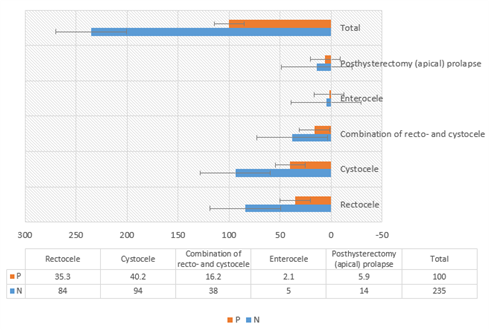
Figure 1: Explain It shows standard deviations for Type of pelvic organ prolapse


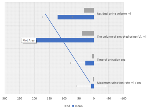
Figure 2: Indicators of uroflowmetry and ultrasound of the bladder before surgical treatment


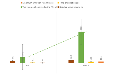
Figure 3: Indicators of uroflowmetry and ultrasound of the bladder after surgical treatment

CONCLUSION

At the same time, traumatic injuries of the pelvic floor - during childbirth or as a result of gynecological operations - according to our data, are the main cause of genital prolapse in at least 35-40% of cases. With age, the role of this factor increases and reaches 67 - 75% in the category of women over 50 who suffer from genital prolapse, according to some authors.


At the same time, as studies show, the syndrome of increased intra-abdominal pressure, as the only influencing factor, in most cases is insufficient to trigger the mechanisms of disease development. As a rule, this requires one or another degree of insufficiency or weakness of the fascial and ligamentous muscular structures of the pelvic cavity.

REFERENCES
  1. Haylen, B.T. et al. (2010). “An International Urogynecological Association (IUGA)/International Continence Society (ICS) joint report on the terminology for female pelvic floor dysfunction”. Neurourology and Urodynamics, 29(1), 4–20.

  2. Handa, V.L. et al. (2004). “Progression and remission of pelvic organ prolapse: A longitudinal study of menopausal women”. American Journal of Obstetrics and Gynecology, 190(1), 27–32.

  3. Wu, J.M. et al. (2014). “Lifetime risk of stress urinary incontinence or pelvic organ prolapse surgery”. Obstetrics and Gynecology, 123(6), 1201–1206.

  4. Swift, S. et al. (2005). “Pelvic Organ Support Study (POSST): The distribution, clinical definition and epidemiologic condition of pelvic organ support defects”. American Journal of Obstetrics and Gynecology, 192(3), 795–806.

  5. Barber, M.D. et al. (2009). “Defining success after surgery for pelvic organ prolapse”. Obstetrics and Gynecology, 114(3), 600–609.

  6. Lee, U. and Raz, S. (2011). “Emerging concepts for pelvic organ prolapse surgery: What is the cure?” Current Urology Reports, 12(1), 62–67.

  7. Nguyen, J.N. and Burchette, R.J. (2008). “Outcome after anterior vaginal prolapse repair: A randomized controlled trial”. Obstetrics and Gynecology, 111(4), 891–898.

  8. Carey, M. et al. (2009). “Vaginal repair with mesh versus colporrhaphy for prolapses: A randomized controlled trial”. BJOG: An International Journal of Obstetrics and Gynaecology, 116(10), 1380–1386.

  9. Nieminen, K. et al. (2010). “Outcomes after anterior vaginal wall repair with mesh: A randomized, controlled trial with a 3-year follow-up”. American Journal of Obstetrics and Gynecology, 203(3), 235.e1–235.e8.

  10. Altman, D. et al. (2011). “Anterior colporrhaphy versus transvaginal mesh for pelvic-organ prolapse”. New England Journal of Medicine, 364(19), 1826–1836.

  11. Chmielewski, L. et al. (2011). “Reanalysis of a randomized trial of 3 techniques of anterior colporrhaphy using clinically relevant definitions of success”. American Journal of Obstetrics and Gynecology, 205(1), 69.e1–69. e8.

  12. Menefee, S.A. et al. (2011). “Colporrhaphy compared with mesh or graft-reinforced vaginal paravaginal repair for anterior vaginal wall prolapse: A randomized controlled trial”. Obstetrics and Gynecology, 118(6), 1337–1344.

  13. Vollebregt, A. et al. (2011). “Primary surgical repair of anterior vaginal prolapse: A randomized trial comparing anatomical and functional outcome between anterior colporrhaphy and trocar-guided transobturator anterior mesh”. BJOG: An International Journal of Obstetrics and Gynaecology, 118(12), 1518–1527.

  14. El-Nazer, M.A. et al. (2012). “Anterior colporrhaphy versus repair with mesh for anterior vaginal wall prolapse: A comparative clinical study”. Archives of Gynecology and Obstetrics, 286(4), 965–972.

  15. de Tayrac, R. et al. (2013). “Comparison between trans-obturator transvaginal mesh and traditional anterior colporrhaphy in the treatment of anterior vaginal wall prolapse: Results of a French RCT”. International Urogynecology Journal, 24(10), 1651–1661.

  16. Delroy, C.A. et al. (2013). “The use of transvaginal synthetic mesh for anterior vaginal wall prolapse repair: A randomized controlled trial”. International Urogynecology Journal, 24(11), 1899–1907.

  17. Turgal, M. et al. (2013). “Anatomical and functional assessment of anterior colporrhaphy versus polypropylene mesh surgery in cystocele treatment”. European Journal of Obstetrics and Gynecology and Reproductive Biology, 170(2), 555–558.

  18. Rudnicki, M. et al. (2014). “Anterior colporrhaphy compared with collagen-coated transvaginal mesh for anterior vaginal wall prolapse: A randomized controlled trial”. BJOG: An International Journal of Obstetrics and Gynaecology, 121(1), 102–110.

  19. dos Reis Brandão da Silveira, S. et al. (2015). “Multicenter, randomized trial comparing native vaginal tissue repair and synthetic mesh repair for genital prolapse surgical treatment”. International Urogynecology Journal, 26(3), 335–342.

  20. Tamanini, J.T. et al. (2015). “A prospective, randomized, controlled trial of the treatment of anterior vaginal wall prolapse: Medium-term follow-up”. Journal of Urology, 193(4), 1298–1304.

  21. Dias, M.M. et al. (2016). “Two-year results of native tissue versus vaginal mesh repair in the treatment of anterior prolapse according to different success criteria: A randomized controlled trial”. Neurourology and Urodynamics, 35(4), 509–514.

  22. Rudnicki, M. et al. (2016). “A 3-year follow-up after anterior colporrhaphy compared with collagen-coated transvaginal mesh for anterior vaginal wall prolapse: A randomized controlled trial”. BJOG: An International Journal of Obstetrics and Gynaecology, 123(1), 136–142.

  23. Glazener, C.M. et al. (2017). “Mesh, graft, or standard repair for women having primary transvaginal anterior or posterior compartment prolapse surgery: Two parallel-group, multicentre, randomized, controlled trials (PROSPECT)”. The Lancet, 389(10067), 381–392.

  24. Maher, C. et al. (2016). “Surgery for women with anterior compartment prolapse”. Cochrane Database of Systematic Reviews, 2016(11), CD004014.

  25. Sung, V.W. et al. (2008). “Graft use in transvaginal pelvic organ prolapse repair: A systematic review”. Obstetrics and Gynecology, 112(5), 1131–1142.

  26. Feiner, B. et al. (2009). Efficacy and safety of transvaginal mesh kits in the treatment of prolapse of the vaginal apex: A systematic review. BJOG: An International Journal of Obstetrics and Gynaecology, 116(1), 15–24.

License
CC BY-NC-ND
Creative Commons Attribution-NonCommercial-NoDerivatives 4.0 International License
Surgical Treatment of Patients with Pelvic Organ Prolapse © 2026 by Alyaa Ajeel Najeeb Al Galiby, Basma Mohammed Abdulsaid, Awatif Najm Kareem licensed under CC BY-NC-ND 4.0
All papers should be submitted electronically. All submitted manuscripts must be original work that is not under submission at another journal or under consideration for publication in another form, such as a monograph or chapter of a book. Authors of submitted papers are obligated not to submit their paper for publication elsewhere until an editorial decision is rendered on their submission. Further, authors of accepted papers are prohibited from publishing the results in other publications that appear before the paper is published in the Journal unless they receive approval for doing so from the Editor-In-Chief.
Himalayan Journal of Medicine and Surgery open access articles are licensed under a Creative Commons Attribution-Share A like 4.0 International License. This license lets the audience to give appropriate credit, provide a link to the license, and indicate if changes were made and if they remix, transform, or build upon the material, they must distribute contributions under the same license as the original.
Recommended Articles
Research Article
Phenotypic Characterization and Antimicrobial Resistance Patterns of Clinical Klebsiella pneumoniae Isolates from Al-Najaf Teaching Hospital
Published: 30/06/2026
Download PDF
Research Article
Impact of Gut-Liver Axis: Hepatic Biochemical and Metabolic Changes Associated with Chronic Gastritis in Iraqi Patients with Helicobacter Pylori
Published: 20/02/2026
Download PDF
Research Article
Effect of Land Degradation on Livelihood
Published: 04/01/2024
Download PDF
Research Article
Awareness and Practices among Professional Healthcare Workers towards COVID-19 in Iraq
...
Published: 30/03/2024
Download PDF
Flowbite Logo
Najmal Complex,
Opposite Farwaniya,
Kuwait.
Email: support@himjournals.com

Useful Links
Order Hard Copy
Privacy policy
Terms and Conditions
Refund Policy
Others
About Us
Team Members
Contact Us
Online Payments
Join as Editor
Join as Reviewer
Subscribe to our Newsletter
Follow us
MOST SEARCHED KEYWORDS
scientific journal
 | 
business journal
 | 
medical journals
 | 
Scientific Journals
 | 
Academic Publisher
 | 
Peer-reviewed Journals
 | 
Open Access Journals
 | 
Impact Factor
 | 
Indexing Services
 | 
Journal Citation Reports
 | 
Publication Process
 | 
Impact factor of journals
 | 
Finding reputable journals for publication
 | 
Submitting a manuscript for publication
 | 
Copyright and licensing of published papers
 | 
Writing an abstract for a research paper
 | 
Manuscript formatting guidelines
 | 
Promoting published research
 | 
Publication in high-impact journals
Copyright © Himalayan Journals . All Rights Reserved.