To assess hepatic metastases between traditional non-contrast phase (NCP), portal venous phase (PVP), and virtual non-contrast phase (VNC) reconstructed from Dual Energy Computed Tomography (DECT) performed in subjects with breast cancer. Methods: Hepatic metastatic lesions from breast cancer in consecutive, pre and post-contrast DECT examinations were retrospectively assessed by 2 experienced readers. Lesions were measured in single longest dimension (SLD) for each phase (NCP, PVP, VNC) and the mean difference was assessed by Kruskal-Wallis test. One reader also assessed contrast to noise ratio (CNR) of the lesions compared to the liver for each phase with difference also assessed by Kruskal-Wallis test. Results: 87 hepatic metastatic lesions were assessed in 23 eligible subjects. SLD was greater for NCP (mean 2.14 cm) compared to PVP (mean 1.94 cm)(p<.001). The difference between VNC and PVP was also significant (2.09 cm vs. 1.94 cm)(p<.001), however, the difference between NCP and VNC was not significant (p=.25). Mean lesion CNR was 3.19 on PVP, 2.65 on VNC, and 1.21 for NCP, all of which were significantly different. Conclusions: VNC may be an adequate substitute to NCP in accurately assessing and measuring hepatic metastases from breast cancer.
Dual energy CT (DECT) uses attenuation values obtained from different energies to make inferences about the composition of tissue, thereby allowing for the production of virtual non-contrast (VNC) images without radiation cost [1,2]. Although there are differences in both qualitative and quantitative image quality between VNC images and traditional non-contrast phase (NCP), VNC has been shown to have the potential to replace NCP in clinical protocols [3]. However, most assessments of VNC have focused primarily on identification of lesions and not accuracy of lesion measurement [4]. While lesion visualization is a component of tumor assessment, lesion size is essential for accurate RECIST classification [5], of which there is not an abundance of literature regarding VNC specifically.
Accurate determination of lesion size can be inconsistent on imaging but is essential, as changes in lesion size between pre-treatment and post-treatment imaging will affect treatment decisions [6]. Because most common hepatic metastases are hypo-vascular relative to normal liver parenchyma, it has become common practice to acquire only portal venous phase images for most suspected hepatic metastases [7]. However, many of the studies that have formed the basis of accepted clinical practice were performed prior to establishment of RECIST criteria for tumor response [5]. As such, many studies which only reported identification of hepatic metastases (and not the exact number of metastases) may have underreported the value of additional imaging phases. The role of non-contrast images is greater in the setting of breast cancer metastases in the liver since some of these may be relatively hyper-vascular and therefore not as conspicuous on post contrast images due to the lack of a significant difference in enhancement from liver parenchyma. [8]. In a recent study, the inclusion of non-contrast phase (NCP) images in addition to PVP images lead to larger measured tumor size, more consistent measurement, and improved detection of hepatic metastases relative to PVP alone [9]. In spite of this, use of PVP-only scanning for hepatic metastases has become increasingly common, likely furthered by cost-containment and radiation dose limitation efforts. It however may be possible to achieve roughly equivalent performance with DECT via VNC in one scanning phase as NCP without additional radiation exposure.
The purpose of this study was to assess for potential differences in measured lesion size as well as quantitative and qualitative visualization assessments on NCP, PVP, and VNC in patients with liver metastases from breast cancer.
This retrospective, single site study was approved by our local Institutional Review Board (IRB) with a waiver of informed consent. It was conducted in compliance with the Health Insurance Portability and Accountability Act.
Study population
A retrospective review identified 23 consecutive subjects with CT examinations meeting the study inclusion criteria:
Breast cancer diagnosis
CT abdomen requested and performed with and without contrast using DECT technique
One or more confirmed liver metastases
An independent study team member (who was not a reader) assessed the eligible studies and chose up to 5 lesions per study for assessment. Two subjects had 2 DECT scans which were eligible. Once assessed, all four scans were included in the study, as there was clear evidence of progressive disease between the scans. Hence, the lesions chosen for assessment were different in sizes despite being in the same subject. Additionally, two subjects had one or more liver lesions that were not visible on PVP but were visible on NCP and VNC. These lesions were not included in the assessments given the lack of visibility on all three phases.
Dect Technique
All CT studies had been performed on a third-generation dual-source CT scanner (Somatom Force; Siemens Healthineers, Forchheim, Germany) in dual-energy mode. As per clinical routine, acquisitions were cranio-caudal from the diaphragmatic dome to the iliac crests before contrast administration (NCP) and subsequently from the diaphragm thru the pubic symphysis following intravenous (IV) contrast administration (Omnipaque 350 mgI/ml, GE Healthcare, Milwaukee, WI, USA) for the PVP. None of the studies included any additional arterial or delayed phase contrast phases, as this was the clinically requested protocol. Contrast material was injected at 4 ml/s, followed by 30-ml saline using a dual-syringe power injector (Stellant D CT Injection System, Medrad, Inc., Warrendale, PA, USA) through 18-gauge IV access placed in a vein at the antecubital fossa.
For the NCP, single energy technique was utilized at 120 kVp with a pitch of 0.7. For PVP, DECT parameters included: tube settings 100/150Sn kVp and 180/90 mAs, respectively, with integrated tin filter (Selective Photon Shield II, SPS II, Siemens); pitch 0.7; collimation 2 × 64 × 0.6 mm for both detectors; field-of-view 350 mm. Also, automated real-time anatomical tube current modulation (CARE Dose 4D, Siemens) was selected for all acquisitions.
Image Reconstruction
For assessment, NCP images were reconstructed using an abdominal soft tissue convolution kernel (Qr40) with a section thickness of 3.0 mm and an increment of 2.0 mm. VNC image sets were reconstructed from the PVP DECT data, as no other post-contrast phases had been performed clinically. PVP image series was designed to simulate the traditional clinical routine single-energy 120 kVp by using vendor-recommended settings to create linearly-blended images (M_0.6), combining 60% of low tube voltage and 40 % of high tube voltage data. An iterative reconstruction was applied (ADMIRE, Siemens Healthcare USA) but only at a strength level of 1 (as this has been the institutionally established clinically preferred level). VNC images were generated with a three material decomposition algorithm. In addition to iodine, the soft tissue and fat attenuation values were from default settings with soft tissue and fat attenuation of 58 and −108 Hounsfield units (HU) at 100 kVp and 56 and −84 HU at Sn150 kVp.
Image Assessment
Two (2) readers, blinded to any available comparison imaging or clinical information, assessed each of the studies. Each reader assessed up to five (5) predetermined liver lesions for each subject in single longest dimension (SLD) on all 3 phases. As each reader read independently and was free to choose the SLD. To reduce variability, the average (mean) of the two measurements for each reader was rendered for this study to represent the SLD of each lesion for each phase (NCP, VNC, PVP).
The same two readers, in concert, also assessed each lesion for 1) lesion visualization and 2) edge conspicuity on each phase (NCP, VNC, PVP) by ranked order (1-3, 1 = highest). Visualization was defined as “most readily distinguished from the liver parenchyma” while edge conspicuity included “edge definition between the lesion and the liver.” One of the readers also assessed each lesion for contrast to noise (CNR) by the following formula:
Lesion CNR = (HU lesion −HU liver ) SD fat
Statistical Assessments
All statistics were performed using a generally available statistical software (SPSS v24, IBM Armonk NY, USA). To
evaluate the measured differences in SLD between PVP, VNC, and NCP image phases, Kruskal-Wallis test was used with Wilcoxon signed rank tests as post-hoc testing. Cohen’s kappa was used to compare the difference between measurements between readers. For the image quality assessments, differences in CNR were compared with Kruskal-Wallis test. For the ranked qualitative assessments, Wilcoxon signed rank test and Spearman’s rank order correlations were used with Bonferroni corrections for multiple comparisons applied to the Wilcoxon tests, resulting in a significance threshold set at p<.017. All other tests were set at a statistical significance threshold of p<.05. Two-tailed p-values are reported for the Spearman and Wilcoxon tests.

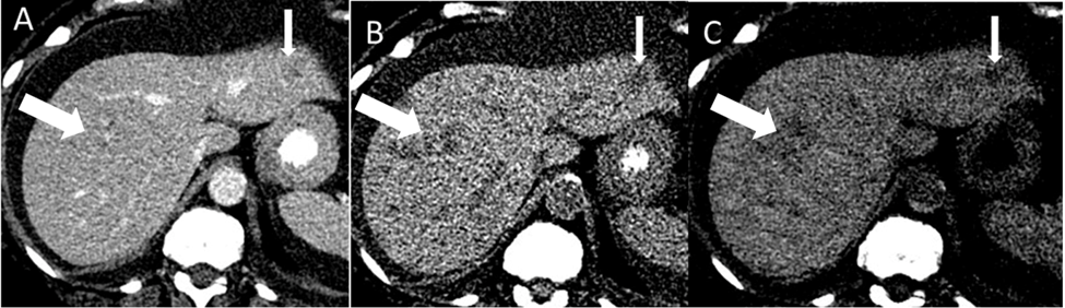
In total, 87 hepatic metastatic lesions were assessed in 23 subjects on all three phases. The subjects were all female with a mean age of 58.2 years (range 51-68 years). One subject had innumerable lesions not visible on PVP (but these were visible on both NCP and VNC) but had a single lesion that was visible on all three phases. See Figure 1. A second subject also had one lesion not visible on PVP but which was identified on both NCP and VNC. Lesions not visible on all three phase were not included in the subsequent SLD measurement assessments.
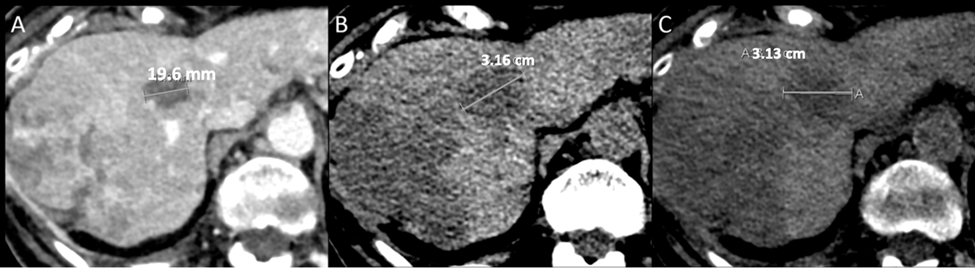
The mean SLD on NCP, VNC and PVP was 2.14 cm, 2.09 cm, and 1.94 cm respectively. See Table 1. Mean PVP SLD was significantly smaller than both NCP (p<.001) and VNC (p<.001), but VNC and NCP were not different (p=0.25). There was very good reader correlation at 0.78. 32 lesions measured greater in SLD on NCP than PVP, with an average of 28% difference in mean SLD. For these lesions, the difference in SLD between VNC and PVP was also greater but there was a lower average difference of 15% Figure 2.
Image quality assessments of the lesions revealed a mean CNR of 3.19 for PVP, 2.65 for VNC and 1.15 for NCP.
Table 1: SLD of Liver Metastases
Phase | Mean (cm) | Range | Standard Deviation |
PVP | 1.94 | 0.50-8.65 | 1.38 |
VNC | 2.09 | 0.40-9.00 | 1.41 |
NCP | 2.14 | 0.20-9.00 | 1.39 |
Table 2: Cnr of Liver Metastases
Phase | Mean (no units) | Range | Standard Deviation |
PVP | 3.19 | 0.21-10.56 | 1.70 |
VNC | 2.65 | 0.05-6.04 | 1.77 |
NCP | 1.15 | 0.10-9.27 | 1.87 |
Table 3: Qualitative Visualization of Liver Metastases Based on Comparative Ranked Assessments
Phase | Mean (no units) | Standard Deviation |
PVP | 1.69 | 0.83 |
VNC | 2.08 | 0.75 |
NCP | 2.23 | 0.79 |
Table 4: Edge Conspicuity of Liver Metastases Based on Comparative Ranked Assessments
Phase | Mean (no units) | Standard Deviation |
PVP | 1.69 | 0.83 |
VNC | 2.08 | 0.75 |

Figure 1: Subject With One Visible Lesion on All Three Phases (Small Arrow) But Innumerable Metastatic Lesions Not Visible on A) Pvp, But Were Visible on Both B) Ncp And C) Vnc (Large Arrow Identifying One of The Many Lesions)

Figure 2: Subject with Liver Metastases with A Single Lesion Measured IN SLD On All Three Phases A) Pvp: 19.6 Mm B) Ncp: 3.16 Mm And C) Vnc: 3.13 Mm
See Table 2. Each mean CNR was significantly different from each of the others: PVP-NCP (p<.001), PVP-VNC (p =.005), NCP-VNC (p<.001). For rank-order assessments, lesion visualization was highest for PVP, which was significantly higher than VNC (p =.009) or NCP (p =.002). However, there was no statistically significant differencebetween visualization rankings for VNC and NCP (p =.25). Similarly, when assessed as a paired “virtual” protocol the combination of PVP and NCP and PVP and VNC was not ranked significantly different (p =.26). See Table 3. Visualization was also not impacted by lesion size for PVP (rho= -.08, p=.49), VNC (rho= .04, p =.84), nor NCP (rho = -.21, p=.06). For edge conspicuity, significant differences were found based on lesion size for all the phases. PVP (rho = .35, p =.001), VNC (rho = .24, p=.03), and NCP (rho = .37, p=.001) Table 4.
The results of this study serve to validate that in a clinical population of breast cancer patients VNC can be an adequate substitute for traditional NCP when considering SLD tumor measurements in liver metastases. Further, VNC had a higher CNR than NCP and ranked similarly to NCP with regard to lesion visualization and edge conspicuity. A “virtual” paired assessment of PVP with VNC and PVP with NCP were also was rated not significantly differently. VNC and NCP were also able to identify several lesions not visible on PVP.
Differences between measured liver lesion sizes is particularly relevant in this specific population given the known propensity for SLD differences to be present between PVP and NCP [9]. Although there is a large history of literature on liver metastases, there is a relative lack of studies aimed at comparing the differences in lesion measurements for different CT phases. However, SLD measurements are essential to therapeutic assessments such as in RECIST version 1.1. At least as far as the authors are aware, there is no prior study looking specifically at measurement differences for breast cancer metastases, and further not with Dual Energy CT. Importantly, mean SLD on VNC was similar to NCP in both cases, demonstrating the potential for VNC to approximate lesion measurements on NCP. VNC was also able to identify the metastatic lesions visible on NCP but not identifiable on PVP, a known limitation of viewing PVP alone [5,8,9]. This may have implications for breast cancer staging and tumor response assessment, although future studies are needed in this regard.
This study has a small population, which is a limitation. However, given that the use of pre and post contrast imaging is rare compared to single post contrast phase, it may be difficult to acquire sufficient data from clinical patients to be able to assess in much greater numbers. However, as DECT becomes more common in clinical practice, a future study could more readily be designed to assess the impact of tumor measurement differences between VNC and PVP, without radiation cost. Further, such a study could include the effect of chemotherapy treatment on measured lesion sizes for each phase.
Although the study in this manuscript was designed to assess lesions from the same primary tumor to ensure homogeneity of the study population, significant differences in the tumor biology and thus imaging appearance likely exist. However, we believe this limitation is one that would be commonly encountered in clinical practice such that the results of this study are likely still applicable.
In conclusion, this study demonstrates that in regard to lesion visualization and measurement, VNC may be an adequate substitute for NCP.
De Cecco, C.N. et al. "Virtual unenhanced images of the abdomen with second-generation dual-source dual-energy computed tomography: image quality and liver lesion detection." Invest. Radiol., vol. 48, no. 1, 2013, pp. 1–9. doi:10.1097/RLI.0b013e31826e7902.
De Cecco, C.N. et al. "Virtual unenhanced imaging of the liver with third-generation dual-source dual-energy CT and advanced modeled iterative reconstruction." Eur. J. Radiol., vol. 85, no. 7, 2016, pp. 1257–64. doi:10.1016/j.ejrad.2016.04.012.
Grajo, J.R., Sahani, D.V. "Dual-energy CT of the abdomen and pelvis: radiation dose considerations." J. Am. Coll. Radiol., vol. 15, no. 8, 2018, pp. 1128–32. doi:10.1016/j.jacr.2017.08.012.
Parakh, A. et al. "Dual-energy CT images: pearls and pitfalls." Radiographics, vol. 41, no. 1, 2021, pp. 98–119. doi:10.1148/rg.2021200102.
Schwartz, L.H. et al. "RECIST 1.1-update and clarification: from the RECIST committee." Eur. J. Cancer, vol. 62, no. 0, 2016, pp. 132–7. doi:10.1016/j.ejca.2016.03.081.
Lee, G. et al. "Measurement variability in treatment response determination for non-small cell lung cancer: improvements using radiomics." J. Thorac. Imaging, vol. 34, no. 2, 2019, pp. 103–15. doi:10.1097/RTI.0000000000000390.
Soyer, P. et al. "Detection of hypovascular hepatic metastases at triple-phase helical CT: sensitivity of phases and comparison with surgical and histopathologic findings." Radiology, vol. 231, no. 2, 2004, pp. 413–20. doi:10.1148/radiol.2312021639.
Sheafor, D.H. et al. "Comparison of unenhanced, hepatic arterial-dominant, and portal venous-dominant phase helical CT for the detection of liver metastases in women with breast carcinoma." AJR Am. J. Roentgenol., vol. 172, no. 4, 1999, pp. 961–8. doi:10.2214/ajr.172.4.10587129.
Flemming, B. et al. "Non-contrast and portal venous phase computed tomography in breast cancer hepatic metastases: comparison of tumor measurements and impact on response assessment." Acta Radiol. Open, vol. 10, no. 2, 2021, pp. 1–10. doi:10.1177/2058460121998015.