Right ventricular pacing is considered a non-physiological modality; the wavefront of depolarization slowly spreads from the lead tip to the rest of the myocardium, creating morphological alterations of the QRS complexes that make it difficult an electrocardiographic diagnosis of myocardial infarction. During His bundle pacing the electrical impulse travels through the normal conduction system, as a result, paced ventricular complexes show morphological features that are completely superimposable to basal ECG. This peculiarity allows to appreciate the electrocardiographic alterations consequent to a previous heart attack or to an acute coronary syndrome. Below, we introduce a clinical case of PMK recipient in which the upgrade to HB pacing unmasked a previous infero-lateral myocardial infarction.
Keywords
His Bundle Pacing
Conduction System Pacing
Myocardial Infarction
Upgrade
Acute Coronary Syndrome
INTRODUCTION
Resting 12-lead ECG is the first-line diagnostic tool in the evaluation of patients with suspected Acute Coronary Syndrome (ACS) and old myocardial infarction [1]; however, in Pacemaker (PMK) recipients, ECG morphological changes related to Right Ventricular (RV) pacing, even in intermittent paced forms, make it difficult to correctly diagnose myocardial ischaemia, also due to the phenomenon known as cardiac memory [2,3], delaying medical therapy.
In recent years we have witnessed a progressive interest of the scientific community in His Bundle (HB) pacing as an alternative to RV pacing [4]. HB pacing is rightly considered physiological, since, unlike RV pacing, the electrical impulse travels through the normal conduction system and, therefore, does not result in ventricular electrical desynchrony. As a result, paced ventricular complexes show morphological features that are completely superimposable to those evident in the baseline ECG in the absence of pacing [3-6]. Although HB pacing, in contrast to RV pacing, should not affect the ventricular repolarisation phase, there are only few cases in the literature about electrocardiographic diagnosis of myocardial ischaemia during HB pacing.
Therefore, below, we introduce a clinical case of PMK recipients in which the upgrade to HB pacing unmasked a previous infer-o-lateral myocardial infarction.
CASE REPORT
We report the clinical case of a 71-year-old man who was implanted with a dual-chamber PMK for complete atrioventricular block in 2012. Suffering from chronic ischemic heart disease, in 2013 he was treated with two Percutaneous Coronary Intervention (PCI) and stenting of the Left Anterior Descending coronary artery (LAD) and Right Coronary Artery (RCA); in 2014 he underwent, successfully, atrioventricular node ablation for the appearance of a high ventricular frequency atrial fibrillation despite optimal, medical therapy for rate control. In March 2021, he was referred to our hospital for acute heart failure. Medical therapy included acetylsalicylic acid, bisoprolol, furosemide, mineralocorticoid receptor antagonist and sacubitril/valsartan. On admission he presented dyspnoeic, with peripheral edema, pulmonary congestion on clinical examination and chest radiography; transthoracic echocardiography showed diffuse hypokinesis, with a Left Ventricular Ejection Fraction (LVEF) of 28% (46% six months earlier).
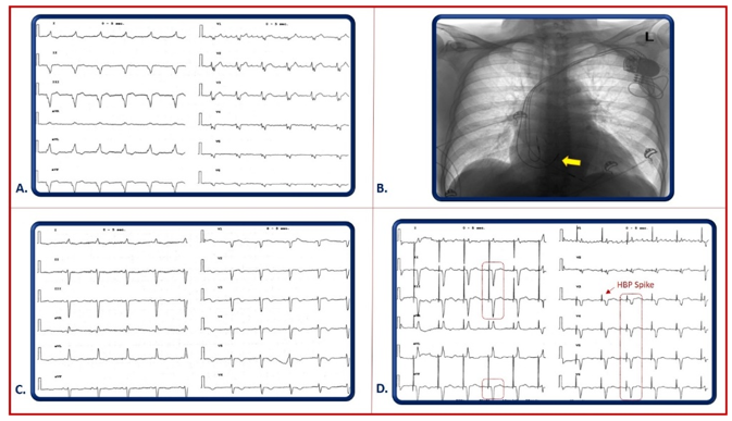
Figure 1: A) Post Dual Chambre PMK, Surface 12 leads ECG: It Shows Atrial Flutter, Ventricular Paced QRS Morphology From Apical Site (Explanation in the text), B) Chest X-Ray Post Upgrade To His Bundle Pacing; Yellow Arrow: His lead, C) Basal Surface 12 leads ECG: It Shows Sinus Rhythm, Left Anterior Fascicular Block, D) Post Upgrade To His Bundle Pacing, Surface 12 leads ECG: It Shows Atrial Flutter And Selective His Bundle Pacing; Note, Highlighted In Red, The Qs Complexes As For Previous Inferior And Anterolateral Transmural Necrosis (leads V3-V6, II, III, aVF). Red Arrow: PMK spike; HBP: His Bundle Pacing.
ECG showed atrial flutter and paced ventricular rhythm with notch of the QRS complex in all 12 leads (Figure A). After achieving good clinical compensation with intravenous diuretics, the patient underwent coronary angiography with evidence of an unchanged coronary situation compared with 2013. Given the constant right ventricular apical pacing, we considered the LVFE decline as a consequence of Pacemaker-Induced Cardiomyopathy (PICM) [7] then, after obtaining the patient's informed consent, a successful upgrade to HB pacing was performed (Figure B). The surface ECG showed selective HB pacing with QRS width comparable to the native ECG (Figure C) but different morphology, because of evidence of QS complexes in V3-V6 and II, III, aVF (Figure D); physiological pacing obtained with selective HBP had made evident electrocardiographic signs of a previous Myocardial Infarction (MI).
DISCUSSION
RV pacing typically produces a Left Bundle-Branch Block (LBBB)-like pattern in septal and anterior precordial leads, with a predominantly negative QRS complex followed by ST elevation and positive T-waves [7,8]. Sgarbossa criteria can help identify myocardial infarction with ST-Segment Elevation (STEMI) in LBBB patients, allowing at least in some cases, timely access to reperfusion therapy. However, the low sensitivity of these criteria (between 18% and 55%) results in a low negative predictive value to exclude an acute coronary event [7-10].
Even more difficult is the diagnosis of a previous MI during RV pacing [3,8,10]; to date, although considered questionable, we have only the criteria of Kochiadakis and colleagues [9] (Figure A):
Notch of 0.04-second duration on the ascending limb of the S-wave of leads V3, V4, or V5 (Cabrera's sign)
Notching of the ascending stroke of the R wave in leads I, aVL, or V6 (Chapman's sign)
Q waves of 0.03 second duration in leads I, aVL, or V6
Notch of the first 0.04 seconds of the QRS complex in leads II, III and aVF
Q wave of 0.03 seconds duration in leads II, III and aVF
Occurring through the normal conduction system, HB pacing should generate in addition to a depolarization also a physiological myocardial repolarization resulting in a paced QRS complex with morphology and duration equal to the basal ECG, without alterations in the ventricular repolarization phase. As proof of the above, it was possible to appreciate that the transition from RV pacing to selective HB pacing allowed to unmasked the signs of a previous infero-lateral IMA, showing clear QS complexes in II, III, a VF, V3-V6. There are only 2 similar cases in the literature, the first case described how, in a patient with Left Bundle Brunch (LBB) block and old inferior infarction, LBB pacing corrected the conduction defect and made evident pathological Q waves in the inferior leads [11]; the second one described how the para Hissian area pacing made evident a Brugada pattern in a patient with left bundle branch block [12]. In our case, for the first time, the upgrade from RV pacing to selective HB pacing has made the conduction physiological in all the surface ECG leads, unmasking a previous infero-lateral myocardial infarction. Finally, in a recent case report Marinaccio et al. described for the first time how physiological stimulation allows to appreciate even minimal electrocardiographic alterations during non-ST Elevation Myocardial Infarction (NSTEMI), speeding up the diagnosis and therapy [13].
CONCLUSION
This clinical case reinforces the limited evidence in the literature that the restoration of cardiac physiological conduction by HB pacing can unmask ECG abnormalities, not evident during LBBB, such as signs of a previous myocardial infarction.
Therefore, in light of the current data and evidence on the feasibility and safety of HBP, it seems reasonable to consider HB pacing also in patients at high risk of ischemic heart disease who require pacing.
REFERENCES
Collet, Jean-Pierre et al. "2020 ESC guidelines for the management of acute coronary syndromes in patients presenting without persistent st-segment elevation." European Heart Journal, vol. 42, 2021, pp. 1289–1367.
Shvilkin, Alexander et al. "Cardiac memory: Diagnostic tool in the making." Circulation: Arrhythmia and Electrophysiology, vol. 8, 2015, pp. 475–482.
Abraham et al. "ECG diagnosis: acute myocardial infarction in a ventricular-paced rhythm." The Permanente Journal, vol. 23, 2019, p. 19-001.
Vetta, Francesco et al. "Alternative sites of ventricular pacing: his bundle pacing." Monaldi Archives for Chest Disease, vol. 90, 2020, pp. 218–223.
Vijayaraman, Pugazhendhi et al. "Permanent his bundle pacing: recommendations from a multicenter his bundle pacing collaborative working group for standardization of definitions, implant measurements and follow-up." Heart Rhythm, vol. 15, 2018, pp. 460–468.
Catanzariti, Davide et al. "Permanent his-bundle pacing maintains long-term ventricular synchrony and left ventricular performance, unlike conventional right ventricular apical pacing." Europace, vol. 15, 2013, pp. 546–553.
Khurshid, Shaan et al. "Incidence and Predictors of Right Ventricular Pacing-Induced Cardiomyopathy." Heart Rhythm, vol. 11, 2014, pp. 1619–1625.
C. Browning, Andrew and Stefan Schaefer. "What lies beneath: posterior st elevation myocardial infarction with underlying right ventricular-paced rhythm." Experimental and Clinical Cardiology, vol. 18, 2013, pp. e55–e56.
Barold, S. Serge et al. "Electrocardiographic diagnosis of myocardial infarction and ischemia during cardiac pacing." Cardiology Clinics, vol. 24, 2006, pp. 387–399.
Nestelberger, Thomas et al. "Diagnosis of acute myocardial infarction in the presence of left bundle branch block." Heart, vol. 105, 2019, pp. 1559–1567.
Ponnusamy, S. Senthil and Pugazhendhi Vijayaraman. "Unmasking of pathologic q waves by left bundle branch pacing." Journal of Interventional Cardiac Electrophysiology, vol. 60, no. 3, 2021, pp. 555–556.
S. Guandalini, Giovanni and Francis E. Marchlinski. "Parahisian Pacing to unmask brugada pattern with concomitant left bundle branch block and to document epicardial ablation endpoint in brugada syndrome." HeartRhythm Case Reports, vol. 7, no. 6, 2021, pp. 382–385.
Marinaccio, Luigi et al. "Electrocardiographic diagnosis of acute and remote myocardial ischemia during his bundle pacing." The Anatolian Journal of Cardiology, accepted for publication.
License
Creative Commons Attribution-NonCommercial-NoDerivatives 4.0 International License
All papers should be submitted electronically. All submitted manuscripts must be original work that is not under submission at another journal or under consideration for publication in another form, such as a monograph or chapter of a book. Authors of submitted papers are obligated not to submit their paper for publication elsewhere until an editorial decision is rendered on their submission. Further, authors of accepted papers are prohibited from publishing the results in other publications that appear before the paper is published in the Journal unless they receive approval for doing so from the Editor-In-Chief.
Himalayan Journal of Medicine and Surgery open access articles are licensed under a Creative Commons Attribution-Share A like 4.0 International License. This license lets the audience to give appropriate credit, provide a link to the license, and indicate if changes were made and if they remix, transform, or build upon the material, they must distribute contributions under the same license as the original.
Recommended Articles
Research Article
Primordial Prevention of Coronary Artery Diseases
Ahmed Adil Ali,
...
Hassan Ali Noori
Published: 26/07/2026
Download PDF
Cite
x
APA
Ali, A. A., Hussien, H. A., Kareem, H. B., Sarhan, S. R. & Noori, H. A. (2026). Primordial Prevention of Coronary Artery Diseases. Himalayan Journal of Medicine and Surgery, 7(2), 1-6.
MLA
Ali, Ahmed Adil, et al. "Primordial Prevention of Coronary Artery Diseases." Himalayan Journal of Medicine and Surgery 7.2 (2026): 1-6.
Chicago
Ali, Ahmed Adil, Hamza Ali Hussien, Hassan Baqer Kareem, Sarhan Raad Sarhan and Hassan Ali Noori. "Primordial Prevention of Coronary Artery Diseases." Himalayan Journal of Medicine and Surgery 7, no. 2 (2026): 1-6.
Harvard
Ali, A. A., Hussien, H. A., Kareem, H. B., Sarhan, S. R. and Noori, H. A. (2026) 'Primordial Prevention of Coronary Artery Diseases' Himalayan Journal of Medicine and Surgery 7(2), pp. 1-6.
Vancouver
Ali AA, Hussien HA, Kareem HB, Sarhan SR, Noori HA. Primordial Prevention of Coronary Artery Diseases. Himalayan Journal of Medicine and Surgery. 2026 Jul;7(2):1-6.
Download PDF
Research Article
Impact of Gut-Liver Axis: Hepatic Biochemical and Metabolic Changes Associated with Chronic Gastritis in Iraqi Patients with Helicobacter Pylori
Noor Zuhair Bakheet N. Z.
Published: 20/02/2026
Download PDF
Cite
x
APA
N. Z., N. Z. B. (2026). Impact of Gut-Liver Axis: Hepatic Biochemical and Metabolic Changes Associated with Chronic Gastritis in Iraqi Patients with Helicobacter Pylori. Himalayan Journal of Medicine and Surgery, 7(1), 1-4.
MLA
N. Z., Noor Z. B.. "Impact of Gut-Liver Axis: Hepatic Biochemical and Metabolic Changes Associated with Chronic Gastritis in Iraqi Patients with Helicobacter Pylori." Himalayan Journal of Medicine and Surgery 7.1 (2026): 1-4.
Chicago
N. Z., Noor Z. B.. "Impact of Gut-Liver Axis: Hepatic Biochemical and Metabolic Changes Associated with Chronic Gastritis in Iraqi Patients with Helicobacter Pylori." Himalayan Journal of Medicine and Surgery 7, no. 1 (2026): 1-4.
Harvard
N. Z., N. Z. B. (2026) 'Impact of Gut-Liver Axis: Hepatic Biochemical and Metabolic Changes Associated with Chronic Gastritis in Iraqi Patients with Helicobacter Pylori' Himalayan Journal of Medicine and Surgery 7(1), pp. 1-4.
Vancouver
N. Z. NZB. Impact of Gut-Liver Axis: Hepatic Biochemical and Metabolic Changes Associated with Chronic Gastritis in Iraqi Patients with Helicobacter Pylori. Himalayan Journal of Medicine and Surgery. 2026 Jan;7(1):1-4.
Download PDF
Research Article
Effect of Land Degradation on Livelihood
Kemalo Abdulmalik,
Isreal Zewide
Published: 04/01/2024
Download PDF
Cite
x
APA
Abdulmalik, K. & Zewide, I. (2024). Effect of Land Degradation on Livelihood. Himalayan Journal of Medicine and Surgery, 5(1), 1-4.
MLA
Abdulmalik, Kemalo and Isreal Zewide. "Effect of Land Degradation on Livelihood." Himalayan Journal of Medicine and Surgery 5.1 (2024): 1-4.
Chicago
Abdulmalik, Kemalo and Isreal Zewide. "Effect of Land Degradation on Livelihood." Himalayan Journal of Medicine and Surgery 5, no. 1 (2024): 1-4.
Harvard
Abdulmalik, K. and Zewide, I. (2024) 'Effect of Land Degradation on Livelihood' Himalayan Journal of Medicine and Surgery 5(1), pp. 1-4.
Vancouver
Abdulmalik K, Zewide I. Effect of Land Degradation on Livelihood. Himalayan Journal of Medicine and Surgery. 2024 Jan;5(1):1-4.
Download PDF
Research Article
Awareness and Practices among Professional Healthcare Workers towards COVID-19 in Iraq
Yasir H Motlaq,
...
Zainab T. N M. Almuhannah
Published: 30/03/2024
Download PDF
Cite
x
APA
H Motlaq, Y., A Aqebian, E. & N M. Almuhannah, Z. T. (2024). Awareness and Practices among Professional Healthcare Workers towards COVID-19 in Iraq. Himalayan Journal of Medicine and Surgery, 5(1), 1-9.
MLA
H Motlaq, Yasir, Esraa A Aqebian and Zainab T. N M. Almuhannah. "Awareness and Practices among Professional Healthcare Workers towards COVID-19 in Iraq." Himalayan Journal of Medicine and Surgery 5.1 (2024): 1-9.
Chicago
H Motlaq, Yasir, Esraa A Aqebian and Zainab T. N M. Almuhannah. "Awareness and Practices among Professional Healthcare Workers towards COVID-19 in Iraq." Himalayan Journal of Medicine and Surgery 5, no. 1 (2024): 1-9.
Harvard
H Motlaq, Y., A Aqebian, E. and N M. Almuhannah, Z. T. (2024) 'Awareness and Practices among Professional Healthcare Workers towards COVID-19 in Iraq' Himalayan Journal of Medicine and Surgery 5(1), pp. 1-9.
Vancouver
H Motlaq Y, A Aqebian E, N M. Almuhannah ZT. Awareness and Practices among Professional Healthcare Workers towards COVID-19 in Iraq. Himalayan Journal of Medicine and Surgery. 2024 Jan;5(1):1-9.
Marinaccio, L. (2022). An Old Myocardial Infarction Unmasked by Upgrade to His Bundle Pacing: A Case Report. Himalayan Journal of Medicine and Surgery, 3(1), 1-3.
MLA
Marinaccio, Leonardo. "An Old Myocardial Infarction Unmasked by Upgrade to His Bundle Pacing: A Case Report." Himalayan Journal of Medicine and Surgery 3.1 (2022): 1-3.
Chicago
Marinaccio, Leonardo. "An Old Myocardial Infarction Unmasked by Upgrade to His Bundle Pacing: A Case Report." Himalayan Journal of Medicine and Surgery 3, no. 1 (2022): 1-3.
Harvard
Marinaccio, L. (2022) 'An Old Myocardial Infarction Unmasked by Upgrade to His Bundle Pacing: A Case Report' Himalayan Journal of Medicine and Surgery 3(1), pp. 1-3.
Vancouver
Marinaccio L. An Old Myocardial Infarction Unmasked by Upgrade to His Bundle Pacing: A Case Report. Himalayan Journal of Medicine and Surgery. 2022 Jan;3(1):1-3.



 Download PDF
 Download XML