Background: The present study was done to assess the results and diagnostic role of 68Ga-PSMA PET/CT in prostate cancer patients. Material and Methods: Forty four patients with suspected PCa on clinical assessment and/or raised serum PSA levels (>4ng/ml) were prospectively recruited. Patient who underwent prostatic biopsy prior to 68Ga-PSMA PET/CT and mpMRI, deranged renal function tests or coagulogram and refused consent were excluded. Forty four patients underwent 68Ga-PSMA PET/CT and 33 patients underwent biopsy. Any focal tracer uptake on 68Ga-PSMA PET/CT was regarded as pathological. Histopathology was taken as gold standard. Results: Mean age of patients was 66.61±9.3 years and median PSA level was 13.4 ng/ml (IQR 13.9). In this study, malignancy was detected in 16/33 (48.5%) patients on biopsy. In 17/33 (51.5%), no evidence of malignancy was noted. 68Ga-PSMA PET/CT showed that 15 patients had positive results and among these 13 patients were found to have adeno-carcinoma on biopsy. Two patients with positive results on 68Ga PSMA PET/CT but negative biopsy (Considered False positive) were closely followed up with serum PSA values. Three patients had negative 68Ga PSMA PET/CT (homogenous/inhomogeneous uptake) but malignancy was detected on biopsy (False negative on 68Ga PSMA PET). In this study, 15 patients had negative results both on biopsy as well as on 68Ga-PSMA PET/CT. Conclusions: 68Ga PSMA PET/CT has good accuracy in initial diagnosis of patients with suspected prostatic adenocarcinoma with significantly high PSMA uptake in malignant tumors.
Keywords
Assessment
Results
Diagnostic Role
68Ga-PSMA PET/CT
Prostate Cancer
INTRODUCTION
Prostate-specific membrane antigen (PSMA) is a type II transmembrane glycoprotein that has shown considerably raised expression in PCa cells and has been successfully used as target for molecular imaging approaches. The density of PSMA expression in malignant prostate cancer has been found to be 1000 times higher compared to normal prostate tissue. For molecular imaging, PSMA can be targeted either with PSMA ligands or antibodies. PSMA targeting ligands get internalized after binding to receptor and undergo endosomal recycling, resulting in increased uptake and retention in PCa cells which can be imaged [1-4].
68Ga-PSMA PET/CT (Positron Emission Tomography) is a relatively new diagnostic modality which targets PSMA binding on PCa cells and has shown potential in detection of nodal/distant metastasis and recurrence in PCa with high accuracy. A meta-analysis reported pooled specificity of 68Ga-PSMA PET/CT for disease staging and restaging to be 84% and 97% respectively. Many studies have evaluated the role of 68Ga-PSMA PET/CT in biopsy proven PCa (done for staging) and evaluated tracer uptake in the primary tumor and compared it with mpMRI [5-6].
Despite increasing clinical use in these clinical settings, the potential of 68Ga-PSMA PET/CT in initial diagnosis of PCa has not been explored much. According to our knowledge, No prospective study has evaluated the role of 68Ga-PSMA PET/CT in suspected PCa
Aims and Objectives
To assess the results and diagnostic role of 68Ga-PSMA PET/CT in prostate cancer patients
MATERIALS AND METHODS
Study Design: Prospective pilot study
Place of Study: This study have been conducted at the Department of Nuclear Medicine, PGIMER Chandigarh in collaboration with the Department Urology, Radiodiagnosis and Histopathology, PGIMER, Chandigarh
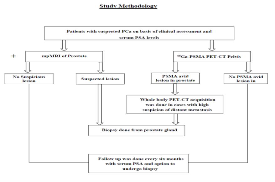
Figure 1: Study Methodology
Period of Study: The study was done during the period between January, 2018 and June, 2019
Inclusion Criteria
Patients having suspicion of carcinoma prostate on clinical assessment and/or raised serum PSA levels (>4ng/ml)
Patient not suffering from bleeding diathesis or renalfailure
Patient consent to participate in thestudy.
Exclusion Criteria
Patient with prior diagnosed PCa or who underwent prostatic biopsy priorto68Ga-PSMA PET/CT and mpMRI
Patients with deranged renal function tests orcoagulogram
Patient refused to participate in thestudy
Study Population
A total of 44patients suspected for PCa on the basis of clinical assessment and/or raised PSA were recruited for the study
Patients suspected for prostate adenocarcinoma and/or serum PSA levels >4ng/ml and willing to participate in the study were recruited. Study was approved by Institutional Ethics Committee, PGIMER Chandigarh. Written informed consent was obtained. After clinical assessment, mpMRI and68Ga-PSMA PET/CT were done. Biopsy was done in cases with high clinical suspicion of PCa.
Statistical Analysis
Qualitative variables are described using number and percentages. Normally distributed continuous quantitative data is described using mean and standard deviation. Skewed quantitative data is described using median and interquartile range. The normality of data will be checked by measures of Kolmogorov-Smirnov tests of normality. The statistical analysis was conducted using the IBM SPSS STATISTICS (version 23.0)
RESULTS
In this prospective study, a total of 44 patients suspected to have PCa were recruited on the basis of clinical assessment and serum PSA levels. All patients were subjected to 68Ga-PSMA PET/CT acquisition. Out of the total 44 patients, 33 underwent biopsy. The remaining 11 patients did not undergo biopsy after revised clinical assessment based on negative. Therefore, 33 patients underwent 68Ga-PSMA PET/CT and biopsy.
The mean and SD of the injected 68Ga PSMA radioactivity was 2.79±0.89 mCi (103.2±32.9MBq).The median time between the injection and scan acquisition was 65 min (IQR 32.5 min). Images were acquired according to standardized institutional protocol (described in methods).In Qualitative (Visual) Analysis, visual interpretation of the PET/CT images was done independently by two Nuclear Medicine Physicians. Focal increased tracer uptake in one or more areas of prostate gland was reported as positive for malignancy. Homogenous or inhomogeneous increased tracer uptake in the prostate was considered as negative.
Table 1: Results of 68Ga-PSMA PET/CT
Total patients analysed (n = 33)
Adenocarcinoma (n = 16)
Negative for malignancy (n = 17)
68Ga-PSMA PET/CT
Positive
(n = 15)
13 (TP)*
2(FP)*
Negative
(n = 18)
3 (FN)*
15 (TN)*
After visual interpretation, 68Ga-PSMA PET/CT data was compared to histopathology (gold standard). Results of 68Ga-PSMA and biopsy findings were shown in Table 1. In this study, malignancy was detected in 16/33 (48.5%) patients on biopsy. In 17/33 (51.5%), no evidence of malignancy was noted.
In the present study 68Ga-PSMA PET/CT showed that 15 patients had positive results and among these 13 patients were found to have adeno-carcinoma on biopsy. Two patients with positive results on 68Ga PSMA PET/CT but negative biopsy (Considered False positive) were closely followed up with serum PSA values. In first patient, serum PSA value was noted to decrease from 5.3 to 4.7ng/ml after 10 months. Repeat biopsy was not done in first patient. In second patient, serum PSA value was noted to increase from 15 to 18.2ng/ml after 12 months. Repeat targeted biopsy in second patient was negative for malignancy.
Three patients had negative 68Ga PSMA PET/CT (homogenous/inhomogeneous uptake) but malignancy was detected on biopsy (False negative on 68Ga PSMA PET). In 2/3 patients, the GS was 3+3 (Grade group 1). mpMRI was not done in both these cases. In both of these, tumor burden was less than 5% on 12 core biopsy. In third case, mass was noted on CT but 68Ga PSMA PET/CT was negative (inhomogeneous tracer uptake). mpMRI showed PIRADS V and GS was 4+4 in this case. In this study, 15 patients had negative results both on biopsy as well as on 68Ga-PSMA PET/CT.
DISCUSSION
68Ga PSMA PET/CT is a relatively new hybrid functional imaging modality which has been extensively used in PCa in the last decade. The basis of 68Ga PSMA PET/CT is targeting of PSMA receptors which are over expressed (100-1000x) in PCa as compared to normal prostate cells. 68Ga PSMA PET/CT has shown encouraging results in staging, restaging, response assessment and evaluation of biochemical recurrence in PCa. In nearly all prior studies which have evaluated role of 68Ga-PSMA PET/CT in detection of PCa and compared it with MRI, PET-CT was done in histopathologically confirmed PCa cases only. Only one recently published retrospective study by Zhang et al has evaluated accuracy of pre-biopsy 68Ga-PSMA PET-CT in suspected PCa (19).To best of our knowledge, this study is first prospective study to assess the diagnostic role of 68Ga-PSMA PET-CT in patients with suspected prostate adenocarcinoma [4,7].
In this study, malignancy was detected in 16/33 (48.5%) patients on biopsy. In 17/33 (51.5%), no evidence of malignancy was noted. 68Ga-PSMA PET/CT showed that 15 patients had positive results and among these 13 patients were found to have adeno-carcinoma on biopsy. In 3 patients, 68Ga-PSMA PET/CT was false negative (homogenous/in-homogenous uptake pattern) but they were found to have PCa on histopathologicalexamination. Final histopathology revealed adenocarcinoma GS 3+3 (n = 2) and GS 4+4 (n = 1). The reason for false negative 68Ga PSMA PET/CT in these cases can be low tumor burden and low grade tumors/low GS are known to have lower PSMA expression (though no correlation was noted in our study) [8]. Tumors other than adenocarcinoma like neuroendocrine tumor show lower PSMA expression too.9 In the third case, GS was 4+4 but PET/CT was False Negative. The cause of low PSMA expression in this case could not be ascertained. In our study, 3/16 (18.8%) cases were false negative on 68Ga PSMA PET/CT which were almost double compared to study earlier by Zhang et al (8.3 %) [8]. Another study by Uprinmy et al on radical prostatectomy cases showed that 8.9% of the PCa patients were having negative 68Ga PSMA PET/CT and were positive on histopathological examination. [7]. Therefore, 68Ga PSMA PET/CT may not pick 100% of the PCa lesions.
Similarly, 2 patients were found to have false positive findings (Focal high tracer uptake) for PCa on 68Ga-PSMA PET/CT. The reason for false positive could be overexpression of PSMA in benign prostatic tissue [9]. However, possibility of missing the lesion on biopsy is also there. As studied by Woythal et al. TRUS biopsy can miss 30-45% of OCs the cases [10]. By the end of this study, one patient underwent repeat targeted biopsy which did not reveal any malignancy.
In cases in whom no malignancy was detected (True negative) and no focal lesion was noted on 68Ga-PSMA PET/CT, different patterns of uptake were noted. Few of the cases had low homogeneous tracer uptake (PSMA expression) and few were having inhomogeneous/heterogeneous radiopharmaceutical uptake on 68Ga-PSMA PET/CT. During analysis of the images, we found that relatively higher physiological tracer uptake was noted in the posterolateral aspect of base of the prostate gland (at 5 and 7’o clock positions) on a homogenous/in-homogenous background pattern in many cases. These patterns should be kept in mind while assessing PSMA PET for suspected PCa.
CONCLUSION
68Ga PSMA PET/CT has good accuracy in initial diagnosis of patients with suspected prostatic adenocarcinoma with significantly high PSMA uptake in malignant tumors.
REFERENCE
Mease, R.C. et al."PET imaging in prostate cancer: Focus on prostate-specific membrane antigen." Curr Top Med Chem, vol. 13, no. 8, 2013, pp. 951–962.
Bostwick, D.G. et al."Prostate-specific membrane antigen expression in prostatic intraepithelial neoplasia and adenocarcinoma: A study of 184 cases." Cancer, vol. 82, no. 11, 1998, pp. 2256–2261.
Donin, N.M., and R. Reiter. "Why targeting PSMA is a game changer in the management of prostate cancer – a urologist’s point of view." J Nucl Med, vol. 59, no. 2, 2018, pp. 177–182.
Demirci, E. et al. "Normal distribution pattern and physiological variants of 68Ga-PSMA-11 PET/CT imaging." Nuclear Med Commun, vol. 37, no. 11, 2016, pp. 1169–1179.
Kallur, K. et al. "Clinical utility of gallium-68 PSMA PET/CT scan for prostate cancer." Indian J Nucl Med, vol. 32, no. 2, 2017, pp. 110–117.
Von Eyben, F.E. et al."68Ga-labeled prostate-specific membrane antigen ligand positron emission tomography/computed tomography for prostate cancer: A systematic review and meta-analysis." Eur Urol Focus, vol. 4, no. 5, 2018, pp. 686–93.
Uprimny, C. et al. "68Ga-PSMA-11 PET/CT in primary staging of prostate cancer: PSA and Gleason score predict the intensity of tracer accumulation in the primary tumour." Eur J Nucl Med Mol Imaging, vol. 44, no. 6, 2017, pp. 941–949.
Zhang, J. et al. "Diagnostic performance of 68Ga-PSMA PET/CT in the detection of prostate cancer prior to initial biopsy: Comparison with cancer-predicting nomograms." Eur J Nucl Med Mol Imaging, vol. 46, no. 4, 2019, pp. 908–920.
Bakht, M.K. et al."Neuroendocrine differentiation of prostate cancer leads to PSMA suppression." Endocr Relat Cancer, vol. 26, no. 2, 2018, pp. 131–146.
Woythal, N. et al. "Immunohistochemical validation of PSMA expression measured by 68Ga-PSMA PET/CT in primary prostate cancer." J Nucl Med, vol. 59, no. 2, 2018, pp. 238–243.
License
Creative Commons Attribution-NonCommercial-NoDerivatives 4.0 International License
All papers should be submitted electronically. All submitted manuscripts must be original work that is not under submission at another journal or under consideration for publication in another form, such as a monograph or chapter of a book. Authors of submitted papers are obligated not to submit their paper for publication elsewhere until an editorial decision is rendered on their submission. Further, authors of accepted papers are prohibited from publishing the results in other publications that appear before the paper is published in the Journal unless they receive approval for doing so from the Editor-In-Chief.
Himalayan Journal of Medicine and Surgery open access articles are licensed under a Creative Commons Attribution-Share A like 4.0 International License. This license lets the audience to give appropriate credit, provide a link to the license, and indicate if changes were made and if they remix, transform, or build upon the material, they must distribute contributions under the same license as the original.
Recommended Articles
Research Article
Primordial Prevention of Coronary Artery Diseases
Ahmed Adil Ali,
...
Hassan Ali Noori
Published: 26/07/2026
Download PDF
Cite
x
APA
Ali, A. A., Hussien, H. A., Kareem, H. B., Sarhan, S. R. & Noori, H. A. (2026). Primordial Prevention of Coronary Artery Diseases. Himalayan Journal of Medicine and Surgery, 7(2), 1-6.
MLA
Ali, Ahmed Adil, et al. "Primordial Prevention of Coronary Artery Diseases." Himalayan Journal of Medicine and Surgery 7.2 (2026): 1-6.
Chicago
Ali, Ahmed Adil, Hamza Ali Hussien, Hassan Baqer Kareem, Sarhan Raad Sarhan and Hassan Ali Noori. "Primordial Prevention of Coronary Artery Diseases." Himalayan Journal of Medicine and Surgery 7, no. 2 (2026): 1-6.
Harvard
Ali, A. A., Hussien, H. A., Kareem, H. B., Sarhan, S. R. and Noori, H. A. (2026) 'Primordial Prevention of Coronary Artery Diseases' Himalayan Journal of Medicine and Surgery 7(2), pp. 1-6.
Vancouver
Ali AA, Hussien HA, Kareem HB, Sarhan SR, Noori HA. Primordial Prevention of Coronary Artery Diseases. Himalayan Journal of Medicine and Surgery. 2026 Jul;7(2):1-6.
Download PDF
Research Article
Impact of Gut-Liver Axis: Hepatic Biochemical and Metabolic Changes Associated with Chronic Gastritis in Iraqi Patients with Helicobacter Pylori
Noor Zuhair Bakheet N. Z.
Published: 20/02/2026
Download PDF
Cite
x
APA
N. Z., N. Z. B. (2026). Impact of Gut-Liver Axis: Hepatic Biochemical and Metabolic Changes Associated with Chronic Gastritis in Iraqi Patients with Helicobacter Pylori. Himalayan Journal of Medicine and Surgery, 7(1), 1-4.
MLA
N. Z., Noor Z. B.. "Impact of Gut-Liver Axis: Hepatic Biochemical and Metabolic Changes Associated with Chronic Gastritis in Iraqi Patients with Helicobacter Pylori." Himalayan Journal of Medicine and Surgery 7.1 (2026): 1-4.
Chicago
N. Z., Noor Z. B.. "Impact of Gut-Liver Axis: Hepatic Biochemical and Metabolic Changes Associated with Chronic Gastritis in Iraqi Patients with Helicobacter Pylori." Himalayan Journal of Medicine and Surgery 7, no. 1 (2026): 1-4.
Harvard
N. Z., N. Z. B. (2026) 'Impact of Gut-Liver Axis: Hepatic Biochemical and Metabolic Changes Associated with Chronic Gastritis in Iraqi Patients with Helicobacter Pylori' Himalayan Journal of Medicine and Surgery 7(1), pp. 1-4.
Vancouver
N. Z. NZB. Impact of Gut-Liver Axis: Hepatic Biochemical and Metabolic Changes Associated with Chronic Gastritis in Iraqi Patients with Helicobacter Pylori. Himalayan Journal of Medicine and Surgery. 2026 Jan;7(1):1-4.
Download PDF
Research Article
Effect of Land Degradation on Livelihood
Kemalo Abdulmalik,
Isreal Zewide
Published: 04/01/2024
Download PDF
Cite
x
APA
Abdulmalik, K. & Zewide, I. (2024). Effect of Land Degradation on Livelihood. Himalayan Journal of Medicine and Surgery, 5(1), 1-4.
MLA
Abdulmalik, Kemalo and Isreal Zewide. "Effect of Land Degradation on Livelihood." Himalayan Journal of Medicine and Surgery 5.1 (2024): 1-4.
Chicago
Abdulmalik, Kemalo and Isreal Zewide. "Effect of Land Degradation on Livelihood." Himalayan Journal of Medicine and Surgery 5, no. 1 (2024): 1-4.
Harvard
Abdulmalik, K. and Zewide, I. (2024) 'Effect of Land Degradation on Livelihood' Himalayan Journal of Medicine and Surgery 5(1), pp. 1-4.
Vancouver
Abdulmalik K, Zewide I. Effect of Land Degradation on Livelihood. Himalayan Journal of Medicine and Surgery. 2024 Jan;5(1):1-4.
Download PDF
Research Article
Awareness and Practices among Professional Healthcare Workers towards COVID-19 in Iraq
Yasir H Motlaq,
...
Zainab T. N M. Almuhannah
Published: 30/03/2024
Download PDF
Cite
x
APA
H Motlaq, Y., A Aqebian, E. & N M. Almuhannah, Z. T. (2024). Awareness and Practices among Professional Healthcare Workers towards COVID-19 in Iraq. Himalayan Journal of Medicine and Surgery, 5(1), 1-9.
MLA
H Motlaq, Yasir, Esraa A Aqebian and Zainab T. N M. Almuhannah. "Awareness and Practices among Professional Healthcare Workers towards COVID-19 in Iraq." Himalayan Journal of Medicine and Surgery 5.1 (2024): 1-9.
Chicago
H Motlaq, Yasir, Esraa A Aqebian and Zainab T. N M. Almuhannah. "Awareness and Practices among Professional Healthcare Workers towards COVID-19 in Iraq." Himalayan Journal of Medicine and Surgery 5, no. 1 (2024): 1-9.
Harvard
H Motlaq, Y., A Aqebian, E. and N M. Almuhannah, Z. T. (2024) 'Awareness and Practices among Professional Healthcare Workers towards COVID-19 in Iraq' Himalayan Journal of Medicine and Surgery 5(1), pp. 1-9.
Vancouver
H Motlaq Y, A Aqebian E, N M. Almuhannah ZT. Awareness and Practices among Professional Healthcare Workers towards COVID-19 in Iraq. Himalayan Journal of Medicine and Surgery. 2024 Jan;5(1):1-9.
Sharma, V., Kumar, J. & Chauhan, A. (2022). Assessment of Results and Diagnostic Role of 68Ga-PSMA PET/CT in Prostate Cancer Patients. Himalayan Journal of Medicine and Surgery, 3(2), 1-4.
MLA
Sharma, Viper, Jeevan Kumar and Abhinav Chauhan. "Assessment of Results and Diagnostic Role of 68Ga-PSMA PET/CT in Prostate Cancer Patients." Himalayan Journal of Medicine and Surgery 3.2 (2022): 1-4.
Chicago
Sharma, Viper, Jeevan Kumar and Abhinav Chauhan. "Assessment of Results and Diagnostic Role of 68Ga-PSMA PET/CT in Prostate Cancer Patients." Himalayan Journal of Medicine and Surgery 3, no. 2 (2022): 1-4.
Harvard
Sharma, V., Kumar, J. and Chauhan, A. (2022) 'Assessment of Results and Diagnostic Role of 68Ga-PSMA PET/CT in Prostate Cancer Patients' Himalayan Journal of Medicine and Surgery 3(2), pp. 1-4.
Vancouver
Sharma V, Kumar J, Chauhan A. Assessment of Results and Diagnostic Role of 68Ga-PSMA PET/CT in Prostate Cancer Patients. Himalayan Journal of Medicine and Surgery. 2022 Jul;3(2):1-4.



 Download PDF
 Download XML