Contents
Download PDF
pdf Download XML
886 Views
441 Downloads
Share this article
Case Report | Volume 4 Issue 1 (Jan-June, 2023) | Pages 1 - 2
Pregnancy in Rudimentary Horn: A Case Report
 ,
 ,
1
MS, Obstetrics and Gynaecology, Medical Officer, Civil Hospital, Nurpur, District Kangra, Himachal Pradesh, India
2
Junior Resident, Department of Pharmacology, Dr. Rajendera Prasad Govt Medical College, Kangra, Tanda, Himachal Pradesh, India
3
MD, Dermatology, Venereology and Leprosy, Medical Officer, Civil Hospital, Nurpur, District Kangra, Himachal Pradesh, India
Under a Creative Commons license
Open Access
Received
Nov. 3, 2022
Revised
Dec. 9, 2022
Accepted
Jan. 19, 2023
Published
Feb. 2, 2023
Abstract

A unicornuate uterus with a rudimentary horn is a uterine anomaly resulting from the incomplete development of one of the Müllerian ducts and an incomplete fusion with the contralateral side. Pregnancy in a rudimentary horn of the uterus is a rare clinical condition with a reported incidence of 1 in 100,000 to 140,000 pregnancies. Usually the diagnosis is missed and may present as an emergency with haemoperitoneum. We described a case of a 26-year-old PGR who had been diagnosed with intrauterine foetal death at 19 weeks of pregnancy and was referred to Dr. RPGMC Tanda.

Keywords
INTRODUCTION

A unicornuate uterus with a rudimentary horn is a uterine anomaly resulting from the incomplete development of one of the Müllerian ducts and an incomplete fusion with the contralateral side. Pregnancy in a rudimentary horn of the uterus is a rare clinical condition that can lead to a catastrophic outcome when it ruptures with a reported incidence of 1 in 100,000 to 140,000 pregnancies. Usually the diagnosis is missed and may present as an emergency with haemoperitoneum. The majority of cases are diagnosed late, after the rupture has occurred [1,2].


The use of ultrasonography helps clinicians to diagnose uterine malformations earlier, which can then be confirmed by a Magnetic Resonance Image (MRI) or a laparoscopy. The standard treatment for a rudimentary horn pregnancy is surgical excision to prevent complications and recurrence [2,3].

CASE DESCRIPTION

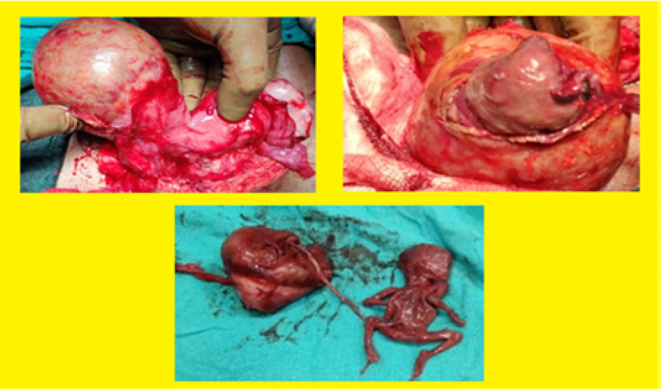
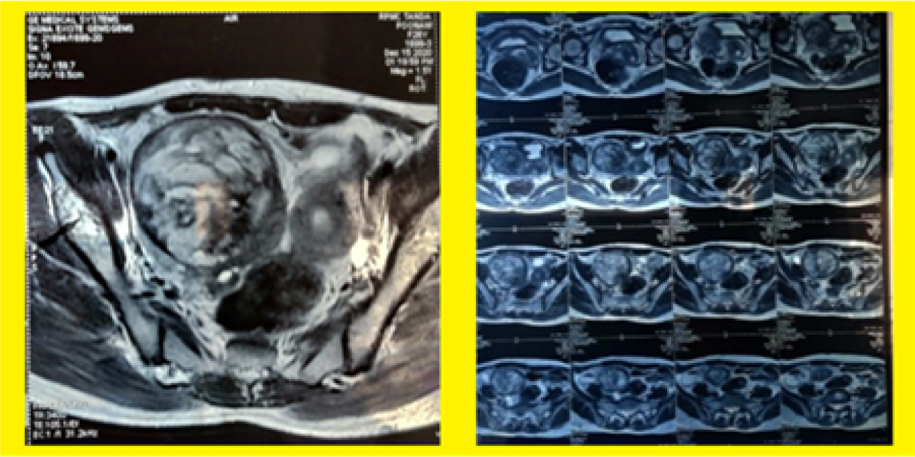
A 26 -year-old PGR was referred to Dr RPGMC Tanda as a case of 19 weeks with diagnosis of intrauterine fetal demise which had been detected on a routine antenatal ultrasound. There was no history of abdominal pain or vaginal blood loss at any time. She had regular menstrual cycles associated with dysmenorrhoea. There was no significant past medical or surgical history. Her vitals were normal. A transabdominal ultrasound showed a single, non-viable, intrauterine fetus with fetal parameters corresponding to 19 weeks of gestation and decision was made to induce abortion after routine investigations. Oral Mifepristone 200 mg was given followed by misoprostol 400 microgram after 48 hours given vaginally every four hours for a maximum of five doses. The patient experienced uterine contractions, with minimal vaginal spotting but no cervical changes. Repeat TVS was done showing a normal cervix leading to a small empty uterus just above it and a non-viable pregnancy was seen above and on the right of the uterus. Pregnancy in a rudimentary horn of the uterus was suspected. An MRI of the pelvis confirmed the diagnosis of pregnancy in the rudimentary horn of the uterus. Exploratory laparotomy with excision of rudimentary horn along with ipsilateral fallopian tube was done. B/l ovaries, contralateral fallopian tubes, kidneys and other abdominal organs were normal. Post-operative period was uneventful (Figure 1,2).


Figure 1: Excision of Rudimentary Horn Along with Ipsilateral Fallopian Tube


Figure 2: MRI of the Pelvis


Intraoperative Findings

Uterus was unicornuate with pregnancy in rudimentary non communicating horn on right side. The myometrium of the rudimentary horn was thin and membranous. Both ovaries and fallopian tubes were grossly normal. Rudimentary horn along the products of conception was resected. No communication with uterus was present. On cut section of the rudimentary horn a single fetus was found along with placenta.

DISCUSSION

Uterine anomalies result from the failure of complete fusion of the Müllerian ducts during embryogenesis. The incidence in the general population is estimated to be 4.3%. A unicornuate uterus with a rudimentary horn is the rarest anomaly and results from the failure of one of the Müllerian ducts to develop completely and an incomplete fusion with the contralateral side [2-4].


The most accepted explanation is the trans peritoneal migration of the sperm cells or a fertilized ovum. These cases usually result in the rupture of the horn in the second or third trimester. 


The rupture occurs because of the underdevelopment of the myometrium and a dysfunctional endometrium. The key for diagnosis prior to the rupture is a high index of clinical suspicion. A history of severe dysmenorrhoea may be a clue for diagnosis [4,5].


A careful pelvic examination in the first trimester showing a deviated uterus with a palpable adnexal mass should provoke suspicion of a Müllerian anomaly. It can be confirmed by an ultrasound or MRI. On USG a variation in the thickness of the myometrium in two horns and a marked distance between them favour the diagnosis of a rudimentary horn pregnancy [1,3].


Approximately 38% of patients have coexisting renal abnormalities. Immediate surgery is recommended whenever a diagnosis of a pregnancy in the rudimentary horn is made. The traditional treatment is a laparotomy and the surgical removal of the pregnant horn to prevent rupture and recurrent rudimentary horn pregnancies [1,4].


CONCLUSION

Even with developments in ultrasound technology, untrained doctors still struggle to make the antenatal diagnosis of a pregnancy with a simple horn. Early intervention combined with a high index of clinical suspicion for uterine abnormalities can lower the mortality rate. The suggested surgical procedure for the best prognosis when a rudimentary horn pregnancy is detected is the removal of the horn with ipsilateral salpingectomy. The requirement for strong clinical suspicion of this uncommon illness is highlighted by this example.

REFERENCES
  1. Ambusaidi, Q. and C.Jha. “Pregnancy in the rudimentary uterine horn: case report of an unusual presentation.” Sultan Qaboos University Medical Journal, vol. 14, no. 1, February 2014, pp. e134–e138.

  2. Saravelos, S.H. et al. “The pattern of pregnancy loss in women with congenital uterine anomalies and recurrent miscarriage.” Reproductive Biomedicine Online, vol. 20, no. 3, March 2010, pp. 416–422.

  3. Buntugu, K. et al. “Rudimentary horn pregnancy: Prerupture diagnosis and management.” Ghana Medical Journal, vol. 42, 2008, pp. 92–94.

  4. Jain, R. et al. “A rare case of intact rudimentary horn pregnancy presenting as hemoperitoneum.” Journal of Human Reproductive Sciences, vol. 3, 2010, pp. 113–115.

  5. Okonta, P.I. et al. “Pregnancy in a noncommunicating rudimentary horn of a unicornuate uterus: A case report.” Cases Journal, vol. 2, 2009, article 6624.

None
License
CC BY-NC-ND
Creative Commons Attribution-NonCommercial-NoDerivatives 4.0 International License
Pregnancy in Rudimentary Horn: A Case Report © 2026 by Komal Sharma, Gaurav Sharma, Jyotshna Sharma licensed under CC BY-NC-ND 4.0
All papers should be submitted electronically. All submitted manuscripts must be original work that is not under submission at another journal or under consideration for publication in another form, such as a monograph or chapter of a book. Authors of submitted papers are obligated not to submit their paper for publication elsewhere until an editorial decision is rendered on their submission. Further, authors of accepted papers are prohibited from publishing the results in other publications that appear before the paper is published in the Journal unless they receive approval for doing so from the Editor-In-Chief.
Himalayan Journal of Medicine and Surgery open access articles are licensed under a Creative Commons Attribution-Share A like 4.0 International License. This license lets the audience to give appropriate credit, provide a link to the license, and indicate if changes were made and if they remix, transform, or build upon the material, they must distribute contributions under the same license as the original.
Recommended Articles
Research Article
Phenotypic Characterization and Antimicrobial Resistance Patterns of Clinical Klebsiella pneumoniae Isolates from Al-Najaf Teaching Hospital
Published: 30/06/2026
Download PDF
Research Article
Impact of Gut-Liver Axis: Hepatic Biochemical and Metabolic Changes Associated with Chronic Gastritis in Iraqi Patients with Helicobacter Pylori
Published: 20/02/2026
Download PDF
Research Article
Effect of Land Degradation on Livelihood
Published: 04/01/2024
Download PDF
Research Article
Awareness and Practices among Professional Healthcare Workers towards COVID-19 in Iraq
...
Published: 30/03/2024
Download PDF
Flowbite Logo
Najmal Complex,
Opposite Farwaniya,
Kuwait.
Email: support@himjournals.com

Useful Links
Order Hard Copy
Privacy policy
Terms and Conditions
Refund Policy
Others
About Us
Team Members
Contact Us
Online Payments
Join as Editor
Join as Reviewer
Subscribe to our Newsletter
Follow us
MOST SEARCHED KEYWORDS
scientific journal
 | 
business journal
 | 
medical journals
 | 
Scientific Journals
 | 
Academic Publisher
 | 
Peer-reviewed Journals
 | 
Open Access Journals
 | 
Impact Factor
 | 
Indexing Services
 | 
Journal Citation Reports
 | 
Publication Process
 | 
Impact factor of journals
 | 
Finding reputable journals for publication
 | 
Submitting a manuscript for publication
 | 
Copyright and licensing of published papers
 | 
Writing an abstract for a research paper
 | 
Manuscript formatting guidelines
 | 
Promoting published research
 | 
Publication in high-impact journals
Copyright © Himalayan Journals . All Rights Reserved.