Contents
Download PDF
pdf Download XML
1598 Views
309 Downloads
Share this article
Research Article | Volume 4 Issue 1 (Jan-June, 2023) | Pages 1 - 4
Prostate Specific Antigen and Gleason Score as Predictors for Bone Metastases in Prostate Cancer Patients
 ,
 ,
 ,
1
Department of Pathology and Forensic Medicine, Faculty of Medicine, University of Kufa, Najaf, Iraq
2
Department of Pathology, Arab Board of Health Specializations, Najaf, Iraq
3
Department of Oncology, Euphrates Cancer Hospital, Najaf, Iraq
Under a Creative Commons license
Open Access
Received
Nov. 3, 2022
Revised
Dec. 9, 2022
Accepted
Jan. 19, 2023
Published
Feb. 28, 2023
Abstract

Background: Prostate Cancer (PCa) is men's second most commonly diagnosed cancer and bone is the most dominant site of metastasis. This study aims to investigate the diagnostic relevance of Bone Metastasis (BM) with Prostate-Specific Antigen (PSA) level and Gleason Score (GS) in Iraqi patients with PCa. Patients and Methods: Eighty-five patients diagnosed with PCa between January 2021 and October 2022 at Euphrates Cancer Hospital, Iraq, underwent Technetium-99 bone scintigraphy and serum PSA tests at the time of diagnosis. Results: Overall, 52 out of 85 (61.2%) patients had bone metastases. PSA and GS were significantly differed between patients with BM and those without BM. PSA and GS were statistically highly significant predictors of BM and their predictive values were (p<0.001) and (p <0.006), respectively, while age was not a predictive factor (p = 0.768). There were 26.7% (4/15) patients who had BM with PSA ≤20 ng/mL and GS <8. In comparison, none (0/7) had BM with GS ≤6 and PSA ≤10 ng/mL. The sensitivity and specificity of PSA level at a cut-off of 34.4 ng/mL were 80.77% and 72.73%, respectively. Conclusions: There was a high incidence of BM in Iraqi patients with newly diagnosed PCa. It was found that patients with PSA >10 ng/mL and GS ≥7 were at higher risk of BM. Therefore, a bone scan is highly recommended for patients with these characteristics. However, a bone scan might not be needed in PCa patients with PSA ≤10 ng/mL and GS ≤6.

Keywords
INTRODUCTION

Prostate Cancer (PCa) is men's second most commonly diagnosed cancer worldwide. The Global Cancer Observatory estimated 1,414,259 new cases and 375,304 deaths globally in 2020 [1]. The American Cancer Society estimates 191,930 new cases and 33,330 deaths in the United States in 2020 [2]. In Iraq, PCa is men's third most common cancer, with 1,117 new cases and 416 deaths in 2020 [3].


Bone Metastasis (BM) is the most frequent site of metastasis in PCa, seen in 90% of patients with metastatic PCa. It is, in fact, the only site of metastasis in the majority of patients (86%) [4]. Once BM occurs in a PCa patient, treatment is restricted and the overall survival rate and quality of life are significantly reduced [5]. Currently, the technetium 99m methylene diphosphonate (Tc 99m MDP) bone scans are accepted as the standard imaging modality for identifying bone metastasis in PCa [6]. Serum Prostate-Specific Antigen (PSA) levels correlate strongly with the risk of BM in patients with PCa [7]. Gleason Score (GS) is based solely on the architectural features of prostate cancer cells and correlates closely with clinical behaviour. A higher GS score (8 to 10) indicates a greater likelihood of having bone metastasis [7,8].


This study aimed to investigate the diagnostic relevance of bone metastasis with PSA and Gleason score in Iraqi patients with newly diagnosed prostate cancer. Consequently, we established a discriminant analysis model to identify the clinical profiles of patients who did not require staging bone scans.                

MATERIALS AND METHODS

This case-control study is based on eighty-five newly diagnosed prostate cancer patients between January 2021 and October 2022 at Euphrates Cancer Hospital, Najaf, Iraq. Age, total serum PSA level, bone scan and Gleason score results were obtained from medical records. Inclusion criteria were patients with histopathologically proven prostate cancer. In addition, total serum PSA level and Technetium-99 MDP whole-body bone scan were performed at the time of diagnosis.


The age of all patients in this study was distributed into four categories: <60 years, 60-69 years, 70-79 years and ≥ 80 years. Total serum PSA levels are distributed into four categories: ≤10 ng/mL, 10.1-20 ng/mL, 20.1-100 ng/mL and >100 ng/mL. Prostate tissue for biopsy examination was collected in various ways: Trans-Rectal Ultrasound guided prostate biopsy, Trans-Urethral Resection of Prostate and radical prostatectomy. The tumours were graded according to Gleason's grading system [9]. The histopathologic findings based on the Gleason score were divided into four groups: GS ≤6, GS 3+4, GS 4+3 and GS ≥8. Technetium 99m MDP whole-body bone scanning was performed and expert physicians judged the results. We sought a consult from a physician with extensive experience when necessary. The bone scan results were initially recorded as negative or positive for the presence of metastasis. According to the results, all patients were categorized into patients with BM and patients without BM.


All statistical analysis was done by IBM SPSS Statistics version 26. Descriptive statistics are expressed as frequencies, percentages, mean and standard deviation according to the variable types. Chi-square and Fisher's Exact tests were used for analyzing categorical data and showing the association between variables. Multivariate logistic regression analysis was used to compare the relevance of bone scan findings with PSA and GS. The receiver operating characteristics (ROC) curve was used to evaluate the accuracy of bone metastasis with serum PSA level. Results were considered significant at p <0.05.

RESULTS

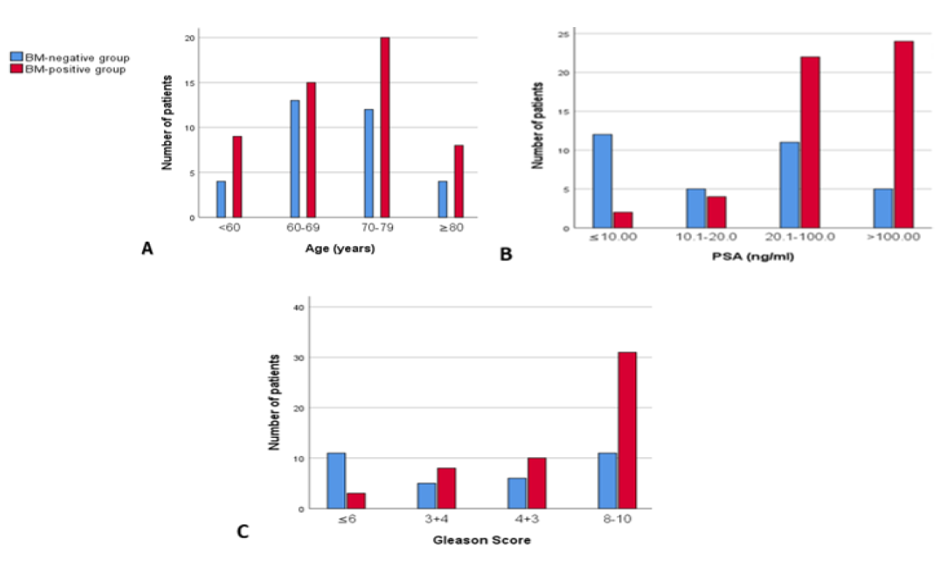
We identified 85 patients diagnosed with prostate cancer. The mean age of all patients was 69.1±9.7 years (range 47-98 years) and the mean serum PSA was 71.50±80.56 ng/mL (range 0.05-500 ng/mL). Overall, 52 out of 85 (61.2%) patients had bone metastases and the remaining 33 (38.8%) patients without bone metastases. Of all patients with bone metastases, 43 patients had multiple and nine patients had solitary bone metastases. The comparative patients’ characteristics of the two groups with different bone metastasis status were listed in (Table 1 and Figure 1). No difference between ages was detected in the two groups (p = 0.768).


Table 1: Patients’ Characteristics

VariablesAll patients (n = 85)BM-negative group (n = 33)BM-positive group (n = 52)p-value
Age/years, n (%)0.768

Average/Median

(Range)

69.16±9.7/70

47-98

69.27±9.9/69

52-98

69.1±9.7/70

47-89


<6013 (15.3)4 (12.1)9 (17.3) 
60-6928 (32.9)13 (39.4)15 (28.8) 
70-7932 (37.7(12 (36.4)20 (38.5) 
≥8012 (14.1)4 (12.1)8 (15.4) 
PSA (ng/ml), n (%)   <0.001

Average/Median

(Range)

71.50±80.56/52.60

0.05-500

34±35.45/17.72

0.05-107.50

95.31±91.72/89.89

1.06-500

 
≤10.014 (16.5)12 (36.4)2 (3.8) 
10.1-20.09 (10.6)5 (15.2)4 (7.7) 
20.1-100.033 (38.8)11 (33.3)22 (42.3) 
>100.029 (34.1)5 (15.2)24 (46.2) 
GS, n (%)<0.006
≤614 (16.5)11 (33.3)3 (5.8) 
3+413 (15.3)5 (15.2)8 (15.4) 
4+316 (18.8)6 (18.2)10 (19.2) 
8-1042 (49.4)11 (33.3)31 (59.6) 

BM: Bone Metastasis, PSA: Prostate-Specific Antigen, GS: Gleason Score


Figure 1: Patients’ Characteristics with and Without Bone Metastasis. A: Age, B: PSA. C: Gleason Score. BM: Bone Metastasis, PSA: Prostate-Specific Antigen


The proportion of bony metastases increased progressively with PSA level. Bone metastasis was present in 2 (2/14, 14.3%) patients with PSA ≤10 ng/mL, 4 (4/9, 44.4%) with PSA 10.1-20 ng/mL, 22 (22/33, 66.7%) with PSA 20.1-100 ng/mL and (24/29, 82.8%) with PSA>100 ng/mL. For Gleason score, a larger prevalence of bone metastasis was detected in patients with higher GS. 21.4% (3/14), 61.5% (8/13), 62.5% (10/16) and 73.8% (31/42) patients were found with bone metastasis in GS ≤6, 3+4, 4+3 and 8-10, respectively. However, the incidence of BM in prostate cancer patients increased dramatically with increasing PSA level and upgrading GS and their predictive values were (p <0.001) and (p <0.006), respectively.


Bone scan results were analyzed per subgroup of PSA level combined with Gleason score. This resulted in a high rate of BM in PSA >20 ng/mL combined with the GS ≥8 group. Low values of the GS and PSA were also combined to see the proportion of BM. These combinations were PSA ≤20 ng/mL plus GS <8 and PSA ≤10 ng/mL plus GS ≤6. This resulted in 4 out of 15 (26.7%) patients having BM in the first combination group. In comparison, none (0/7) had BM in the second combination group (negative predictive value of 100% for BM in the second group) (Table 2).


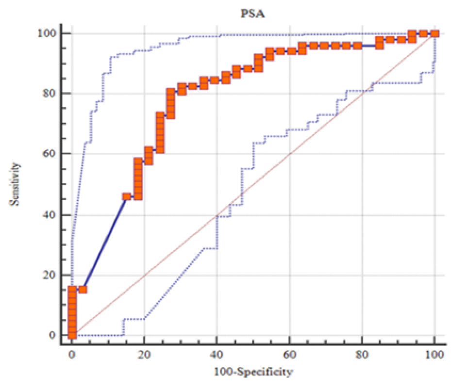
The receiver operating characteristic (ROC) curve for accuracy of PSA level for predicting bone metastasis on bone scan has an area under the curve 0.787 (95% CI: 0.685 - 0.869, p <0.0001). The PSA level's sensitivity and specificity at a 34.4 ng/mL cut-off point were 80.77% and 72.73%, respectively (Figure 2).


Table 2:Bone Scan Results for Combined Prostate-Specific Antigen and Gleason Score

ParametersGS ≤6GS ≤6

GS = 7

3+4                                                                       4+3

GS 8-10GS 8-10
PSA (ng/ml)CountBM, n (%)Count                          BM, n (%)                      Count                   BM, n (%)CountBM, n (%)
≤1070 (0.0)10 (0.0)11 (100)51 (20)
10.1–2021 (50)11 (100)31 (33.3)31 (33.3)
20.1–10041 (25)96 (66.7)64 (66.7)1411 (78.6)
>10011 (100)21 (50)64 (66.7)2018 (90)
           

BM: Bone Metastasis, PSA: Prostate-Specific Antigen, GS: Gleason Score


Figure 2: Receiver Operating Characteristic Curve of Prostate-Specific Antigen Level for Predicting Bone Metastasis in Prostate Cancer Patients

DISCUSSION

The incidence of prostate cancer differs significantly between the world regions and countries. The highest incidence of PCa is in western countries, representing 23.1% and 17.1% of all male malignancies in the United Kingdom and the United States, respectively, in 2020 [10, 11]. In Iraq, PCa represented 7.9% of all male malignancies in 2020 [3]. Environmental, genetic factors and the absence of PSA screening programs influence this incidence difference [12-14].


Bone metastasis significantly affects the patient’s treatment and prognosis. In our study, we observed bone metastasis in 61.2% of Iraqi patients with PCa, which is significantly higher than that in the United States (18%–29%) and the United Kingdom (8%) [8,15]. This could be partly attributed to the adoption of patients who presented with urological symptoms rather than population PSA-based screening. The second explanation is that 83.5% of patients in our study had GS ≥7, whereas 61.5% in McArthur et al. [8]. However, the incidence of BM in our study is comparable to other Asian studies: Zhuo et al. 47.5%, Sanjaya et al. 53.6% and Singh et al. 69.1% [16-18].


The proportion of bone metastasis in patients with PSA levels ≤10 ng/mL is 14.3%, which is comparable to Zaman et al. 14.3% but higher than Wang et al. 9.5%, Chen et al. 5%, Lin et al. 4% and McArthur et al. 2.7% and lower than Sanjaya et al. 23.8% [8, 18-22]. The proportion of BM in patients with PSA levels 10.1–20 ng/mL is 44.4%, higher than Singh et al. 38.46% and Sanjaya et al. 31.6% [17, 18]. The rate of bony metastases with PSA levels of 20.1-100 ng/mL is 66.7%, comparable to Chen et al. 61.6% and Singh et al. 60.9% [17, 19]. The rate of BM in patients with PSA levels >100 ng/mL is 82.8%, comparable to Sanjaya et al. 80.4% and Zaman et al. 82%, higher than Chen et al. 74% and less than Singh et al. 100% [17-19,22]. This discrepancy most likely reflects the fact that 16.5% of our patients' overall population had a PSA level ≤10 ng/mL, which is a significantly lower proportion than in other studies: McArthur et al. 38.4%, Lin et al. 21.3% and Wang et al. 20.9% [8,20,21].


Gleason score is one of the strongest prognostic predictors of prostate cancer. The rate of positive bone scans in patients with a GS ≤6 is 21.4%, higher than Chen et al. 13.2% but lower than Sanjaya et al. 35.5% [18, 19]. The rate of positive bone scans in patients with GS 3+4 and GS 4+3 are 61.5% and 62.5%, respectively, which is higher than Sanjaya et al. 44.2% [18]. The proportion of bony metastases with a GS ≥8 is 73.8%, comparable to Sanjaya et al. 64.6% [18].


Furthermore, the results of this study indicated that patients with PSA levels ≤10 ng/mL combined with GS ≤ 6 had a negative predictive value for BM of 100%, which is a similar conclusion to studies carried out by McArthur et al. and Lin et al. [8, 20]. Thus, this research suggests that bone scans might not be needed for newly diagnosed PCa patients with these characteristics. These characteristics support the application of European Society for Medical Oncology (ESMO) guidelines and this would lead to a reduction in staging bone scans of 8.2% in our sample [23].


Another essential fact is the significantly increased incidence of bone metastasis in patients with PSA ≤20 ng/mL and Gleason score <8. This is inconsistent with many published studies showing a high negative predictive value of PSA <20 ng/mL and GS <8 [8]. In this study, if bone scans were omitted for patients with PSA <20 ng/mL and GS <8, result in 4 (4/15, 26.6%) patients would have missed the diagnosis of bone metastasis. The last result is similar to a study from India by Singh et al., which revealed bony metastases in 29% of patients with PSA <20 ng/mL and a study from Indonesia by Sanjaya et al. that reported an incidence of bone metastasis in 18.6% of patients with PSA <20 ng/mL and Gleason score <8 [17,18]. However, it seems imperative to use bone scans in bone staging, even in patients with PSA <20 ng/mL and GS <8, to avoid under-staging in a significant number of patients.

.

These results are in keeping with previous works like McArthur et al. and Chen et al., that PSA level and GS are independent predictors of bone metastasis [8, 19]. Age was not a predictor for bone metastasis (p = 0.768), as reported in the literature and by Chen et al. [19].


In Lin et al., the PSA cutoff point was 88 ng/mL, while the PSA cutoff point in our study was 34.4 ng/mL. This discrepancy was due to twenty tests in this study showed a PSA level of more than 100 ng/mL without representing an exact PSA level [20].


The limitations of this study were the performance of serum PSA tests, prostate biopsies and bone scans at different laboratories and institutions, which might affect the standardization of results. In addition, the metastatic lesions detected by bone scans were not histologically confirmed. Moreover, it did not investigate other variables that could predict bone metastasis such as: prostate volume, clinical tumour stage and bony symptoms.


CONCLUSION

This research found a high incidence of bone metastasis in Iraq's newly diagnosed prostate cancer patients. It was found that patients with PSA >10 ng/mL and GS ≥7 were at higher risk of bone metastasis. Therefore, a bone scan is highly recommended for patients with these characteristics. However, PCa patients with PSA ≤10 ng/mL and GS ≤6 might not need a bone scan. On the other side, serum prostate-specific antigen and Gleason score were found as independent predictors for bone metastasis.


Abbreviations


  • AUC: Area Under Curve

  • BM: Bone Metastasis

  • CI: Confidence Interval

  • ESMO: European Society for Medical Oncology

  • GS: Gleason Score

  • PCa: Prostate Cancer

  • PSA: Prostate-Specific Antigen

  • ROC: Receiver Operating Characteristic

  • Tc 99m MDP: Technetium Tc 99m Methylene Diphosphonate

REFERENCES
  1. Sung, H. et al. “Global cancer statistics 2020: GLOBOCAN estimates of incidence and mortality worldwide for 36 cancers in 185 countries.” CA: A Cancer Journal for Clinicians, vol. 71, no. 3, 2021, pp. 209–249.

  2. Siegel, R.L. et al. “Cancer statistics, 2020.” CA Cancer Journal for Clinicians, vol. 70, no. 1, 2020, pp. 7–30.

  3. World Health Organization. Global Cancer Observatory: Iraq Fact Sheet. International Agency for Research on Cancer, 2020.

  4. Hess, K.R. et al. “Metastatic patterns in adenocarcinoma.” Cancer, vol. 106, no. 7, 2006, pp. 1624–1633.

  5. Yang, C. et al. “Clinical features and treatment strategies for older prostate cancer patients with bone metastasis.” Asian Journal of Andrology, vol. 15, no. 6, 2013, pp. 759.

  6. Trabulsi, E.J. et al. “Optimum imaging strategies for advanced prostate cancer: ASCO guideline.” Journal of Clinical Oncology, vol. 38, no. 17, 2020, pp. 1963.

  7. Briganti, A. et al. “Predicting the risk of bone metastasis in prostate cancer.” Cancer Treatment Reviews, vol. 40, no. 1, 2014, pp. 3–11.

  8. McArthur, C. et al. “Changing the referral criteria for bone scan in newly diagnosed prostate cancer patients.” The British Journal of Radiology, vol. 85, no. 1012, 2012, pp. 390–394.

  9. Epstein, J.I. et al. “The 2014 international society of urological pathology (ISUP) consensus conference on gleason grading of prostatic carcinoma.” The American Journal of Surgical Pathology, vol. 40, no. 2, 2016, pp. 244–252.

  10. World Health Organization. Global Cancer Observatory: United States of America Fact Sheet. International Agency for Research on Cancer, 2020.

  11. World Health Organization. Global Cancer Observatory: United Kingdom Fact Sheet. International Agency for Research on Cancer, 2020.

  12. Catalona, W.J. “Prostate cancer screening.” Medical Clinics of North America, vol. 102, no. 2, 2018, pp. 199–214.

  13. Moch, H. et al. “The 2016 who classification of tumours of the urinary system and male genital organs—Part A: Renal, penile and testicular tumours.” European Urology, vol. 70, no. 1, 2016, pp. 93–105.

  14. Zhau, H.E. et al. “Interracial differences in prostate cancer progression among patients from the united states, china and Japan.” Asian Journal of Andrology, vol. 15, no. 6, 2013, pp. 705.

  15. Hernandez, R.K. et al. “Incidence of bone metastases in patients with solid tumors: Analysis of oncology electronic medical records in the united states.” BMC Cancer, vol. 18, 2018, pp. 1–11.

  16. Zhuo, L. et al. “Prostate cancer with bone metastasis in Beijing: An observational study of prevalence, hospital visits and treatment costs using data from an administrative claims database.” BMJ Open, vol. 9, no. 6, 2019, pp. e028214.

  17. Singh, O.P. et al. “Role of serum prostate-specific antigen as predictor for bone metastases in newly diagnosed prostate cancer.” Journal of Cancer Research and Therapeutics, vol. 15, no. 8, 2019, pp. 39.

  18. Sanjaya, I. et al. “Correlation between low gleason score and prostate-specific antigen levels with incidence of bone metastases in prostate cancer patients: when to omit bone scans?” Asian Pacific Journal of Cancer Prevention, vol. 14, no. 9, 2013, pp. 4973–4976.

  19. Chen, S. et al. “Establishing a prediction model for prostate cancer bone metastasis.” International Journal of Biological Sciences, vol. 15, no. 1, 2019, pp. 208.

  20. Lin, Y. et al. “When to perform bone scintigraphy in patients with newly diagnosed prostate cancer? A retrospective study.” BMC Urology, vol. 17, no. 1, 2017, pp. 1–5.

  21. Wang, Y. et al. “Is it safe to omit baseline bone scan for newly diagnosed prostate cancer patients?” Urologia Internationalis, vol. 94, no. 3, 2015, pp. 342–346.

  22. Zaman, M.U. et al. “Metastasis on bone scan with low prostate-specific antigen (≤20 ng/mL) and Gleason’s score (<8) in newly diagnosed Pakistani males with prostate cancer: Should we follow western guidelines.” Asian Pacific Journal of Cancer Prevention, vol. 12, no. 6, 2011, pp. 1529–1532.

  23. Parker, C. et al. “Prostate cancer: ESMO clinical practice guidelines for diagnosis, treatment and follow-Up.” Annals of Oncology, vol. 31, no. 9, 2020, pp. 1119–1134.

None
License
CC BY-NC-ND
Creative Commons Attribution-NonCommercial-NoDerivatives 4.0 International License
Prostate Specific Antigen and Gleason Score as Predictors for Bone Metastases in Prostate Cancer Patients © 2026 by S. J. Ala Alam, Maytham Ahmed Kadhm Kraidi, Salam Salah Jumaah, Haider Jaber Al-Shiblawi licensed under CC BY-NC-ND 4.0
All papers should be submitted electronically. All submitted manuscripts must be original work that is not under submission at another journal or under consideration for publication in another form, such as a monograph or chapter of a book. Authors of submitted papers are obligated not to submit their paper for publication elsewhere until an editorial decision is rendered on their submission. Further, authors of accepted papers are prohibited from publishing the results in other publications that appear before the paper is published in the Journal unless they receive approval for doing so from the Editor-In-Chief.
Himalayan Journal of Medicine and Surgery open access articles are licensed under a Creative Commons Attribution-Share A like 4.0 International License. This license lets the audience to give appropriate credit, provide a link to the license, and indicate if changes were made and if they remix, transform, or build upon the material, they must distribute contributions under the same license as the original.
Recommended Articles
Research Article
Phenotypic Characterization and Antimicrobial Resistance Patterns of Clinical Klebsiella pneumoniae Isolates from Al-Najaf Teaching Hospital
Published: 30/06/2026
Download PDF
Research Article
Impact of Gut-Liver Axis: Hepatic Biochemical and Metabolic Changes Associated with Chronic Gastritis in Iraqi Patients with Helicobacter Pylori
Published: 20/02/2026
Download PDF
Research Article
Effect of Land Degradation on Livelihood
Published: 04/01/2024
Download PDF
Research Article
Awareness and Practices among Professional Healthcare Workers towards COVID-19 in Iraq
...
Published: 30/03/2024
Download PDF
Flowbite Logo
Najmal Complex,
Opposite Farwaniya,
Kuwait.
Email: support@himjournals.com

Useful Links
Order Hard Copy
Privacy policy
Terms and Conditions
Refund Policy
Others
About Us
Team Members
Contact Us
Online Payments
Join as Editor
Join as Reviewer
Subscribe to our Newsletter
Follow us
MOST SEARCHED KEYWORDS
scientific journal
 | 
business journal
 | 
medical journals
 | 
Scientific Journals
 | 
Academic Publisher
 | 
Peer-reviewed Journals
 | 
Open Access Journals
 | 
Impact Factor
 | 
Indexing Services
 | 
Journal Citation Reports
 | 
Publication Process
 | 
Impact factor of journals
 | 
Finding reputable journals for publication
 | 
Submitting a manuscript for publication
 | 
Copyright and licensing of published papers
 | 
Writing an abstract for a research paper
 | 
Manuscript formatting guidelines
 | 
Promoting published research
 | 
Publication in high-impact journals
Copyright © Himalayan Journals . All Rights Reserved.