Contents
Download PDF
pdf Download XML
1615 Views
127 Downloads
Share this article
Case Report | Volume 4 Issue 1 (Jan-June, 2023) | Pages 1 - 3
Canaliform Medial Raphe Cyst -A Rare Entity
 ,
 ,
 ,
1
Post Graduate Student, Department of Radiodiagnosis, DRPGMC, Tanda.Kangra, Himachal Pradesh, 176002 India
2
MD, Department of Biochemistry, Civil Hospital, Kangra, Himachal Pradesh, 176001 India
3
MS, Department of Surgery, Dr RPGMC Kangra at Tanda, HP, 176002 India
4
MD, Department of Dermatology, Civil Hospital, Kangra, Himachal 176001 Pradesh India
Under a Creative Commons license
Open Access
Received
March 10, 2023
Revised
April 17, 2023
Accepted
May 24, 2023
Published
June 6, 2023
Abstract

Median raphe cysts (MRCs) are rare, benign congenital lesions of unknown origin, that can be found anywhere on the ventral side of the genital area, between the urethral meatus and the anus. The canaliform variety is very uncommon but clinically distinct. The rarity of our case is attributed to the canaliform type, the scrotal and perineal localization.

Keywords
INTRODUCTION

Mermet first reported a case of MRC in “Revue de Chirurgie” [1]. MRCs present with a bimodal age distribution at approximately 1–10 and 21–40 years old. MRCs were previously designated with multiple terms such as mucoid cyst of the penile skin, genitoperineal cyst of the median raphe, parameatal cyst, hydrocystoma and apocrine cystadenoma [2].MRCs mostly present at birth and may remain asymptomatic or unrecognized during childhood. 25.5% of all cases are diagnosed within the first decade of life [3]. Due to the fact that the majority of children (up to 75% of all cases) are asymptomatic, it has been suggested that MRCs are more common than it is considered [4].


Infection and trauma are the main causes that lead to rapid development of MRCs or clinical manifestation [5]. Other causes include rupture of a cyst or ulcer’s development on the surface of MRCs. Pain during sexual intercourse or even rarely discomfort during urination, especially when the cyst is located near the urethral meatus, are the main symptoms. Staphylococcus aureus and Neisseria gonorrheae are commonest pathogens that cause infection of MRC [6].


MRCs should be differentially diagnosed from other conditions such as epidermal inclusion cyst, pilonidal cyst, dermoid cyst and urethral diverticulum.Especially in cases of perianal localization, differential diagnosis should include condyloma, viral wart, hemorrhoid, hypertrophied papilla, glomus tumor and steatocystoma.


Case Reports

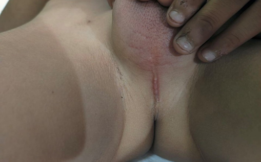
Case 1-A 3 year old boy presented with congenital multiple whitish cysts along a cord-like swelling running in the midline extending from the anterior margin of the anus to the lower pole of the scrotum. (Figure 1) The lesions spread along the perineal raphe. Infant showed no signs of pain, urinary complaints or other symptoms. He had no history of medical disease or congenital anomaly, and the rest of the skin was normal. Biopsy showed a dermal cyst lined by pseudostratified columnar epithelium.


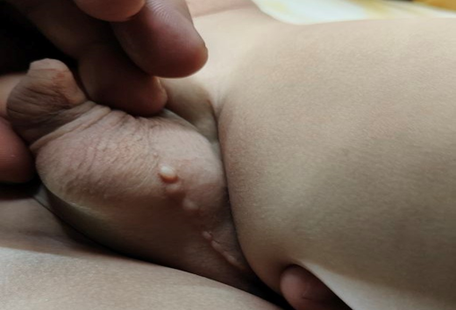
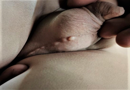
Case 2-A 8-month-old boy presented with linearly arranged, multiple pinhead-sized to rice-sized whitish cysts along the median raphe from the perineum to mid of scrotum (Figure 2-3), which were present from birth and progressively increased in size and number.The lesions spread along the perineal raphe and had a cordlike appearance. The infant showed no signs of pain, tenderness, pruritus, or other symptoms. He had no history of medical disease or congenital anomaly, and the rest of the skin was normal. Ultrasound was performed showing cystic formations with hyperechoic content was documented, without evidence of expansion deeper to the endoscrotal structures. A skin biopsy was performed at the upper cystic portion of the lesion and the biopsy specimen showed the cysts located in the dermis with variable lining. Some parts were lined by stratified squamous epithelium andother parts consisted of cuboidal urothelium-like epithelium with mucinous cells. Thus, a diagnosis of mixed-type median raphe cysts was made.


DISCUSSION

Median raphe cysts are rare, benign congenital lesions that can develop anywhere along the midline of the ventral side of the male genital area. They can form from the urethral meatus to the anus and the perineum along the perineal raphe. Although present since childhood, they are sometime reported by young adults when they increase in size, get infected or cause sexual discomfort histologically, they are classified into four types. The urethral type, the most common, consists of an urothelium-like epithelium, with a layer of columnar cells overlaid with several stratified layers of uniform small cells. The epidermoid type consists of stratified squamous cell epithelium. The glandular type consists of a well-formed intraepithelial glandular structure in the lining of the urethral epithelium. The mixed type, the second most common and the type of this case, consists of more than one type of epithelium, including the urethral epithelium with squamous metaplasia, urethral epithelium with mucinous cells, or a combination of these three [3].


The pathogenesis is unclear, but several hypotheses have been proposed. First, the 'tissue trapping' theory states that median raphe cysts are caused by either a defective fusion of the urethral folds or an anomalous outgrowth of the epithelium during the development of the urethra [7]. Second, median raphe cysts may result from the anomalous developmental rest of the periurethral glands of Littre. This hypothesis may help explain glandular type median raphe cysts and the involvement of mucinous cells in some cases [8]. A third hypothesis suggests that blockage of the paraurethral ducts may underlie the development of median raphe cysts [9]. MRCs may regress spontaneously. Shibagaki et al. believe that small and asymptomatic MRCs can be observed without excision, because there is the possibility of regression [10]. Therapeutic intervention may be indicated due to secondary infection leading to pain or cosmetic reasons [11]. Optimal treatment of MRCs is surgical excision and primary closure, which prevents infection or cosmetic sequelae [12]. Aspiration alone is not recommended as a treatment option for MRCs. Asymptomatic lesions are left untreated .Complete local excision is recommended for the symptomatic lesions to prevent possible complications. Furthermore, whether associated congenital anomalies are present should also be assessed.

CONCLUSION

The canaliform median raphe cysts demonstrated in the present cases are an uncommon presentation of this rare condition, and reports of these cysts are rare. Most patients present with a single isolated from or a few cysts. The patient described here presented with multiple, continuous cysts. This unique morphology makes diagnosis confusing due to its rarity and particular clinical features. Thus, histologic confirmation is often required.

Conflict of interest and financial funding–none


Figure 1: Congenital Multiple Whitish Cysts


Figure 2: Canaliform Medial Raphe Cyst


Figure 3: Congenital Multiple Whitish Cysts Extending from Anus to Mid Scrotum

REFERENCE
  1. Mermet P. et al. "Congenital cysts of the genitoperineal raphe." Revue de Chirurgie, vol. 15, 1895, pp. 382–435.

  2. Betül Ü. et al. "Perianal median raphe cyst: a rare lesion with unusual histology and localization." Case Rep Dermatol Med, 2015, Article ID 487814, 3 pages.

  3. Shao I.H. et al. "Male median raphe cysts: serial retrospective analysis and histopathological classification." Diagn Pathol, vol. 7, 2012, Article 121.

  4. Krauel L. et al. "Median raphe cysts of the perineum in children." Urol., vol. 71, 2008, pp. 830–831.

  5. Soendjojo A. and Pindha S. "Trichomonas vaginalis infection of the median raphe of the penis." Sex Transm Dis, vol. 8, 1981, pp. 255–257.

  6. Ark C.O. et al. "Median raphe cyst on the scrotum and perineum." J Am Acad Dermatol, vol. 55, no. 5, 2006, pp. 114–115.

  7. Nagore E. et al. "Median raphe cysts of the penis: a report of five cases." Pediatr Dermatol, vol. 15, 1998, pp. 191–193.

  8. Dini M. et al. "Median raphe cyst of the penis: a report of two cases with immunohistochemical investigation." Am J Dermatopathol, vol. 23, 2001, pp. 320–324.

  9. Shiraki I.W. et al. "Parametal cysts of the glans penis: a report of 9 cases." J Urol., vol. 114, 1975, pp. 544–548.

  10. Shibagaki N. et al. "Spontaneous regression of congenital multiple median raphe cysts of the raphe scrota." Br J Dermatol, vol. 134, 1996, pp. 376–378.

  11. Deliktas H. et al. "Median raphe cyst of the penis." J Urol., vol. 12, no. 4, 2015, pp. 2287–2288.

  12. Soyer T. et al. "Scrotal pearl is not always a sign of anorectal malformation: median raphe cyst." Turk J Pediatr, vol. 55, no. 6, 2013, pp. 665–666.

None
None
License
CC BY-NC-ND
Creative Commons Attribution-NonCommercial-NoDerivatives 4.0 International License
Canaliform Medial Raphe Cyst -A Rare Entity © 2026 by Archita Gupta, Shelja Rawat, Rohit Kumar, Rohit Verma licensed under CC BY-NC-ND 4.0
All papers should be submitted electronically. All submitted manuscripts must be original work that is not under submission at another journal or under consideration for publication in another form, such as a monograph or chapter of a book. Authors of submitted papers are obligated not to submit their paper for publication elsewhere until an editorial decision is rendered on their submission. Further, authors of accepted papers are prohibited from publishing the results in other publications that appear before the paper is published in the Journal unless they receive approval for doing so from the Editor-In-Chief.
Himalayan Journal of Medicine and Surgery open access articles are licensed under a Creative Commons Attribution-Share A like 4.0 International License. This license lets the audience to give appropriate credit, provide a link to the license, and indicate if changes were made and if they remix, transform, or build upon the material, they must distribute contributions under the same license as the original.
Recommended Articles
Research Article
Phenotypic Characterization and Antimicrobial Resistance Patterns of Clinical Klebsiella pneumoniae Isolates from Al-Najaf Teaching Hospital
Published: 30/06/2026
Download PDF
Research Article
Impact of Gut-Liver Axis: Hepatic Biochemical and Metabolic Changes Associated with Chronic Gastritis in Iraqi Patients with Helicobacter Pylori
Published: 20/02/2026
Download PDF
Research Article
Effect of Land Degradation on Livelihood
Published: 04/01/2024
Download PDF
Research Article
Awareness and Practices among Professional Healthcare Workers towards COVID-19 in Iraq
...
Published: 30/03/2024
Download PDF
Flowbite Logo
Najmal Complex,
Opposite Farwaniya,
Kuwait.
Email: support@himjournals.com

Useful Links
Order Hard Copy
Privacy policy
Terms and Conditions
Refund Policy
Others
About Us
Team Members
Contact Us
Online Payments
Join as Editor
Join as Reviewer
Subscribe to our Newsletter
Follow us
MOST SEARCHED KEYWORDS
scientific journal
 | 
business journal
 | 
medical journals
 | 
Scientific Journals
 | 
Academic Publisher
 | 
Peer-reviewed Journals
 | 
Open Access Journals
 | 
Impact Factor
 | 
Indexing Services
 | 
Journal Citation Reports
 | 
Publication Process
 | 
Impact factor of journals
 | 
Finding reputable journals for publication
 | 
Submitting a manuscript for publication
 | 
Copyright and licensing of published papers
 | 
Writing an abstract for a research paper
 | 
Manuscript formatting guidelines
 | 
Promoting published research
 | 
Publication in high-impact journals
Copyright © Himalayan Journals . All Rights Reserved.