Contents
Download PDF
pdf Download XML
1331 Views
370 Downloads
Share this article
Case Report | Volume 4 Issue 1 (Jan-June, 2023) | Pages 1 - 3
Discoid Lupus Erythematosus leading to Squamous Cell Carcinoma – A Case Report
 ,
 ,
 ,
1
Post Graduate Student, Department of Radiodiagnosis, DRPGMC, Tanda. Kangra, Himachal Pradesh, India
2
MD Biochemistry, Civil Hospital, Kangra, Himachal Pradesh, India
3
MS Surgery, Department of Surgery, Kangra at Tanda, HP, India
4
MD Dermatology, Civil Hospital, Kangra, Himachal Pradesh, India
Under a Creative Commons license
Open Access
Received
March 9, 2023
Revised
April 14, 2023
Accepted
May 28, 2023
Published
June 6, 2023
Abstract

Discoid lupus erythematosus (DLE) is a benign disorder of the skin, clinically characterized by red scaly patches which heal with atrophy, scarring and pigmentary changes. DLE is subdivided into a localized form in which lesions are confined to the face and neck or a disseminated form in which lesions also occur elsewhere on the body. Malignant transformation is a rare complication of this condition. We report a case of squamous cell carcinoma (SCC) developing over lesions of disseminated DLE. A 60 years male diagnosed as a case of Discoid lupus erythematosus (DLE) developed Squamous cell carcinoma from the lesions over a period of 5 years due to treatment gap and late follow up. Diagnosis was based on physical and histopathological examination, the absence of systemic involvement and routine laboratory parameters, which were all within normal range. It is every essential to counsel the diagnosed cases of DLE and warn all patients about all the possible outcomes and compliance with medications should be ensured

Keywords
INTRODUCTION

Discoid lupus erythematosus (DLE) is a benign disorder of the skin, clinically characterized by red scaly patches which heal with atrophy, scarring and pigmentary changes and histo- pathologically by vacuolar degeneration of basal cell layer of epidermis and patchy dermal lymphocytic infiltrate. DLE is subdivided into a localized form in which lesions are confined to the face and neck or a disseminated form in which lesions also occur elsewhere on the body [1]. Disease commonly affects the sun-exposed areas of the skin. Malignant transformation is a rare complication of this condition. Photosensitivity and the long-standing immune mediated chronic inflammation and dysregulated healing characterized by atrophy, hypopigmentation or scarring inherent to DLE are risk factors for progression to SCC [2]. We report a case of squamous cell carcinoma (SCC) developing over lesions of disseminated DLE.


Case Report 

A 60-year male was diagnosed with localised type DLE 10 years ago. He had lesions only over the face initially. He was undergoing treatment with oral hydroxychloroquine, topical corticosteroid and sunscreen. However, He was lost to follow up 5 years back. After a late follow up period of 5 years and treatment gap, patient was having DLE lesions over the right side of face, ear, nose and neck [figure 1]. There was lesion of size 5*6 cm on left side of the face, which was ulcerated and progressed to left ear lobule to form a fungating growth with non-foul-smelling discharge since 6 month. History of photosensitivity and loss of appetite were present. There was no history of trauma, prior drug intake, and exposure to ionizing radiation, fever, weight loss, joint pain, fatigue, urinary disturbances, diabetes mellitus, oral ulcers, thrombophlebitis, chilblains, Raynaund’s phenomenon or bleeding tendencies. Patient was chronic alcoholic and was chronic smoker.  Regional lymph nodes were not palpable. Oral cavity, nasal mucosa, eyes, nails, palms, soles, external genitalia, perianal region, and joints were normal.Systemic examination was normal. Haematological investigation, chest X-ray, ECG was unremarkable.
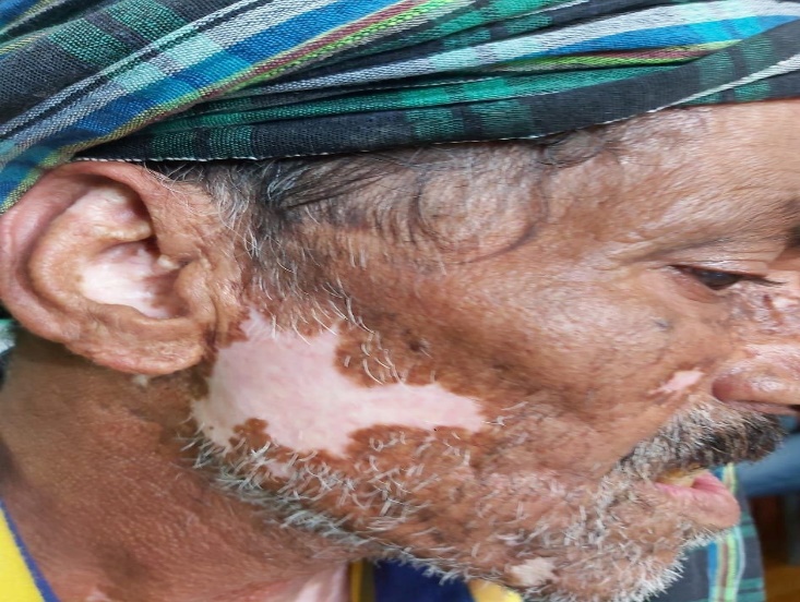


Figure 1: DLE Lesion on Right Side Check, Ear, Nose and Neck
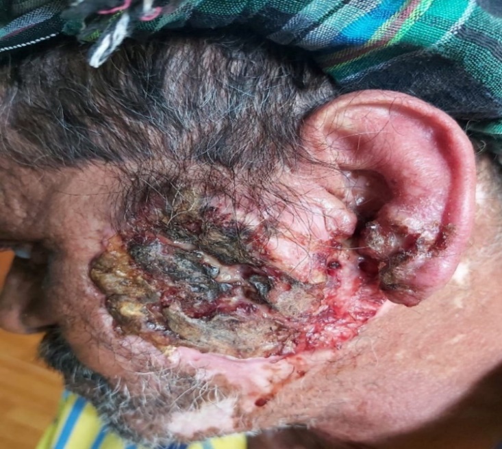


Figure 2: Tumour Growth over the Left Side of Face over Previous DLE Lesion Involving the Left Ear Lobule
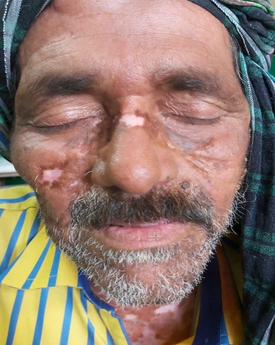


Figure 3: DLE Lesion on Right Cheek, Nose and Neck


Based on clinical examination, the lesions on left side of face was suspected to have malignant transformation, hence, a skin punch biopsy for histopathological evaluation was done. The histopathological reading was Squamous cell carcinoma, moderately differentiated. Histopathology showed acanthotic epidermis with papillomatosis and parakeratosis. The epidermis shows loss of polarity involving full thickness. The squamous cells were pleomorphic having hyperchromatic nucleus with nuclear rim irregularity. Mitosis constituted 1-3/HPF. Nests of atypical squamous cells surrounded by large number of lymphocytes infiltrated the upper dermis was seen.


Patient underwent a thorough workup to see for metastasis. CBC (complete blood count), LFT (liver function test), RFT (renal function test), TFT (thyroid function test), Thyroid Ultrasound, ECG (electrocardiogram), ANA, Anti -single-stranded DNA done were unremarkable.


Oral hydroxychloroquine and topical steroids along with sun protection were exhibited for management of DLE while SCC was managed by the surgeons with wide excision of the tumour followed by skin grafting.


DISCUSSION

DLE is a benign disorder, rarely associated with the development of SCC [3]. Reports of SCCs arising in the lesions of DLE are limited from India. DLE is more common in females. Although SCC developing over DLE is more among males, as in our patient. The mean age at presentation is 49.85±12.06 years, which is in accordance with our patient. According to the studies, disseminated type of DLE developing SCC is more common compared to localized type of DLE [4,5]. But in contrast, Tao et al found more patients of localized DLE developing SCC which is similar to our case report [6].


In a review by Sherman et al, the interval between development of DLE and SCC has varied from 4-20 years [7]. However, there have been reports of shorter duration of up to four years between the onset of the disease and development of SCC. Precipitating factors for SCC are age more than 50 years, female sex, and sun/ultraviolet ray exposure. The most common site for SCC developing over DLE is sun exposed and heavily scarred areas of the face, scalp and forearm [8]. The lips were the most commonly affected area in DLE related SCC. We are reporting a case of SCC over DLE presenting over left side of face. In our patient, SCC occurred in a DLE lesion over a period of 5 years. Continuous sun exposure and history of heavy smoking have contributed to malignant transformation in the DLE lesion. Thus, it is vital to look for malignant transformation in cases of DLE, especially in presence of risk factors like a photo exposed area, smoker and non-compliant patient.

CONCLUSION

Even though the incidence of malignant transformation of DLE lesions is rare, a high degree of suspicion for malignant changes is still necessary. DLE patients with risk factors should be followed closely, and expert histopathologic evaluation of biopsy specimens from doubtful lesions is required to make an early, accurate diagnosis of SCC. It is every essential to counsel and warn all patients about all the possible outcomes and compliance with medications should be ensured


Conflict of interest

None 


Funding

None


Ethical approval

Not required


REFERENCE
  1. Goodfield M.J.D. et al. “The connective tissue diseases (discoid lupus erythematosus).” In: Burns T et al. (eds), Rook's Textbook of Dermatology. 7th ed. Oxford: Blackwell Science, 2004, pp. 5–24.

  2. Molomo E.M. et al. “Discoid lupus erythematosus-related squamous cell carcinoma of the lip in an HIV-seropositive black male.” J Can Ther, vol. 11, 2015, pp. 1036. http://dx.doi. org/ 10.4103/0973-1482.146107

  3. Cassarino D.S. et al. “Cutaneous squamous cell carcinoma: a comprehensive clinicopathologic classification.” J Cutan Pathol, vol. 33, 2006, pp. 191–206.

  4. Grover S. et al. “Discoid lupus erythematous leading to squamous cell carcinoma.” Med J Armed Forces India, vol. 63, 2007, pp. 184–185.

  5. Bhat M.R. et al. “Disseminated discoid lupus erythematous leading to squamous cell carcinoma.” Indian J Dermatol, vol. 57, 2012, pp. 158–161.

  6. Tao J. et al. “Squamous cell carcinoma complicating discoid lupus erythematosus in Chinese patients: review of the literature 1964–2010.” J Am Dermatol, vol. 66, 2012, pp. 695–696.

  7. Sherman R.N. et al. “Cutaneous squamous cell carcinoma in black patients with chronic discoid lupus erythematosus.” Int J Dermatol, vol. 32, 1993, pp. 677–679.

  8. Gupta U. et al. “Squamous cell carcinoma complicating an untreated chronic discoid lupus erythematosus (CDLE) lesion in a black female.” J Dermatol, vol. 32, 2005, pp. 1010–1013.

None
None
License
CC BY-NC-ND
Creative Commons Attribution-NonCommercial-NoDerivatives 4.0 International License
Discoid Lupus Erythematosus leading to Squamous Cell Carcinoma – A Case Report © 2026 by Archita Gupta, Shelja Rawat, Rohit Kumar, Rohit Verma licensed under CC BY-NC-ND 4.0
All papers should be submitted electronically. All submitted manuscripts must be original work that is not under submission at another journal or under consideration for publication in another form, such as a monograph or chapter of a book. Authors of submitted papers are obligated not to submit their paper for publication elsewhere until an editorial decision is rendered on their submission. Further, authors of accepted papers are prohibited from publishing the results in other publications that appear before the paper is published in the Journal unless they receive approval for doing so from the Editor-In-Chief.
Himalayan Journal of Medicine and Surgery open access articles are licensed under a Creative Commons Attribution-Share A like 4.0 International License. This license lets the audience to give appropriate credit, provide a link to the license, and indicate if changes were made and if they remix, transform, or build upon the material, they must distribute contributions under the same license as the original.
Recommended Articles
Research Article
Phenotypic Characterization and Antimicrobial Resistance Patterns of Clinical Klebsiella pneumoniae Isolates from Al-Najaf Teaching Hospital
Published: 30/06/2026
Download PDF
Research Article
Impact of Gut-Liver Axis: Hepatic Biochemical and Metabolic Changes Associated with Chronic Gastritis in Iraqi Patients with Helicobacter Pylori
Published: 20/02/2026
Download PDF
Research Article
Effect of Land Degradation on Livelihood
Published: 04/01/2024
Download PDF
Research Article
Awareness and Practices among Professional Healthcare Workers towards COVID-19 in Iraq
...
Published: 30/03/2024
Download PDF
Flowbite Logo
Najmal Complex,
Opposite Farwaniya,
Kuwait.
Email: support@himjournals.com

Useful Links
Order Hard Copy
Privacy policy
Terms and Conditions
Refund Policy
Others
About Us
Team Members
Contact Us
Online Payments
Join as Editor
Join as Reviewer
Subscribe to our Newsletter
Follow us
MOST SEARCHED KEYWORDS
scientific journal
 | 
business journal
 | 
medical journals
 | 
Scientific Journals
 | 
Academic Publisher
 | 
Peer-reviewed Journals
 | 
Open Access Journals
 | 
Impact Factor
 | 
Indexing Services
 | 
Journal Citation Reports
 | 
Publication Process
 | 
Impact factor of journals
 | 
Finding reputable journals for publication
 | 
Submitting a manuscript for publication
 | 
Copyright and licensing of published papers
 | 
Writing an abstract for a research paper
 | 
Manuscript formatting guidelines
 | 
Promoting published research
 | 
Publication in high-impact journals
Copyright © Himalayan Journals . All Rights Reserved.








